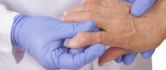
Salt deposition in the knee joint is an excessive accumulation of inorganic compounds of micro- and macroelements, which leads to a decrease in its functional activity. The pathological condition is manifested by moderate pain, crunching, clicking, and inflammatory swelling. It is possible to cleanse the knee joint of salt deposits with the help of pharmacological preparations and folk remedies. Physiotherapeutic procedures, massage, and regular physical therapy exercises also cope well with this.
What is osteophyte
An osteophyte is a bone outgrowth. It appears due to the fact that the surfaces of the joint begin to come into contact with each other during movement. This creates additional stress on the cartilage, and the cartilage tissue begins to increase in volume.
Then such a cartilaginous outgrowth undergoes ossification, that is, the cartilage tissue is replaced by bone. In this way, the body tries to stabilize the joint surfaces and prevent possible injury. Osteophytes can be multiple or single and have different shapes (tubercles, spines, growths).
Cleansing joints with walnuts
How to remove salts from joints using folk remedies? This question interests many. And not everyone knows that this can be done using a walnut. In fact, this fruit has excellent ability to cleanse the body. A medicine based on it can be prepared in two ways.
1. Infusion of leaves. To prepare it, take 1 tbsp. spoon of dry nut leaves, pour 250 ml of boiling water and leave for at least an hour in a warm place under a lid. Drink 80 ml of medicine 3-4 times a day.
2. Alcohol tincture of nut partitions. It is no less effective for removing salts. To prepare the tincture, you need to pour vodka (0.5 l) into 1 glass of partitions and leave for three weeks. Reception is carried out in 1 tbsp. spoon morning and evening for a month.
Causes of salt deposits
Osteophytes begin to form in a joint when excessive pressure is applied to the articular surfaces. This can happen for various reasons: excess weight, consequences of injuries (including microtraumas), inflammation in the joint. Salt deposition in the knee joint is often a consequence of a violation of general metabolic processes:
- Unbalanced diet.
- Diabetes.
- Gout.
- Excessive alcohol consumption.
- Smoking.
- Sedentary lifestyle.
- Degenerative changes in joint tissues in old and senile age.
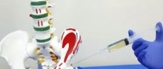
LOCAL GLUCOCORTICOID THERAPY
In 1951, J. Hollander et al. For the first time, hydrocortisone was injected into the knee joint of a patient with RA, which led not only to a decrease in the severity of arthritis, but also to an improvement in the general condition of the patient. After 10 years, J. Hollander et al. (1961), having analyzed the results of more than 100 thousand intra-articular and periarticular injections of GC carried out in 4000 patients, confirmed the anti-inflammatory and analgesic effect of local therapy and the safety of repeated injections of drugs. Recent magnetic resonance imaging studies have shown that in RA, the clinical effectiveness of intra-articular GC is associated with a decrease in synovial volume and signs of inflammation. Currently, the effectiveness of local GC therapy is beyond doubt and depends on a number of factors, which include a correct assessment of indications and contraindications, the correct choice of drug, its dosage, and, finally, manipulation techniques and strict adherence to the rules of asepsis and antisepsis. Intra-articular injections and soft tissue infiltration of HA are used to treat a wide range of musculoskeletal diseases.
| Indications for local GC therapy: • Rheumatoid arthritis (in adults or juveniles). • Microcrystalline arthropathy (gout and pseudogout). • Arthritis with DBST • Acute traumatic arthritis • Osteoarthritis • Synovitis of the knee joint, occurring after arthroplasty of the hip joint on the opposite side • Seronegative spondyloarthropathies • Other diseases occurring with inflammation of the joints • Periarthritis of the shoulder joint (adhesive capsulitis, frozen shoulder) • Tietze syndrome ) • Fibromyalgia |
Characteristics of drugs
The first drug that was used in clinical practice for local therapy and continues to be used to this day is hydrocortisone. However, at present there are GCs, the use of which can significantly increase the effectiveness and safety of local therapy. Table 1. Local administration of GC for PA (Guidelines for the management of rheumatoid arthritis, 1996)
| Target | Treatment of active synovitis at the onset of the disease. Treatment of exacerbations of synovitis in one or more joints. Improving joint function. |
| Restrictions | Impact only on local process. Only temporary improvement. Efficiency mark. Disease progression despite symptomatic relief. Avoid repeated injections into the same joint, more often than once every 3 months. |
GCs used for intra-articular injections are divided into short-acting (hydrocortisone) and long-acting drugs. The latter, in turn, include intermediate-acting drugs (methylprednisolone acetate and triamsinolone acetate) and long-acting ones (betamethasone acetate + betamethasone sodium phosphate and betamethasone propionate + betamethasone sodium phosphate). Hydrocortisone is used mainly to relieve synovitis of small joints of the hands and feet, tenosynovitis and inflammation of periarticular soft tissues, and for inflammation of large joints, long-acting drugs are usually used. The minimum acceptable time interval between administrations of long-acting drugs is 1 month, for betamethasone derivatives - more than 1.5 - 2 months. It is especially necessary to emphasize that since intra-articular injection of prolonged GCs provides a pronounced anti-inflammatory effect for 6 weeks or more, with properly planned treatment tactics this period coincides with the development of the anti-inflammatory effect of a number of basic antirheumatic drugs. Thus, the need for repeated HA injections should be decided individually. Unreasonably frequent administration can lead to the development of GC dependence, the progression of secondary osteoarthritis, and even reduce the patient’s functional ability. Table 2. Controlled studies of the effectiveness of GCs in OA
| Authors | A drug | Number of patients | Nature of the study | Duration (weeks) | Efficiency |
| Friedman, 1980 | TG 20 mg vs placebo | 34 | parallel | 8 | TG placebo for 1 week only |
| Dieppe, 1980 | TG 20 mg vs placebo | 12+16 | parallel/cross | 6+2 | Placebo TG only for 2 weeks |
| Valtonen, 1981 | TG 20 mg vs betamethasone 6 mg | 42 | parallel | 24 | TG betamethasone |
| Gaffney, 1995 | TG 20 mg vs placebo | 84 | Same | 6 | TG placebo for 1 week only |
| Jones, 1995 | MP vs placebo | 59 | cross | 8 | MP placebo for 3 weeks only |
It must be borne in mind that the effectiveness of GCs in different patients may vary significantly. If after 1 - 2 injections there is no clinical improvement or is minimally expressed, further local GC therapy is inappropriate in most cases. To increase the effectiveness of local therapy, it is necessary to provide rest to the joints into which GCs were injected for a period of at least 48–72 hours. This is best achieved with the help of so-called “working” orthoses, which allow movement in the joint. Some authors recommend that patients use crutches or a stick for the next three weeks after injection of GC into load-bearing joints. Of particular importance is the drug diprospan, which consists of two salts of betamethasone. The first component, the sodium phosphate salt of betamethasone, has high solubility, is quickly hydrolyzed and quickly absorbed, which ensures a rapid onset of effect (within several hours). The other component (betamethasone acetate or betamethasone propionate, on the contrary, is characterized by poor solubility, slow hydrolysis and absorption. The combination of these substances provides, on the one hand, a rapid, on the other hand, a very long-term effect of the drug (up to 30 - 35 days), and the total duration the effect can reach 6 weeks. This distinguishes diprospan from other long-acting drugs (kenalog or depomedrol), the duration of the effect is about 3 weeks. Another important feature of diprospan should also be mentioned - the low concentration and small size of the crystals. Thus, the concentration of crystals in 1 ml of kenalog is 40.0; and in 1 ml of diprospan - 6.4, and their approximate value is 12.0 microns and 2 - 6 microns, respectively. These unique properties expand the indications for the use of diprospan in rheumatology. In addition, this particular drug can be used to relieve the attack of microcrystalline arthritis.
Indications for local therapy of RA
The ability of GCs, when administered intra-articularly, to suppress the development of synovitis in RA has been confirmed in many studies. In PA, the competent use of injection therapy can improve the quality of life of patients, maintain the functional activity of joints, prevent their deformation and is an important component of the treatment and rehabilitation program for patients with this disease. The introduction of slow-acting drugs makes it possible to suppress rheumatoid inflammation for three or more months. It is especially advisable to use injection therapy in the early stages of the disease, in the presence of contraindications to therapy with “basic” antirheumatic drugs and NSAIDs. Data regarding the effect of intra-articular injections on the progression of joint destruction are contradictory. According to J. Hollander (1969), intra-articular steroids have a more symptomatic than “basic” effect. When analyzing the results of a 17-year follow-up of patients who received GCs into the knee joints, signs of radiographic progression occurred in more than half of the cases. It is noteworthy that similar results were obtained in patients with coxarthrosis. At the same time, according to D. McCarty (1972), intra-articular administration of HA (triamsinolone hexacetonide) in combination with splinting the joints for three weeks (the splint was removed once a day during physical therapy) allows to slow down the radiographic progression in the joints of the hands and wrist joints. Table 3. Soft tissue diseases requiring local GC therapy
| Shoulder | Biceps tendonitis Subacromial bursitis Infraspinatus tendonitis Periarthritis (adhesive capsulitis, frozen shoulder) |
| Elbow | Lateral epicondylitis (tennis elbow) Medial epicondylitis (golfer's elbow) Elbow bursitis Cubital tunnel syndrome |
| Wrist and hand | Gangliitis De Courvain's disease (stenosing tenosynovitis of the abductor pollicis brevis and abductor pollicis longus tendons) Snapping fingers (stenosing ligamentitis) Carpal tunnel syndrome |
| Hip area | Trochanteric bursitis Bursitis in the m. ileopsoas |
| Knee area | Bursitis in the crow's foot area. Prepatellar bursitis |
| Pelvis | Sciatic bursitis Iliopsoas bursitis Maralgia paraestetica |
| Back, torso | Trigger points for fibromyalgia? Stockmann's nodes (hernial presacral fat pads) Copemann's nodes (fibrotic nodules) |
| Foot | Achilles tendinitis Achilles bursitis Calcaneal bursitis Morton's neuroma Tarsal tunnel syndrome |
The main indications and contraindications for intra-articular administration of GCs in RA (Table 1) (and to a certain extent for other diseases) are the following.
Indications:
• Mono-, oligoarthritis of moderate or high local activity • Predominant damage to one or two joints in polyarthritis • In RA at the beginning of basic therapy with high local activity of 1 - 2 joints • In the presence of contraindications for basic therapy, as a temporary palliative method • Prevention of deformities , as a component of a rehabilitation program
Contraindications:
• Local or systemic infection • Severe bone destruction or uncorrectable static deformation of the joint (increasing the risk of joint infection or aggravation of destruction) • Severe periarticular osteoporosis • Difficult access to the joint • Transarticular fracture • Blood coagulation pathology • Ineffectiveness of the previous intra-articular therapy (taking into account the timing of development and duration of action of various drugs).
Microcrystalline arthropathy
In microcrystalline arthropathy, intra-articular injection of HA is rarely used. Indications for this procedure may include resistance to NSAIDs and colchicine, as well as the presence of contraindications for the use of these drugs. The drugs of choice are betamethasone derivatives (diprospan), which, due to their properties, do not cause exacerbation of arthritis.
DBST (SLE and mixed connective tissue disease, etc.)
Indications for local therapy of GC are such variants of the course of diseases in which the clinical picture is dominated by the phenomena of arthritis, the severity of which does not correspond to the general activity of the pathological process.
Traumatic joint damage
The main method of treating traumatic joint injuries is the use of cold, immobilization with a gradual expansion of motor activity. However, prolonged immobilization (especially of the shoulder and ankle joints) can lead to dysfunction. Intra-articular administration of HA allows to shorten the period of immobilization and thereby prevent dysfunction of the joints.
Osteoarthritis
Intra-articular administration of GC for osteoarthritis is widely used in clinical practice. According to a recent US survey, 95% of rheumatologists have ever used intra-articular HA for OA, and 53% use this procedure frequently. However, the results regarding the use of GCs in OA are contradictory. This is primarily due to the heterogeneity of OA and the different role of the inflammatory component in the development and progression of OA in different patients and in different phases of the disease. According to dynamic X-ray and scintigraphic studies, the disease is characterized by an undulating course; in some patients, stabilization or even spontaneous positive dynamics of the articular process, in particular pain syndrome, is observed. Treatment effectiveness
depends partly on the type of joint affected.
For example, with damage to the metatarsophalangeal joint of the first toe, in which there is virtually no thickening of synovial tissue and synovial effusion, but there is a significant narrowing of the radiographic “joint space,” the administration of GC is painful. However, after a few hours there is usually a significant reduction in pain, and the effect can last for many months. On the contrary, in case of coxathrosis, intra-articular administration of GC is very rarely effective. With proper administration of the drug into the joint cavity, a decrease in pain is observed after 12 - 14 hours, but the effect is usually short-lived (2 - 7 days), which is associated with mechanical overload of the joint. Repeated injection of HA into the hip joint is not advisable. The results of placebo-controlled studies of intra-articular GC for OA are summarized in Table 2. As can be seen from Table 2, in most cases the effectiveness of GC in reducing pain was greater than placebo, but the duration of the analgesic effect was relatively short (no more than three weeks). of predicting
is widely discussed .
There is evidence that the presence of joint effusion is associated with higher GC effectiveness. However, other authors have not identified a definite relationship between the effectiveness of GC and the clinical features of OA, including the presence of crystals or the content of leukocytes in the synovial fluid. If a single administration of GC led to only a short-term effect, repeated administration of the drug requires special indications. This is due to the fact that the cause of increased pain may not be an exacerbation of synovitis, but cartilage damage, which can progress with repeated administration of GC. Thus, the lack of effect with a single (maximum two-time) administration of GC is a contraindication for further intra-articular injections of the drug. Some results indicate the high effectiveness of HA administration for OA of the thumb. The mechanisms
that determine the effectiveness of GCs in OA are not completely clear. The most likely factor is the presence of an inflammatory component in OA. When studying synovial tissue, synovial hyperplasia, infiltration with mononuclear cells, indistinguishable from that in RA, overexpression of oncoproteins and NF-kB, migration of leukocytes into the joint cavity in vivo, and an increase in the concentration of CRP, which correlates with the progression of the disease, are revealed. In addition, as with RA, with OA there is an increase in the serum concentration of hyaluronic acid, the synthesis of which in vivo by synovial cells is inhibited by HA. In experimental OA, the severity of inflammation correlates with the rate of cartilage loss, and the results of clinical studies indicate that the presence of inflammation in the knee joint is associated with a more unfavorable clinical outcome and radiographic progression of OA. Early studies showed that administration of GC to patients with OA leads to a decrease in synovial permeability, as measured by the clearance of 99Tc-labeled albumin. It is also believed that the synthesis of proinflammatory cytokines (for example, IL-1) plays an important role in the development of pain in OA. It is known that IL-1 induces the release of proteolytic enzymes (collagenase, stromelysin), prostaglandins and plasminogen activator, which are involved in the development of pain. Mediators such as bradykinin and histamine have the ability to directly stimulate primary afferent nociceptor fibers, and prostaglandins, leukotrienes, IL-1 and IL-6 increase the sensitivity of nociceptors to mechanical and other painful stimuli. Thus, GCs, by suppressing the synthesis of these mediators, can have a direct analgesic effect. Data from experimental studies indicate that GCs can have a certain modifying effect on the course of OA, preventing the formation of osteophytes, cartilage destruction and damage to chondrocytes. According to J. Pellitier et al. (1995), GCs cause a dose-dependent decrease in the level of stromelysin in cartilage, accompanied by a decrease in the synthesis of IL-1 and the expression of proto-oncogenes (c-fos, c-myc) in experimental OA. Similar data were obtained in patients with OA. However, other studies have shown that GCs do not always have a chondroprotective effect and in some cases their administration may lead to increased loss of cartilage proteoglycan.
Synovitis of the knee joints after hip arthroplasty
Sometimes, after hip arthroplasty, synovitis of the knee joint develops (usually transient), usually on the opposite side. Persistence of synovitis is an indication for local administration of GC and, as a rule, is highly effective.
Seronegative spondyloarthropathy
Lesions of peripheral joints due to seronegative arthropathy (ankylosing spondylitis, psoriatic arthritis, Reiter's disease, arthritis due to inflammatory bowel diseases, etc.) may be an indication for intra-articular injections of GC. However, when carrying out local therapy, it is necessary to keep in mind the possibility of concomitant pathology. For example, the development of Reiter's syndrome in patients with AIDS is a contraindication for intra-articular administration of GC.
Periarthritis of the shoulder joint (adhesive capsulitis, frozen shoulder)
In the treatment of this pathology, the administration of GC intra-articularly or into the synovial tendon sheath (in combination with NSAIDs and physiotherapy) gives a very good clinical effect.
Tietze syndrome
The administration of GC and local anesthetics is the main method of treating Tietze syndrome, which is manifested by pain and swelling of the parasternal areas - the sternocostal joints.
Soft tissue damage
Local administration of GC (sometimes in combination with local anesthetics) is among the most effective methods for treating inflammatory diseases of soft tissues (Table 3).
General recommendations
When performing arthrocentesis or soft tissue infiltration, strict adherence to aseptic
.
The doctor prepares for the arthrocentesis procedure in the same way as for a spinal tap. If the rules of asepsis are observed, iatrogenic infection is observed very rarely, in 1 - 2 cases per 60,000 manipulations. However, in Russia the frequency of septic arthritis for 1988 - 1992. increased approximately 12 times compared to the previous five years, which is associated with the uncontrolled introduction of GC. It is believed that when GC is administered into a joint in patients with intercurrent infection, the prerequisites are created for the formation of foci of infection outside the joint, for example, the occurrence of septic endocarditis. It must be borne in mind that patients with chronic inflammatory rheumatic diseases are especially predisposed to the development of septic hematogenous arthritis. When administering GC, the risk of soft tissue atrophy, necrosis and atrophy of nerve endings should be taken into account. Patients are described in whom needle fragments remained in the soft tissues after intra-articular manipulation. Table 4. Amount of HA preparation for intra-articular injection into various joints
| Knuckle size | Example | Amount of drug |
| Large | Knee/Ankle/Shoulder | 1 - 2 ml |
| Average | Elbow/Wrist | 0.5 - 1 ml |
| Small | Interphalangeal/Metatarsophalangeal | 0.1 - 0.5 ml |
Although the question is how much of the drug
can be injected into joints of various sizes continues to be debated in the literature, there are some general recommendations regarding the relationship between joint size and the amount of drug administered (Table 4).
It is recommended to inject a mixture of HA and an anesthetic
into small joints and tendon sheaths .
This allows you to prevent large amounts of the drug from entering a small volume of inflamed tissue and atrophy of soft tissues. According to some rheumatologists, it is advisable to first inject HA, and then refill the syringe and inject the anesthetic. This helps prevent the appearance of pericapsular calcifications of small joints. In any case, it is necessary to consider the possibility of joint use of drugs. HA should not be mixed with anesthetics containing preservatives. In addition, injection of a mixture of HA and anesthetic into the bursa, tendon sheath, or periarticular tissue usually produces a rapid effect, which is associated precisely with the action of the anesthetic, and not with HA. In this case, patients should be informed about the possibility of a short-term resumption of pain. Finally, the administration of HA and anesthetic reduces the risk of soft tissue atrophy at the injection site. To reduce pain during arthrocentesis, it is also recommended to spray the skin at the site of the intended injection with a local anesthetic (chlorethyl). In this sense, the advantage of betamethasone
(diprospan), which contains a water-soluble steroid component, is clearly manifested. The form of the drug allows you to avoid the use of an anesthetic, since the rapidly developing anti-inflammatory effect eliminates possible manifestations of microcrystalline synovitis. Before treating the joint, the site of arthrocentesis must be marked. This is usually done by pressing on the skin at the desired point with a ballpoint pen that has run out of coloring paste, after which an indentation is left on the skin that lasts for 5 to 15 minutes. This technique is also used for infiltration of soft tissues. The arthrocentesis area must be thoroughly cleaned with an antiseptic (iodine-containing solution); rubber gloves must be used, which will reduce the risk of contracting certain infections (hepatitis B and C, HIV). The set of instruments and material for arthrocentesis includes: ethyl alcohol, antiseptic solution (iodine, iodopirone, etc.), sterile swabs, gauze pads, sterile syringes 3, 10 and 20 ml, needles, sterile glass and plastic tubes, heparinized tubes, clean microscope slides and cover glasses, heparin solution for adding to test tubes if necessary, marker, plates with medium for inoculating material, including for anaerobes, kits for determining glucose concentration using the express method, dressing material, ethyl chloride spray, hemostatic, anesthetic - 1% lidocaine. Puncture of joints (usually large ones) is carried out not only for therapeutic, but also for diagnostic purposes. Therefore, the doctor should have test tubes at his disposal for collecting synovial fluid for sterility and other laboratory tests.
Symptoms
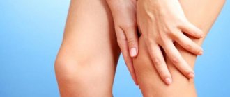
The deposition of salts in the knee joint does not manifest itself in any way at first. As a rule, patients consult a doctor when the pain becomes intense and significantly worsens the quality of life. However, if you pay close attention to yourself, you can notice problems much earlier.
The first sign of the appearance of osteophytes is an unusual crunching sound in the knee joint when moving. The crunch is not accompanied by pain, and a person often ignores this symptom. Pain appears later, first after intense exercise: long walking or working with emphasis on the knees (this happens during repairs or work in the garden).
If you do not start treatment on time, the pain will bother you even with light loads
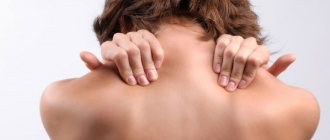
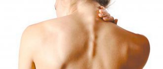
As the osteophyte grows, injury to surrounding tissues occurs and an inflammatory process develops: the knee swells, hurts, and the skin over it becomes hot. When the osteophyte begins to compress the nerve endings in the joint area, the pain becomes longer lasting, and sensitivity disturbances may occur - a feeling of numbness or tingling appears in the knee area.
Pain in the knee joint when walking
Thus, the most common symptoms of salt deposits include the following:
- Crunching in the joint when moving.
- Pain after physical activity, and then at rest, especially at night.
- Joint swelling.
- Violation of the knee configuration.
- Aching pain when weather conditions change (especially cold and humid weather).
- Limitation of range of motion in the knee joint.
How to remove salts from joints
Treatment of this pathological condition is very long and complex. First of all, doctors recommend changing your usual diet. Fatty, spicy, fried foods are excluded from it. As well as foods that provoke excessive formation of urates (meat, fish, spinach, grapes, sorrel). More dairy and plant products are being introduced into the menu. Eating plums, melons, watermelons, persimmons, parsley and dill has a particularly good effect on the body.
It is worth giving up bad habits and playing sports. The level of physical activity is determined individually, but a sedentary lifestyle is under no circumstances allowed.
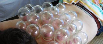
Saline dressings
Similar procedures have been practiced in folk medicine for quite some time. Bandages are used to treat:
- arthrosis;
- arthritis;
- periarthritis;
- bursitis;
- radiculitis;
- as well as from spinal pain.
For the procedure you will need a hypertonic salt solution with a concentration of 8-10%. You should not make it more saturated, otherwise you will not avoid harmful effects on the body. Fabric (cotton, linen) is also needed. It should be loose and hygroscopic, for example, a waffle towel works well.
- You need to mix 100 g of salt (not iodized) in 1 liter of water.
- Fabric folded in several layers is immersed in this solution.
- It is left there for 2-3 hours.
- Then the solution along with the fabric is heated to 50°.
- The flap is removed and easily wrung out.
- It must be applied to the joint immediately.
- A bandage or another piece of cotton fabric is placed on top.
Salt acts locally, effectively drawing out toxins and pathogenic microorganisms from the subcutaneous layers and tissue fluid. It cleanses the sore area, renewing the deep and subcutaneous layers of the lesion, so the bandage must be applied exactly to the sore spot.
May be interesting: Diclofenac - injections: price, reviews, use, dosage
The course of procedures is 12-14 sessions without breaks. It is better to perform them at night. The peculiarity of this compress is that you cannot use paper and polyethylene, since the bandage must “breathe”.
Treatment of joint inflammation with snow and salt
- Take ½ cup of snow out of the freezer.
- Add 1 tsp to it. salt.
- The ingredients are thoroughly mixed and placed on the sore spot.
- This kind of compress with snow is kept for no more than 4 minutes.
- To relieve pain, as a rule, 7-10 procedures are sufficient. These measures are great for relieving signs of inflammation and soreness.
Honey and salt for sore joints
- You need to mix 1 tbsp. l. honey and regular rock salt. It is worth noting that it is better to take natural honey.
- The ingredients are thoroughly mixed and applied to a clean cloth, preferably linen.
- Then it is applied to the inflamed area.
- Compress paper or cellophane must be placed on top.
- After this, a piece of woolen cloth is applied and fixed so that it holds well and warms the sore spot.
- It is recommended to perform this procedure at night.
- In this case, the bandage is not wrinkled, but kept until the morning.
- After waking up, the limb is simply washed with running warm water.
Diagnostics
Symptoms of salt deposits are very similar to those of other musculoskeletal diseases. Therefore, if you experience any discomfort in the joint area, you should first consult a doctor to find out the correct diagnosis, and not self-medicate. The doctor will examine the patient, find out the details of the course of the disease and prescribe the necessary examinations.
To identify joint pathology, the following is prescribed:
- X-ray image in two projections.
- Magnetic resonance imaging. This method allows you to most accurately assess the condition of not only the bone apparatus, but also soft tissues, nerves and blood vessels, as well as determine the presence of tumor formations.
- Ultrasound diagnostics. Its advantage is the absence of radiation exposure to the body with a fairly accurate diagnosis of the condition of the anatomical structures of the joint.
Calcium gout
Inflammation of the joints can occur as a result of the deposition of calcium pyrophosphates. In this case, they talk about such a phenomenon as calcium or false gout. This disease is accompanied by arthritis in the ankles, hips or arms.
The main symptoms of the disease include spontaneously occurring pain (quite severe) and swelling that disappears in the same way.
The causes of false gout lie in the following provoking factors: old age, the presence of hypothyroidism, hemophilia, manifestations of amyloidosis, heredity.
Correct diagnosis requires laboratory testing of joint fluid. The disease is confirmed when calcium poriphosphate is detected in the sample.
Treatment
The deposition of salts in the joint is a consequence of impaired metabolic processes, so treatment should be comprehensive and consist of the following measures:
- Balanced diet.
- Therapeutic exercise and massage.
- Physiotherapeutic procedures.
- Medicines.
- Traditional medicine methods.
The earlier treatment is started, the more effective it is. In the later stages of the disease, conservative treatment methods do not help, and sometimes it is necessary to resort to joint replacement (endoprosthetics).
Diet for salt deposits
Without establishing proper nutrition, we cannot talk about healthy joints. Principles of diet composition:
- Calorie calculation. It is necessary to limit the excess intake of calories through fast carbohydrates and, partly, animal fats. This approach will help normalize weight and reduce stress on your knees.
- Maintaining water balance. Pure water, green tea and herbal infusions should be about 2 liters per day, mainly in the first half of the day. Water is a catalyst for a huge number of biochemical reactions and helps remove toxins and waste from the body. If a person has diseased kidneys, then you should first consult a doctor.
- Eliminating or minimizing confectionery products, baked goods, pickles, marinades, smoked and fried foods.
- Introduction to the diet of fresh vegetables, fruits, whole grains, fish, jelly.
- Additional intake of biologically active substances.
Recommended Products:
- Raw vegetables and fruits (broccoli, apples, citrus fruits, zucchini, beets, carrots).
- Whole grain porridge (buckwheat, rolled oats, brown rice, millet).
- Foods rich in Omega 3 polyunsaturated fatty acids (fish, flaxseed oil).
- Skim cheese.
- Jellied meat, jelly, berry and milk jelly. Contains natural gelatin, beneficial for cartilage.
- Chicken and quail egg.
Fresh vegetables, foods rich in calcium and Omega 3 should form the basis of the daily diet
Drinking regime
In case of purine metabolism disorders, you need to receive at least three liters of fluid per day, as this speeds up the release of urate deposits from the joints. In addition to clean water, it is recommended to drink alkaline mineral waters, vegetable and fruit juices, and infusions of medicinal plants.
Herbalists recommend using rose hips, mint, bearberry, and birch buds. Dried fruit compote is very useful. It is better to postpone taking any liquid for 30-45 minutes after a meal. This will improve the digestion process.
The exception is alkaline mineral water, which should be drunk warm before meals or regardless of meals.