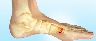
© Dotan — stock.adobe.com
Share:
The supporting functions and mobility of the ankle joint are provided by the distal epiphyses (ends) of the fibula and tibia. This joint bears shock loads when walking, running, jumping, as well as jerking lateral and twisting moments of force when balancing to keep the body in an upright position. Therefore, an ankle fracture is one of the most common injuries of the musculoskeletal system not only among athletes, but also among ordinary people who do not engage in sports (from 15 to 20% of the total).
Causes
Traumatic ankle fractures occur from a strong blow or other excessive external force on the ankle during sports, falls, or traffic accidents. Twisting your foot on a slippery, uneven surface or wearing ill-fitting shoes often leads to this type of injury. Unsuccessful falls can be caused by underdeveloped muscles and poor coordination of movements, especially if you are overweight. Due to disruptions in the normal process of bone tissue restoration, adolescents, pregnant women and older people are at risk.
Congenital or acquired degenerative changes, as well as various diseases: arthritis, osteopathy, osteoporosis, tuberculosis, oncology increase the likelihood of injury. An unbalanced diet, lack of calcium and other microelements reduce the strength of bones and the elasticity of ligaments.
What is the danger
With timely and qualified treatment, even complex fractures, as a rule, heal without complications and the functionality of the ankle is fully restored. In cases of severe displacement or crushing of bones, serious complications and only partial rehabilitation of the functionality of the joint are possible.
In the event of a delayed visit to a medical facility or improper provision of first aid, serious consequences can occur, including disability.
Open fractures and displaced fractures are especially dangerous, when bone fragments can damage surrounding tissues and nerve endings, which can lead to loss of sensation and disruption of the muscles of the foot. Therefore, it is important at the first moment to ensure immobilization of the limb, not to allow any load on the injured leg, and to deliver the patient to the emergency room as quickly as possible.
Sometimes a closed fracture is only bothered by swelling of the joint, minor pain, and the ability to walk remains. Despite this, even in such cases it is necessary to consult a doctor to establish an accurate diagnosis and correct treatment.
Fracture of the outer malleolus
This is the destruction of the lower end of the fibula. The ICD-10 (International Classification of Diseases) code is S82.6. This injury is characterized by mild symptoms - swelling of the ankle joint, sharp pain at the time of injury and tolerable pain even when supporting the leg, since the main load falls on the tibia. This often provokes delays in contacting a traumatologist, which can cause improper bone healing and destruction of ligaments, muscles and nerve fibers. As a result, an easily curable fracture of the lateral malleolus can turn into a serious pathology.
Inner ankle fracture
This is the destruction of the lower end of the fibula (according to ICD-10 - S82.5.). In such cases, oblique or straight (pronation) fractures of the medial ankle occur, which are often complicated by sprains and may be accompanied by acute pain, loss of supporting function of the leg, severe swelling and bruising in the joint area.
Displaced fracture
These are the most dangerous and complex cases of ankle injury, which have pronounced symptoms: sharp unbearable pain, severe swelling, extensive local hemorrhage and a characteristic crunch when the muscles of the leg are tense or the foot moves. Sometimes a bone fragment destroys the surrounding tissue and comes out, causing bleeding and the risk of infection in the wound. This often occurs with an apical fracture (a fracture of the tibia or fibula near the distal epiphysis). In the most severe cases, both ankles are damaged with dislocation and rupture of ligaments.
Fracture without displacement
Such injuries are characterized by destruction of the distal part of the lower leg without acute pain and severe swelling. Only slight discomfort is felt when bending the foot and walking.
A non-displaced ankle fracture can be confused with a sprain, so it is best to confirm the diagnosis with a medical professional.
Strengthening exercises
Once a person's ankle cast is removed, they can begin to perform exercises. Exercise therapy after an ankle fracture includes the following exercises:
- Swing your injured leg to the side. You will need to lean on a chair or any other reliable object and begin swinging your straightened leg. Perform 15–20 repetitions. Swings improve blood supply to the ankle.
- Lying on the bed, you need to stretch the toe of the injured leg. The number of repetitions is from 30. The exercise allows you to put a load on the ligamentous apparatus of the foot and develop atrophied muscles.
- Rotation of feet in the air while sitting. You need to sit on a chair or bed, raise your legs and begin to rotate your feet inward and outward. Rotation is carried out for 1 minute. Every day the rotation time can be increased.
- Seated calf raise. It is necessary to take a stable sitting position with your legs parallel to each other. After this, you need to lift both feet up onto your toes, then return to the starting position. Repeat the lifts 25–40 times. This action strengthens the calf muscle.
Exercises allow you to put stress on joints, ligaments and muscles, and also speed up rehabilitation.
Diagnostics
The exact location and extent of damage is determined by X-ray examination. Several photographs are always taken in different planes (from two or more, depending on the complexity of the injury). To assess the condition of soft tissues and ligaments, as well as to exclude the presence of internal hematomas, magnetic resonance or computed tomography is prescribed.
© richard_pinder — stock.adobe.com
Features of treatment
The main way to restore bone integrity is complete immobilization of the ankle joint. Depending on the type of injury, the correct position of the fragments is ensured by closed or open reduction. After surgery, the necessary procedures are carried out to heal the wound.
Conservative treatment
Such methods are used in cases of closed fractures without displacement or if it can be eliminated by closed reduction and the ligamentous apparatus has minor damage. In addition to immobilization, medications are used to relieve pain, swelling and eliminate inflammatory processes.
The patient's unsatisfactory health condition may be the reason for refusing surgery and using conservative treatment.
Using an immobilizing bandage
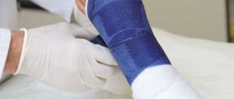
For a simple fracture without displacement or rupture of the ligaments, after the diagnosis has been established and the swelling has been eliminated, an immobilizing U-shaped or long-circular bandage made of plaster, synthetic bandage or low-temperature plastic is applied. Covering part of the foot and the lower part of the leg, it should provide clear fixation of the joint and not interfere with normal blood circulation in the limb. If such immobilization is carried out after closed reposition, a control x-ray must be taken to ensure the correct position of the fragments.
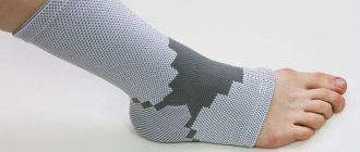
In addition to bandages, various types of plastic and combined bandages and orthoses are used. Such devices are easily adjusted to the size of the limb. With your doctor's permission, you can take them off and put them on yourself.
Depending on the complexity of the fracture, any load on the immobilized limb is excluded for a certain period of time. The length of time you wear a fixation device or bandage also depends on this (from 4-6 weeks to two months or more).
© stephm2506 — stock.adobe.com
Closed manual reduction
This procedure is performed under local anesthesia. The surgeon touches and aligns the displaced bones and ensures their correct anatomical position in the joint and lower leg.
The time and quality of restoration of limb functionality largely depends on the timeliness and accuracy of its implementation.
Surgical treatment
The surgical operation is necessary:
- With an open fracture.
- When the injury is complicated by a complete rupture of the ligaments or there are many fragments.
- With a two- or three-malleolar fracture.
In these cases, under general anesthesia, the joint is opened and the bones and fragments are repositioned openly, as well as their fixation using special medical nails, screws and pins (osteosynthesis). In this case, damaged tendons, ligaments and nerve endings are restored. Then a plaster cast is applied, which does not cover the surgical site and allows for treatment and control of the wound healing process.
Symptoms
Because a sprain can sometimes feel similar to a broken ankle, it is always important to consult a doctor. Assessing the symptoms of an ankle fracture and post-injury pain help in determining how serious the injury is.
An ankle fracture is accompanied by one or all of the following symptoms:
- pain at the fracture site, which in some cases can spread from the foot to the knee;
- significant swelling, which may occur along the length of the leg or be more localized. How long the swelling lasts will depend on the severity of the injury and the volume of damaged tissue;
- a bruise that appears soon after the injury;
- inability to walk while stepping on the entire leg. Therefore, your doctor will tell you how to walk correctly after an injury. However, a person can walk with less severe fractures, so never rely on walking as a test of whether a bone has been broken;
- the appearance of the ankle has been changed - it will be different from the other ankle;
- the bone protrudes from the skin in an open fracture. Fractures that penetrate the skin require immediate attention as they can lead to severe infection and a long recovery time.
Possible complications
If you visit a doctor late, self-medicate, or violate the rules and timing of wearing a fixation device, the bones and their fragments can grow together in an unnatural position, which will interfere with the normal functioning of the joint and provoke dislocations and the development of flat feet.
An improperly formed callus can impinge on nerve fibers and impede or block the innervation of the adductor muscles of the foot and the sensitivity of the skin. Untimely treatment of a postoperative wound can cause the development of an inflammatory process or infectious disease of muscle tissue, bones and blood vessels.
Anatomy of the ankle joint
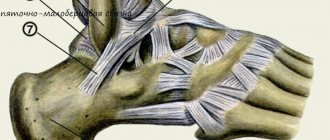
The ankle connects the bones of the foot to the bones of the lower leg. The ankles are the distal portions of the tibia and fibula.
The structure of the ankle is quite complex: it is formed by the talus and tibia bones, which are connected by cartilage and muscle tissue. Each joint has an extensive network of blood vessels and nerves, which provide tissue trophism and coordinated movement of the limb.
In the event of an injury, all this is disrupted and, if not treated correctly, is never fully restored.
How long to walk in a cast if you have a broken ankle
In any case, the plaster cast or other fixation device is removed only after a control x-ray, which confirms the complete and correct fusion of bones and fragments, as well as the normal condition of the ligaments and tendons.
Wearing time
First of all, the timing of wearing the fixation device depends on:
- Timeliness and correctness of first aid.
- Type and complexity of the fracture.
- Individual characteristics of the patient’s body.
A balanced diet and compliance with the recommendations of your doctor will help speed up recovery.
With offset
In this case, the determining factor is the correct preliminary fixation of the joint during first aid and the rapid delivery of the victim to the emergency room. Otherwise, the displacement may become difficult to correct using closed reduction and surgical intervention will be required.
No offset
In most cases, with such fractures, immobilization lasts from one to two months. The time for complete recovery depends on the intensity of rehabilitation measures and the individual characteristics of the patient.
If the outer part is damaged
Such fractures are treated with surgery, so wearing a fixing bandage will take two months or more. As after any surgical operation, in this case the recovery period is also determined by the speed of healing of the postoperative wound.
For a fracture of the lateral malleolus without displacement
This is the mildest case of destruction of the integrity of the ankle, and fixation of the joint is required for a period of one to one and a half months. Within a week, gradual normalized load on the leg is allowed.
Massage
Light massage after removing the bandage can improve blood circulation, relieve swelling and get rid of muscle atrophy. To obtain a noticeable result, massage must be performed by a qualified specialist. The course of treatment must include at least 15 sessions. And also after studying the basic rules, it becomes possible to perform self-massage.
To get the most benefits from self-massage, you need to avoid pressing on the injured area and excessive force when rubbing.
The duration of walking in a cast depends on the complexity of the bone fracture, the person’s condition, age and concomitant diseases. In case of improper fusion and other complications, the plaster will need to be worn longer.
Stages of fusion
At the moment of fracture, local hemorrhage occurs, and for the first five or seven days there is an inflammatory process with the formation of a soft compaction of fibrous tissue (resorption). Then the creation of collagen connecting threads (reversion) from special cells - osteoclasts and osteoblasts - begins. After this, as a result of cell mineralization, a callus is formed between the fragments within a month. Over the next three to four weeks, the formed structure ossifies, due to its saturation with calcium.
Complete restoration of the damaged bone and its surroundings, which ensures the full functioning of the ankle joint, is possible after 4-6 months of rehabilitation.
