- home
- Treatment of cancer (oncology)
- Cancer metastases
- Spinal metastases
—
—
—
Spinal metastases are the most common type of secondary cancer. Most often, the primary focus is located in organs close to the spinal column (lungs, kidneys, prostate, stomach, mammary glands, etc.), from where cancer cells are transported through blood or lymphatic vessels.
Hospitalization of cancer patients. Daily. Around the clock
9,500 patients trust us annually.
Call me back!
There are osteoblastic and osteoclastic metastatic formations - in the first, the shape of the vertebrae and, accordingly, the body changes; in the second, their height decreases. The disease is also classified according to which part is affected - cervical, thoracic or lumbar, since each location has its own characteristics of the course.
With metastases in the spine, the prognosis is unfavorable, since the appearance of secondary tumors indicates that the disease is in advanced stages. However, with well-organized treatment and palliative care, it is possible to prolong the patient’s life and alleviate his condition.
Metastases in the spine: symptoms and diagnosis
In addition to severe fatigue, weakness, low-grade fever, loss of appetite and sudden weight loss characteristic of cancer in the later stages, the following signs are observed:
- severe pain syndrome;
- numbness, pain, tingling in the limbs;
- impaired motor activity, paralysis and loss of sensitivity (if the spinal cord is affected);
- uncontrolled bowel movements and urination.
Depending on the location of the lesions, the symptoms may differ - for example, only one arm or half of the body suffers, the pain is most intense in some parts, etc.
Although metastasis indicates an advanced stage of the disease, it is recommended that if this process is suspected, you immediately contact the oncology center to diagnose the disease, determine the number, size and location of tumors and begin treatment. To diagnose metastases, as well as primary spinal cancer, the following are used:
- examination by an oncologist and neurologist to check reflexes, muscle tone, preservation of the functions of the pelvic organs;
- blood and urine tests, including tumor markers;
- cerebrospinal fluid puncture and histological studies;
- radiography and ultrasound;
- magnetic resonance and computed tomography (MRI and CT);
- scintigraphy.
Our expert in this field:
Ivanov Anton Alexandrovich
Medical director, oncologist-surgeon, candidate of medical sciences
Call the doctor
Call the doctor
2. Symptoms of metastatic lesions of the spine
The main clinical sign of spinal cancer is back pain. Unlike pain associated with a hernia or spinal injury, with malignant formations the pain is very persistent and long-lasting
. Its intensity does not change in different body positions. A night's rest does not bring relief. Painful sensations increase over time and turn existence into a very painful one.
The specificity of pain allows us to assume metastases in the spine
- specific location of the diseased area
- increased pain when lying down
- increase in pain during straining, coughing, sneezing
- no connection between pain and physical activity
- a clear tendency towards increasing pain intensity
- weak effect of analgesics
- forced position of the patient's body
- neurological complications (paresis, paralysis)
- pelvic dysfunction
Visit our Neurosurgery page
Book your room today
6 seats
1 local ward
- 4 meals a day
- Bathroom in the room
- Anti-decubitus mattresses
8200 rub.
Book
13 places
2 local ward
- 4 meals a day
- Bathroom in the room
- Anti-decubitus mattresses
5700 rub.
Book
2 seats
VIP chamber
- a guest room
- 4 meals a day
- Bathroom in the room
- Anti-decubitus mattresses
17700 rub.
Book
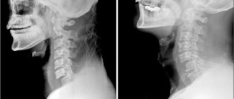
MRI of the spine
This is one of the modern methods for diagnosing metastases and is the safest and most harmless. After which a conclusion is made, and the attending physician can prescribe a further course of treatment based on the results. An MRI of the spine can determine the extent of damage or compression of the spinal cord.
The specialists of our clinic will carry out an accurate diagnosis, and subsequent treatment, if necessary, will take place exclusively under the supervision of doctors. Treatment can take place in several stages, and at each of them one or another method or a combination of several will be used. The qualifications of our clinic’s doctors allow us to effectively eliminate cancer cells and prevent their possible spread throughout the body. You can always get a detailed consultation from the right clinic specialist by making an appointment at a convenient time. Be attentive to yourself and promptly pay attention to the slightest symptoms, identification of which at an early stage saves lives.
Reviews
30.09.2021
Feedback on the treatment of ovarian cancer at the NACFF clinic
07.09.2021
NACFF patient about treatment, quick examinations and attentive attitude
03.09.2021
I have healed again, I have been born again
10.08.2021
Treatment of multiple fractures and traumatic brain injury
View all reviews
Hospital, hospitalization, emergency medical care - 24/7, 7 days a week
+7 (495) 259-44-44
3. Diagnosis of malignant tumors in the spine
Most often, metastases in the spinal column are detected when a patient comes in with complaints of already quite severe back pain. An accurate diagnosis is not difficult to make using MRI, X-ray, scintigraphy, or ultrasound of internal organs.
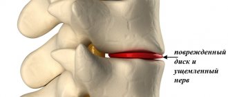
A characteristic symptom of metastases in the spine is a pathological fracture of the vertebral body
. This is a compression destruction of the affected vertebra as a result of changes in the structure of bone tissue.
At the diagnostic stage, to clarify the nature of malignant formations, a percutaneous biopsy of the spine is indicated.
About our clinic Chistye Prudy metro station Medintercom page!
Chemotherapy for liver metastases
Traditional cytostatic drugs for secondary tumor lesions of the liver rarely demonstrate effectiveness. However, in accordance with international treatment protocols, the use of multi-drug chemotherapy is sometimes recommended. Their choice mainly depends on the type of tumor that is the primary one.
In addition, targeted therapy is widely used today. It uses drugs that have a specific “target” of action - a specific molecule necessary for the growth and survival of cancer cells.
The most common targeted drug for metastatic primary liver cancer is sorafenib.
In addition, a number of other modern techniques can be used to treat liver metastases:
- radiosurgery;
- selective internal irradiation of the tumor focus (SIRT);
- proton and thermal effects;
- transcatheter embolization of tumor foci;
- implantation of port systems into liver vessels for infusion of chemotherapy drugs;
- external radiation exposure.
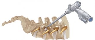
What diagnostics are performed before performing RFA of bone metastases?
For the most accurate localization of the lesion and three-dimensional reconstruction of the operated tissue area, we perform high-resolution computed tomography of the problem area of the spinal column or bone and make its 3D visualization.
Fig. 1 Inserting the needle as close as possible to the spinal cord, bypassing it (front view). Rice. 2 Inserting the needle as close as possible to the spinal cord, bypassing it (rear view).
Causes
Spinal tumors may originate from the spinal cord, within the meninges covering the spinal cord (intradural), between the meninges and bones (extradural) of the spinal column, or they may be located elsewhere.
Most spinal tumors are located extradurally. These may be primary tumors that originate in the spine, or secondary tumors that are the result of cancer spreading (metastasis) from other organs (primarily the lungs, breasts, prostate, kidneys, or thyroid).
Any type of tumor can occur in the spine, including lymphoma, leukemia tumors, multiple myeloma, and others. A small percentage of spinal tumors occur within the location of the spinal cord nerves (most often, these are ependymomas and other gliomas).
The cause of primary tumors of the spinal cord and spine is unknown. Some tumors are associated with genetic defects. Tumors of the spine and spinal cord are much less common than brain tumors.
As the tumor grows, spinal cord tissue, spinal roots, spinal blood vessels and bone tissue are involved. Exposure to the tumor causes symptoms similar to other compression syndromes (spinal injuries). In addition, tissue ischemia occurs due to invasion of tumor cells or due to pressure on blood vessels.
Risk factors
Most of us are aware of some of the risk factors that are associated with cancer. Smoking, poor diet, chemical and radiation exposure, a family history of cancer such as breast or ovarian cancer and hyperinsolation are common risk factors for cancer. These types of cancer generally occur in various organs and metastasize to the spine only after long-term development in the primary site. The spine has a well-developed circulatory system, and tumor cells can metastasize to the spine from other organs hematogenously (through the bloodstream). Low back pain is not usually the first symptom of malignant cancer coming from another part of the body. And so, doctors evaluate a patient for the potential development of a primary cancer site, but not for the presence of cancer in a patient with low back pain. Regular breast examinations (mammograms), smear tests (to detect cervical cancer), chest x-rays (to detect lung cancer), stool occult blood tests (to detect bowel cancer).
Complications of metastatic liver cancer
The most common complication of secondary tumor foci in the liver tissue is the growth of the tumor into the gallbladder and biliary tract, which manifests itself:
- pain in the upper and right parts of the abdomen, which is dull and constant;
- increased formation of gases in the intestines;
- a slight increase in body temperature, which stubbornly remains at the same level;
- appetite disturbances and weight loss;
- nausea and sometimes vomiting that does not bring relief;
- icteric coloration of the skin and whites of the eyes;
- increased sweating, especially at night.
What is the survival prognosis for bone metastases?
The median life expectancy is known for different types of bone metastases This indicator indicates the time during which 50% of patients die:
- For breast cancer - 19–25 months.
- For prostate cancer - 12–53 months.
- For thyroid cancer - 48 months.
- For kidney cancer - 12 months.
- For bladder cancer - 6–9 months.
- For lung cancer - 6–7 months.
The international clinic Medica24 employs experienced doctors who know how to maximize the life of patients with stage 4 cancer, how to deal with bone metastases and cope with painful symptoms. We use advanced diagnostic and treatment methods. Contact us.
The material was prepared by a member of the international society of oncological surgeons EESG, candidate of medical sciences Sergeev Petr Sergeevich.
Diagnostic methods
Bone metastases help to visualize diagnostic methods such as radiography, CT, and MRI. Currently, PET scanning has become the “gold standard” for searching for secondary lesions. During this test, a radiopharmaceutical is injected into the body and accumulates in cancer cells. Then they take pictures with a special device, and all the metastases are clearly highlighted and “highlighted”.
As bones break down, levels of calcium and the enzyme alkaline phosphatase . These changes can be detected by performing a biochemical analysis.
If in a patient suffering from cancer it was possible to visualize formations in the bones on photographs, while the level of calcium in the blood is elevated, this highly likely indicates the presence of bone metastases. But in order to confirm this definitively, a biopsy must be performed; it helps to directly detect tumor cells.
You can obtain a fragment of tumor tissue using a needle. But this is often very difficult, since metastases are located in hard-to-reach places. The doctor risks damaging the tissue surrounding the bone. The needle may enter a vessel or nerve, which will lead to bleeding and pain. If the needle position is incorrect, a false positive or false negative result will be obtained.
The task is greatly simplified when the biopsy is performed under CT guidance - the current gold standard. During the procedure, the doctor makes a layer-by-layer marking of the tumor using CT, then inserts a needle into the bone. This allows you to accurately “get” into the tumor, even if it has a complex localization.
The procedure lasts approximately 30 minutes and does not require anesthesia. At the same time, the radiation load on the body is low: the body receives the same dose of X-ray radiation as when flying from Novosibirsk to Moscow. The patient can leave the clinic immediately after the doctor finishes collecting the material. After 3-5 days, the doctor will invite you for a second consultation based on the results of histological examination. Due to high accuracy, the probability of false positive and false negative results is minimized.
Why do bone metastases occur?
Metastasis is a complex process, its mechanisms are not fully understood. As the primary tumor grows into the surrounding tissue, some cells may break away from it and penetrate into the lumen of the blood or lymph vessels. They migrate with the blood or lymph flow, then, reaching small vessels in the bone, they exit into the tissue and give rise to new lesions. As the metastasis grows, the cancer cells release substances that stimulate the formation of new blood vessels.
The main problem with metastases is that they are usually not one or two nodes that can be removed. As a rule, there are many secondary lesions, some of them very small. Therefore, it is difficult to fight them.
Most often, cancer cells settle in bones, which have a rich blood supply. In most cases, they are found in the vertebrae, pelvic bones, femurs, ribs, and arm bones.
How to treat liver metastases?
The choice of the optimal treatment regimen for this pathology depends on:
- size of the secondary lesion;
- the exact location of its location;
- patient's age;
- its general condition and other parameters.
But the most radical and effective way to get rid of liver metastases is surgical intervention. The operation in this case involves removal of the lobe of the liver in which the secondary lesion was identified. In some cases, when the tumor is large enough, resection of half the liver may be required. Sometimes the decision on the amount of tissue to be removed is made by the surgeon directly during the intervention - based on the results of a biopsy, which is performed directly in the operating room.
The technique of performing interventions can be either traditional, with wide access through the abdominal wall, or laparoscopic. The latter method is preferable due to less tissue trauma, but it can only be used for single small metastases.