The shoulder joint is the most mobile joint in the human body. It has the form of a ball-and-socket joint, which allows movement in different directions - inward and outward rotation, abduction/adduction, and flexion/extension. A special feature of this joint is the absence of a ligamentous apparatus inside it, which increases the risk of injury to the periarticular tissues that bear the main load. The specific structure of the joint explains the high frequency of pathological processes both inside and around the joint. Thus, according to various sources, up to 26% of all people consult a doctor with shoulder pain. The problem is somewhat more common in women.
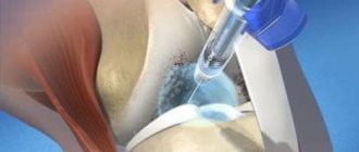
Injection into the shoulder joint
Diseases of the shoulder joint are divided into several groups and include:
- damage to the rotator cuff (impingement syndrome, tendon ruptures, tendinitis, bursitis);
- adhesive capsulitis (inflammation of the joint capsule, accompanied by “sticking together” of its membranes), which is associated with diabetes and thyroid disease;
- arthritis;
- damage to the coracoid and acromioclavicular joints;
- joint instability, for example, with hereditary connective tissue pathologies.
Diseases of the shoulder joint are accompanied by pain and dysfunction, which significantly reduces performance and worsens the quality of life of patients. Therefore, the main focus of therapy is pain relief and restoration of normal mobility.
An effective treatment method is joint injections, which use several groups of medications.
Injections can be given in:
- the cavity of the joint itself (intra-articular);
- surrounding tissues (periarticular);
- pain points sub- or intradermally (periarticular).
The choice of drug and the site of its administration is determined by the nature of the disease and the severity of symptoms.
Causes of development of arthrosis of the shoulder joint
- Excessive prolonged load on the shoulder, including of a professional nature (for example, builders, plasterers, loaders).
- An acquired or congenital defect of cartilage, ligaments and other structures, against which the joint quickly wears out even under normal load.
- Hand injuries - dislocations, fractures, bad falls.
- Deposition of salts in the joint cavity due to metabolic disorders.
- Constant microtrauma over a long period of time, for example due to regular sports activities.
- Chronic and acute inflammation of the joints.
Shoulder arthrosis is a hereditary disease: if your parents were ill, be vigilant
Principle of the method
The PRP method in the treatment of knee joints is based on enhancing the regeneration of intra-articular tissues. Thanks to platelet-rich plasma, cartilage, menisci, ligaments and other tissues are restored faster.
Even in ancient times, doctors drew attention to the fact that traumatic injuries accompanied by the formation of a hematoma heal much faster. The reasons for this have been established only in recent decades. It turned out that platelets secrete a number of substances that accelerate regeneration. These are the growth factors:
- epidermal;
- epithelial growth;
- endothelium;
- fibroblasts;
- insulin-like;
- transformative.
These substances have a polypeptide (protein) structure. They have different purposes. But all of these compounds take part in tissue regeneration. They stimulate the mitotic activity of cells, accelerate their growth and division.
Plasmolifting (PRP therapy) of the knee joint makes it possible to significantly increase the intensity of reparative processes of both soft and bone tissues. In medicine, platelet-rich plasma is increasingly used. It is used for any injuries: soft tissue damage, muscle and ligament ruptures, bone fractures. All types of tissues regenerate faster if a sufficient number of platelets are present.
Degree of development of arthrosis of the shoulder joint
Aching pain in the shoulder is an initial sign that cannot be ignored. If arthrosis is not treated, the load on the problem joint is not reduced, and you generally believe that “it will hurt and stop,” events will develop according to this scenario.
- The first degree of the disease is accompanied by aching pain that intensifies at night. The person can still move the arm freely, but already notices a limitation in the amplitude of movement, especially when moving the limb back. An x-ray shows a “ring symptom” - the glenoid cavity resembles an oval ring.
The first stage of shoulder arthrosis can last several years
- The second degree makes itself felt with much more vivid symptoms. The pain intensifies and does not stop, hand movements are accompanied by a characteristic crunch, and the amplitude is significantly reduced. The image shows a narrowing of the joint space, bone growths and thickening of the bone surfaces. In some cases, atrophy of the shoulder muscles is also found.
At the second stage, joining your hands behind your back is a real feat!
- The third degree is always accompanied by severe joint deformation and constant pain. At this stage, it is almost impossible to move your hand - only pump with a small amplitude. In the area of the junction of the scapula and shoulder, bone outgrowths are visually visible. The position of the hand becomes unnatural: the patient reflexively looks for the least painful position.
Do you keep putting pressure on your sore joint? There is a high risk of developing arthrosis of the third degree
Symptoms/red flags
- History of malignancy or symptoms/signs consistent with neoplasia, such as weight loss, deformity, swelling or swelling, abdominal discomfort/swelling.
- Skin erythema may indicate a tumor or infection.
- Symptoms/signs of systemic disease that may indicate polymyalgia rheumatica/giant cell arteritis.
- Fever may indicate a malignant disease or infection.
- A history of trauma or recent convulsion/electrical injury may indicate unreduced dislocation.
- A change in shoulder contour with loss of range of rotation suggests dislocation.
- The presence of significant sensory or motor deficits suggests neurological involvement.
How is arthrosis of the shoulder joint treated?
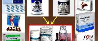
Classical medicine offers a traditional treatment regimen for arthrosis, aimed at reducing symptoms and preventing complications. It includes:
- non-steroidal anti-inflammatory drugs to relieve pain and tissue swelling plus painkillers;
- local anti-inflammatory ointments;
- chondroprotectors that restore the structure of cartilage.
To improve the trophism of the affected joint, doctors recommend starting its dosed development with the help of physical therapy, and to consolidate the effect, regularly undergoing physiotherapeutic procedures. In different cases, therapeutic baths, dry heating, magnetic therapy, mud therapy and other manipulations are indicated. All this is allowed only during the period of remission.
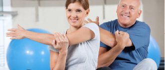
Physical therapy for second or third degree arthrosis should be performed only under the supervision of a specialist. But for preventive purposes, in order to strengthen the shoulder joint, you can exercise even before alarming symptoms appear. For example, according to this simple scheme:
Side effects
This is often the result of poor technique, too much dosage, too little dosage, or improper mixing and dissolution of medications.
Local effects
- Infection (1/10000)
- Post-injection increase in pain (2-5%)
- Skin discoloration that goes away over time
- Atrophy of subcutaneous fat tissue
- Bleeding (rare).
- Soft tissue calcification with repeated injections into one area of the skin
- Joint damage; Cartilage damage and osteoporosis: It is advisable to avoid repeated injections (no more than four injections at each site per year).
- Tendon atrophy and rupture (<1%): Avoid direct tendon injection.
- Pericapsular calcification (>40%).
- Avascular necrosis.
What other recommendations are given for shoulder arthrosis?
The patient is recommended to limit the consumption of spicy and salty foods, and increase collagen-containing foods: fresh herbs, poultry, seafood, salmon fish. Diet helps to eliminate pain more quickly, but is by no means an independent treatment method. An integrated approach helps reduce symptoms, eliminate inflammation and put the disease into remission.
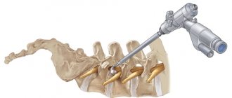
However, traditional treatment does not affect the root causes of degenerative intra-articular changes. One of them is the lack of synovial fluid, which acts as an intra-articular lubricant. Against the background of its deficiency, excessive friction of the joint tissues and their rapid wear begin. The problem can be eliminated by intra-articular injections.
Mechanism of action of intra-articular injections "Noltrex"
To restore the viscosity of synovial fluid, a special artificial endoprosthesis Noltrex with the addition of silver ions has been developed. The gel is injected into the joint cavity in a medical office at intervals several times. The composition is completely biocompatible with tissues and therefore does not cause allergic or other reactions.
By introducing the drug into the joint cavity, the following results can be achieved:
- protect cartilage - the gel covers the synovial membrane and articular surfaces with an even layer, softens mechanical stress and stops further destruction;
- restore the viscosity of the synovial fluid - a drug with viscosity characteristics close to natural normalizes the properties of the joint environment;
- dilute the rubbing surfaces - the product restores the normal viscosity of the synovial fluid, as a result of which the joint spaces widen and friction stops.
Noltrex relieves pain in the shoulder joint for a year and a half
Literature
- Bruyère O, Cooper C, Pelletier JP et al. An algorithm recommendation for the management of knee osteoarthritis in Europe and internationally: A report from a task force of the European Society for Clinical and Economic Aspects of Osteoporosis and Osteoarthritis (ESCEO) // Semin Arthritis Rheum. 2014; 44(3):253–63.
- Hochberg MC, Altman RD, April KT et al. American College of Rheumatology 2012 recommendations for the use of nonpharmacologic and pharmacologic therapies in osteoarthritis of the hand, hip, and knee // Arthritis Care Res. (Hoboken). 2012; 64(4):465–74.
- Jordan K.M. EULAR Recommendations 2003: an evidence based approach to the management of knee osteoarthritis: Report of a Task Force of the Standing Committee for International Clinical Studies Including Therapeutic Trials (ESCISIT). Ann Rheum Dis 2003; 62:1145–55
- McAlindony TE, Bannuruy RR, Sullivany MC et al. OARSI guidelines for the non-surgical management of knee osteoarthritis // Osteoarthritis Cartilage. 2014; 22(3):363–88.
- Zhang W, Moskowitz RW, Nuki G et al. OARSI recommendations for the management of hip and knee osteoarthritis, Part II: OARSI evidence-based, expert consensus guidelines. Osteoarthritis Cartilage. 2008; 16(2):137-62.
- Zhang W, Doherty M, Leeb BF, Alekseeva L et al. EULAR evidence based recommendations for the management of hand osteoarthritis: report of a Task Force of the EULAR Standing Committee for International Clinical Studies Including Therapeutics (ESCISIT). Ann Rheum Dis. 2007; 66(3):377-88.
- Di Giacomo G, de Gasperis N. Hyaluronic Acid Intra-Articular Injections in Patients Affected by Moderate to Severe Glenohumeral Osteoarthritis: A Prospective Randomized Study. Joints. 2017; 5(3):138-42.
- Funk L. HA for inoperable arthritis of the schoulder. 9th World Congress Osteoarthritis Research Society International, 2004 Chicago, USA.
- Meloni F et al. Clinical evaluation of sodium hyaluronate in the treatment of patients with sopraspinatus tendinosis under echographic guide: experimental study of periarticular injections. Eur J Radiol. 2008; 68(1):170-3.
- Merolla G et al. US-guided subacromial injections of HA for the management of rotator cuff tendinopathy: a prospective comparative study with rehabilitation therapy. Musculoskelet Surg 2013; 97:49-56.
- Russo A et al. Conservative integrated treatment of adhesive capsulitis of the shoulder. Joints. 2014; 2(1):15-19.
- Strakhov MA, Skoroglyadov AV et al. The use of low-molecular preparations of bound hyaluronic acid in athletes with pain syndrome of extra-articular localization // Polyclinic. 2013, no. 2, 54-60.
How often to take a course of injections
Unlike drugs with hyaluronic acid, Noltrex has a much more prolonged effect. It is recommended to administer it at intervals of 9 months to 2 years, depending on the degree of damage to the shoulder joint. The medicine is of synthetic origin, therefore it is not detected and is not rejected by phagocytes - the immune cells of the body. This explains the long-term therapeutic effect.
Of course, any treatment for arthrosis will be effective only if the load on the joint is reduced. In the case of a shoulder, the prognosis is always favorable, especially if you seek help in a timely manner, without waiting for critical symptoms. “It will hurt and stop” - with this diagnosis, such an option, unfortunately, is excluded.
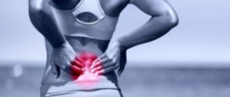
Shoulder pain does not always indicate the onset of arthrosis. Sometimes the reason is, indeed, a joint overwork, an uncomfortable position during sleep, or being in a draft. But much more often, pain is caused by other serious diseases, such as cervical osteochondrosis or glenohumeral periarthritis. Do you feel discomfort? Consult a specialist!
What is BCG?
The name BCG vaccine comes from its Latin name bCG, which is short for bacillus Calmette-Guérin. The drug, which is injected into newborn children in the absence of contraindications, consists of weakened and dead bovine tuberculosis bacteria. These bacteria are grown in laboratory conditions in a nutrient medium.
The BCG vaccine has been used for more than 80 years and has a proven protective effect against tuberculous meningitis and disseminated tuberculosis among children. The microorganisms that make up the BCG vaccine do not pose a danger to the child. They do not provoke a person to become infected with tuberculosis, but stimulate his immune system to produce antibodies to the tuberculosis bacillus.
As a result of the use of this vaccine, if pathogenic microorganisms Mycobacterium tuberculosis enter the human body, the body easily deals with them. In cases where a person who was given BCG as a child still gets tuberculosis, the disease is milder compared to people who did not receive such a vaccination. BCG cannot protect a person from becoming infected with tuberculosis, but it is an excellent preventive measure for severe forms of pulmonary tuberculosis and tuberculous meningitis, which are one of the leading causes of premature death in developing countries.
After vaccination in the maternity hospital, the BCG vaccination can be given to the child again at 7 years of age. The second time the BCG vaccination is prescribed if the child shows a negative reaction to the Mantoux test, which means that his body’s resistance to tuberculosis is very low. Revaccination at the age of 7 should be mandatory for a child if one of his family members suffers from tuberculosis.