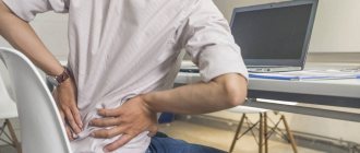
The wedge-shaped bone at the base of the spine is called the sacrum. Pain in the sacrum is considered one of the most severe among those occurring in the spine. It becomes more intense when sitting, standing up suddenly, bending forward and lifting heavy objects.
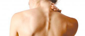
Pain in the sacrum in men is much less common than in representatives of the fair half of humanity. This is explained by the fact that women's vertebrae are not as strong as men's, and therefore it is much harder for them to bear loads. We should not forget about the additional loads on the female sacrum during pregnancy and menstruation. All these factors determine the more frequent occurrence of pain in the sacrum in women.
The term “sacrodynia” is used to refer to pain in the sacral area that occurs as a result of pathologies in the pelvis and adjacent areas.
At CELT you can get advice from a specialist algologist.
- Initial consultation – 4,000
- Initial consultation with the head of the Pain Clinic - 4,500
Make an appointment
general description
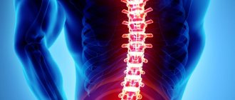
The sacrum is a bone formation consisting of five fixedly connected vertebrae and connecting the lumbar spine, coccyx and pelvic bones. The sacrum has a pyramidal shape, the base of which faces upward and forms a joint with the fifth lumbar vertebra, the sides form the sacroiliac joints, and the apex faces down and connects to the coccyx. The anterior surface of the sacrum has a concave shape and faces the pelvic cavity, while the posterior surface faces outward and is curved. The sacrum plays a significant role in the process of childbirth, so in women it is shorter, wider and less curved.
Lumbosacral radiculitis: symptoms
The leading symptom is pain of varying intensity. It can be aching and chronic, or it can be burning and sharp. Sometimes a painful attack occurs suddenly when a person tries to straighten his back, but it is not possible to do this completely, and new attempts only intensify the pain. An attack can begin after hypothermia, heavy physical labor, or staying in one position for a long time. The pain often radiates to the leg. In severe cases, the function of the pelvic organs is impaired.
Development mechanism
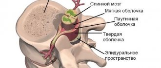
The spine is formed by vertebrae separated by intervertebral discs. In the middle is the spinal canal with the spinal cord inside: spinal nerves extend from it on opposite sides.
Some of them are responsible for motor functions, others for sensitivity. All of them pass through the foraminal openings and innervate different parts of the body and internal organs. If one or another nerve is compressed by pathological structures, a whole set of motor and sensory disturbances arise, which are typical signs of radiculitis.
Diagnosis of pain in the sacrum
- X-ray (allows you to assess the condition of bone structures, joints, articular cartilage along the width of the joint space)
- CT/MRI (allows for a more detailed assessment of the condition of bone structures and joints in cases where radiography is not enough; MRI visualizes soft tissue well)
- laboratory diagnostics (detects pathological changes in the blood, including markers of rheumatic diseases)
- Ultrasound (diagnosis of abdominal and pelvic organs)
Prevention
Preventive measures to prevent radiculitis are simple:
- competent organization of sleeping space. A comfortable bed is an elastic mattress of medium hardness and a small pillow of medium thickness. The ideal choice is orthopedic bedding;
- a complete and balanced diet will not only provide the body with healthy vitamins and minerals, but will also help maintain normal weight;
- adequate physical activity. Hard work is bad for your back, so if possible, you should reduce the load. Physical inactivity also threatens with negative consequences for the spine, and you should exercise at least once a week.
Doctors at our center have been working with patients suffering from radiculitis for many years. With timely treatment, the prognosis is always favorable. With us you will receive complete treatment, starting from examination and diagnosis, and ending with advice on lifestyle correction.
Minimally invasive techniques
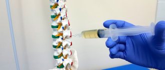
Therapeutic blockade
Therapeutic blockades are performed by injecting an anesthetic into the area of a peripheral nerve or spasmodic muscle. The blockade not only relieves pain, but also helps to relax muscles, normalize blood flow and improve tissue trophism.
Radiofrequency denervation (RFD)
Radiofrequency denervation (RFD) is a modern and safe non-surgical method suitable for patients for whom other treatment methods are ineffective or impossible.
When is this technique used?
- If conservative drug therapy is ineffective - when painkillers, even the most powerful ones, do not help enough or cannot be prescribed due to side effects;
- With a long wait for surgery, the patient often finds himself in a situation where he has to wait more than several months for surgery. In this case, RFD can reduce the intensity of pain and make the wait for the operation more comfortable;
- If surgery is not possible - when the presence of severe concomitant diseases or other reasons do not allow radical surgery to be performed, RFD is the most effective and safe alternative;
How is radiofrequency denervation performed?
After a standard examination by an algologist, additional examination and diagnostics if necessary, a decision is made to perform radiofrequency denervation.
- The procedure is performed on an outpatient basis. Under sterile conditions, the doctor, under X-ray control, inserts special needles into the area that needs to be treated. After the doctor is sure that the tip of the needle is exactly in the right place, a local anesthetic is injected so that the denervation is painless. After this, a thin electrode is inserted through the needle channel, which is connected to a radio frequency generator and the tip of the needle is heated to a predetermined temperature. The patient, as a rule, does not experience discomfort; the most painful moment of the procedure is the usual injection.
In some cases, a therapeutic and diagnostic blockade is first performed in order to determine how effective the technique will be for this particular case. Under sterile conditions, under the guidance of an x-ray system, the needles are precisely positioned in the area to be treated. To reduce discomfort, all manipulations are performed using local anesthesia. After installing the needles, a small amount of anesthetic is injected, interrupting the pain impulse.
Treatment
Pre-hospital assistance
For bruises and hematomas, rest should be ensured. Suspicion of a sacral fracture is an indication for immediate hospitalization. The patient must be placed on a backboard and given an anesthetic. For non-traumatic pathologies of the spine and neurological disorders, warming, anti-inflammatory and local anesthetics are effective.
For dysmenorrhea, analgesics are allowed. If you suspect other gynecological pathologies, especially those that suddenly arise and are accompanied by increasing pain, self-medication can be dangerous. An urgent examination by a gynecologist is required.
Conservative therapy
The plan of conservative measures is determined by the nature of the pathology. The list of treatment methods used includes:
- Protective mode
. Recommended for injuries, diseases of the spine and nervous tissue. May include bed rest, load limitation, and the use of orthopedic devices. - NSAIDs
. Indicated for rheumatic pathology, chronic pain caused by damage to the musculoskeletal system. Prescribed in the form of tablets, injections and topical medications. - Chondroprotectors
. Used for degenerative diseases to restore cartilage tissue. - Physiotherapy
. Physiotherapeutic measures are carried out for pain in the sacrum that is not associated with volumetric processes. Electrophoresis, heat therapy, magnetotherapy, and laser therapy are used. For many pathologies, the treatment regimen includes massage, exercise therapy, and acupuncture. - Chemotherapy, radiation therapy.
Necessary for oncological lesions. They can be carried out as an independent treatment or as an addition to surgery.
Surgery
The tactics of surgical treatment depend on the localization of the pathological process:
- Spinal diseases
: nucleoplasty, microdiscectomy, discectomy, facetectomy, laminectomy, sequestrectomy, tumor removal. - Women's diseases
: removal of paraovarian cyst, hysterectomy, panhysterectomy, supravaginal amputation of the uterus, amputation of the cervix, instrumental removal of the ovum, etc. - Urological and andrological pathologies
: tumor removal, bladder resection, cystectomy, prostatectomy, cryoablation and FUS ablation of the prostate gland.
In the postoperative period, restoration measures are carried out. For some pathologies, reconstructive interventions are subsequently performed.
Pathologies of the urinary system
A number of diseases of the urinary system are characterized by chronic pelvic pain
.
| Disease | Clinical manifestations | Diagnostics | Treatment |
| Nephroptosis | Moderate pain that is aching in nature at the initial stage of the disease. As it progresses, it spreads to the abdominal area and intensifies, becomes permanent, and is accompanied by the appearance of blood in the urine. |
|
|
| Chronic cystitis | A symptom of pelvic pain, which is accompanied by the urge to urinate frequently, accompanied by unpleasant sensations and blood in the urine. |
|
|
| Congenital malposition of the kidneys - dystopia |
|
|
|
| Urolithiasis disease |
|
|
|
Possible complications and prognosis for recovery
If severe pain in the sacrum occurs, it is necessary to determine its root cause. You cannot ignore symptoms or self-medicate. Lack of timely treatment can lead to the following complications:
- progression of the inflammatory/degenerative process;
- disruption of the functional activity of individual organs;
- problems with the reproductive system.
With timely treatment of the inflammatory process, the prognosis for recovery is usually positive. It is also almost always possible to contain degenerative-dystrophic pathologies. Delaying a visit to the doctor in some cases leads to a person’s disability.
Gastrointestinal pathologies
There are many diseases of the gastrointestinal tract that require treatment for pelvic pain
, along with eliminating the root cause that caused them.
| Disease | Clinical manifestations | Diagnostics | Treatment |
| Adhesions due to surgery, inflammatory processes, injuries | Painful pulling sensations in the abdomen, radiating to the pelvis and lower back. |
|
|
| Irritable bowel syndrome |
|
|
|
| Chronic colitis |
|
|
|
Pathologies of the musculoskeletal system
One of the clinical manifestations of diseases of the musculoskeletal system is pain in the pelvic area.
.
| Disease | Clinical manifestations | Diagnostics | Treatment |
| Osteochondrosis of the sacrolumbar region |
|
|
|
| Herniated discs |
|
|
|
| Neoplasms of the pelvic bones, metastases to the spinal column |
|
|
|
| Arthrosis of the sacroiliac joint |
|
|
|
| Tuberculosis ODA |
|
|
|