Author: Zemereva N.Yu., physiotherapist. March, 2021.
Synonyms: thoracodynia, thoracalgia - chest pain.
ICD-10 code: M42, M54.6.
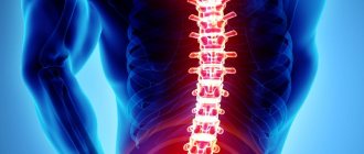
Thoracic osteochondrosis is a degenerative lesion of the spine (depletion and destruction of the bone structure of the vertebra). It begins with damage to posture, the appearance of vegetative symptoms (shortness of breath, weakness, sweating, malaise) and the development of severe pain. Thoracic osteochondrosis imitates cardiovascular diseases, and therefore requires accurate differential diagnosis. Therapy involves a wide range of interventions: medications, physical therapy, physiotherapy and massage.
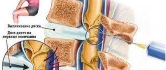
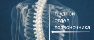
Thoracic osteochondrosis is less common than cervical or lumbar osteochondrosis. This is due to the specifics of the anatomical structure. The vertebral discs in the thoracic region occupy two-thirds of the entire spine, and they are also larger in diameter, but inferior in size to the lumbar region. This area is strong and has low mobility, and is also protected by the rib cage and ribs. The physiological curvature is directed towards the back. This leads to increased stress on the front of the spinal column. Next, the formation and growth of pathological bone structures on the vertebral bodies (osteophytes) occurs. Peripheral nerve endings are located among ligaments and muscle tissue, their tension leads to compression with the development of pain.
There are also polysegmental lesions of the spine due to osteochondrosis. This combines degeneration of the cervical, thoracic and lumbar spine with corresponding clinical symptoms. The clinical symptoms of thoracic osteochondrosis among women and men are approximately the same and do not have significant differences.
Classification
Thoracic pain syndrome is characterized by severe pain in the chest of an intense nature. The syndrome is associated with damage to peripheral nerves. The damage is due to compression of the nerves by muscles and ligaments.
Degrees of thoracic osteochondrosis:
- The first degree is characterized by the absence of pronounced clinical manifestations. There is a loss of elasticity in the intervertebral discs, and their protrusions form.
- The second degree is marked by a further loss of elasticity of the intervertebral discs and a decrease in their height. The likelihood of a hernia increases. Pain syndrome appears, and concomitant pain symptoms are possible.
- In the third degree, the pain syndrome intensifies. A herniated disc located between the vertebrae may occur. The severity of symptoms depends on the location of the hernia.
- Fourth degree with complete disruption of elasticity and loss of function of the intervertebral discs, destruction of the bone structure of the vertebrae. Neurological disorders are most pronounced.
According to the types of pain symptoms:
- Vertebrogenic thoracalgia is based on spinal pathology.
- Nonvertebrogenic thoracalgia is caused by the formation of pathologies of internal organs: cardiovascular diseases, gastroduodenal reflux, traumatic and inflammatory lesions of the musculoskeletal system.
- Psychogenic thoracalgia is caused by panic attacks and damage to organs of neural origin.
Causes and risk factors
Osteochondrosis does not form without damage factors. A number of reasons or a combination of them lead to the development of disease in the thoracic region.
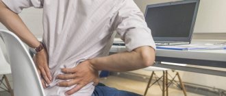
- Sedentary lifestyle. Lack of physical activity leads to weakness of the back muscles and intervertebral segment. Sedentary work and improper organization of the workplace are additional factors for thoracic osteochondrosis.
- Improper lifting of weights and various injuries. Excessive loads that disrupt the functioning of the spine. In this situation, the muscles and intervertebral discs cannot withstand the load.
- Acquired lesions and curvatures of the spine. Against the background of these pathologies, the functioning of the spinal column is disrupted and the likelihood of developing osteochondrosis increases. Destruction increases if the doctor’s recommendations are not followed.
- Lack of required minerals and vitamins. With insufficient calcium concentration in bone tissue, bones become less strong and the likelihood of damage to the musculoskeletal system increases.
- Pregnancy as a combination of main factors: increased load on the spine and lack of minerals and vitamins.
Important! Hereditary predisposition plays a significant role. If there are lesions of the musculoskeletal system in a family line, then you should be attentive to your health and the prevention of lesions. A competent system of preventive measures can prevent massive destruction of bone tissue.
Who's at risk
Often, factors in the formation of degenerative changes in the spine are combined.
- Decreased immune status associated with greater susceptibility to infections that can enhance the clinical manifestations of osteochondrosis due to muscle inflammation.
- Stressful influences that can cause psychogenic thoracalgia. This occurs due to a large release of catecholamines, which provoke increased pain.
- Damage to the nervous system of non-infectious and infectious etiology.
- Physical overload.
- Failure to comply with ergonomic principles (carrying heavy objects).
- Spinal injuries of various origins.
- Muscle spasms.
- Osteoporotic degeneration of the musculoskeletal system.
Spinal fractures
The vertebrae have great strength and can withstand a lot of pressure, while at the same time the spine does not lose flexibility. But like other bones in the body, they can break under extreme excess pressure, injury or disease. In such cases, damage or fractures to the vertebrae can range from minor to severe.
Compression fractures
As the name suggests, compression fractures occur from excessive axial loads that compromise the integrity of the vertebral body. Osteoporosis is one of the leading causes of compression fractures, as there is a decrease in the ability of the vertebrae to withstand stress. In such cases, even a slight fall or even a cough can lead to a compression fracture. People often perceive back pain as a normal part of aging, and sometimes compression fractures go unnoticed. Repeated compression fractures can lead to a decrease in spinal height. Another common cause of a compression fracture is trauma, such as a fall.
Often, vertebral compression fractures will eventually heal on their own (without treatment). NSAIDs (for example, aspirin) may be prescribed to relieve pain. For severe fractures, surgical methods (vertebroplasty and kyphoplasty) may be used.
Burst fractures
Blowout fractures usually occur after severe trauma (for example, a car accident or a fall from a height). Blast fractures are significantly more dangerous than compression fractures because the anterior and middle portion of the vertebral body is fractured into multiple fragments and is more likely to result in spinal cord injury. Additionally, as the vertebral body loses its integrity, the spine becomes unstable. In some cases, with burst fractures, if there is no impact on the spinal cord, conservative treatment can be performed. If there are loose fragments or damage to the nerve structures, then surgical treatment is necessary.
Flexion-extension fractures
Such fractures are sometimes called Chance fractures and occur during sudden flexion-extension. Most often, this type of injury occurs in car accidents, in people wearing a seat belt, and not only fractures of the vertebrae, but also ligaments, discs, and sometimes internal organs. Such fractures are usually unstable and require surgical treatment. This type of fracture occurs in 5-10% of cases of spinal fractures.
Vertebral fracture with dislocation. Such fractures occur when exposed to great force, and not only the integrity of the vertebral body is violated, but also its displacement (due to rupture of ligaments and discs). Such fractures often require surgical intervention.
Fractures are also divided into stable and unstable. Compression fractures are generally considered stable and do not require surgery. In contrast, unstable fractures (eg, burst or Chance fractures) typically require surgical treatment, often as an emergency.
Symptoms
Leading symptoms of thoracic osteochondrosis
- A burning sensation that occurs in the intercostal spaces.
- Paroxysmal and constant pain in the chest, predominantly stabbing in nature.
- With thoracalgia, the pain syndrome is stabbing, squeezing and aching in nature.
- Surrounding nature of the pain.
- Pain in one half of the body.
- During movement there is a crunching of the vertebrae.
- Painful symptoms increase significantly with movement, deep breathing, coughing and sneezing, which is the main difference between thoracic osteochondrosis and angina pectoris.
- The affected areas are palpable, that is, they can be felt, and are located along the affected nerves.
- Numbness of the skin along the intercostal spaces.
- The patient's condition worsens when exposed to low temperatures or prolonged exposure to an uncomfortable position.
Types of pain syndromes in osteochondrosis of the thoracic spine:
- Damage to the lower neck. There is pain in the upper chest, which can radiate to the neck, arms, and the left half of the body.
- Damage to the upper thoracic spine. The pain is aching in nature and affects the central part of the chest. Frequent combination with pain in the shoulder blades.
- Damage to the scapular-costal zone. Painful symptoms are cutting, aching and stabbing in nature. It has the form of attacks, both long and short. It occupies the lateral area and is also concentrated in the area of the shoulder blades.
- The appearance of pain in the anterior chest wall, varying in duration. Occur between the perithoracic and frontal axillary lines.
In addition to the leading signs, there are two types of pain syndromes for osteochondrosis of the thoracic region:
- Dorsago - intense but short-term pain at the location of the affected intervertebral discs. Disturbance of normal breathing.
- Dorsalgia is mild but prolonged pain in the area of the affected intervertebral discs.
Spondylogenic thoracalgia , associated with damage to the musculoskeletal system, is often accompanied by severe pain and instability of the vertebrae in the thoracic spine (their increased mobility). The lesion is expressed in impaired mobility of the thoracic spine, stabbing and cutting pain in the intercostal spaces.
Vertebrogenic thoracalgia can provoke the following symptoms:
- radicular (pain symptoms);
- disturbance of the innervation of the thoracic zone (visceral manifestations: a number of patients have pain symptoms of a stabbing nature in the gastrointestinal tract or cardiovascular system.);
- radicular syndrome with vegetative signs (pain in the intercostal spaces).
When diagnosing a problem, it is necessary to distinguish symptoms from cardiovascular diseases and myalgia. Heart damage of ischemic etiology is distinguished by the regularity of its appearance during physical or psycho-emotional stress and the relief of the attack by taking nitroglycerin.
A psychogenic attack of thoracalgia is accompanied by the appearance of panic, anxiety, suffocation and mental state disorder. It turns out that the disease is a consequence of problems with psychological stability.
Clinical signs of osteochondrosis are divided into two main parts:
1. Neuralgic symptoms:
- With thoracic osteochondrosis, numbness and tingling can occur both in the upper extremities and along the intercostal spaces, spreading to the anterior surface of the chest.
- The latissimus dorsi and chest muscles are constantly tense.
- There is high emotional lability, attacks of tearfulness and irritability.
- In rare situations, the disease manifests itself as severe intercostal neuralgia.
2. Various types of pain:.
- Dorsago: sharp, acute pain in the thoracic spine, sometimes making breathing difficult. Movement in the cervical and thoracic spine is limited. It appears or worsens when sitting in a twisted position.
- Dorsalgia: the formation of pain symptoms takes from two to three weeks, so the first time proceeds without clinical manifestations for the patient. There is mild discomfort in the chest. The pain intensifies when turning the body to the sides and deep breathing. With the final stabilization of the pathological process, a persistent pain syndrome is formed.
- Intercostal neuralgia: radiating pain along the intercostal spaces. When taking a sharp breath, a stabbing pain appears in the heart area. As a result, the pathology is often confused with damage to the cardiovascular system.
- Cardiac or pseudocoronary syndrome is formed when there is damage at the level of ThI segments with the development of reflex angina. The difference from organ damage to the cardiovascular system is the appearance of pain when tilting or rotating the spine. They intensify with prolonged stay in a forced position. There is pain on palpation of the spinous processes in the thoracic spine.
- Radicular syndrome: pain in the intercostal spaces (Erb's points).
- Visceral syndrome: dysfunction of the abdominal organs with damage at the level of the V-XII thoracic vertebrae. Expressed in girdle pain, heaviness in the right hypochondrium, heartburn.
Clinical symptoms depending on the level of damage to the thoracic spine:
*Damage to the nerve processes in thoracic osteochondrosis occurs in cases of the appearance of osteophytes - bone outgrowths on the vertebrae. This is due to the rate of destruction. Therefore, the symptoms listed below are not an integral part of the disease.
- Deformation of the nerve process at the Th2 and Th3 levels. Damage to the cardiovascular system occurs with the appearance of attacks of arrhythmia and coronary heart disease. Thus, chronic pain symptoms during thoracalgia can provoke organ dysfunction of the cardiovascular system.
- Damage at the Th4-Th5 level. Organs with damaged nerve fibers: pleurisy and bronchitis, pneumonia, bronchial asthma.
- Th5-Th6: the bile ducts and gallbladder are affected. The absorption of fats in the body is reduced.
- Th6-Th7: the liver and solar plexus area are affected. The functioning of the hepatobiliary tract is disrupted.
- Th7-Th8: the stomach is affected. Main pathologies: ulcerative lesions of the duodenum and stomach, dyspepsia and gastritis.
- Th8-Th9: the functioning of the duodenum and pancreas changes. Manifestations: duodenitis, pancreatitis and loose stools.
- Th9-Th10: damage to nerve cells of internal organs (spleen and diaphragm). Hiccups and breathing problems occur.
- Th10-Th11: the adrenal glands are affected. The activity of the immune system decreases and allergies appear.
- Th11-Th12: kidney function is disrupted, which leads to the formation of pyelonephritis and urolithiasis.
- Th12-L1 (level of the first lumbar vertebra). The kidneys and ureters are damaged. This leads to dysuria - problems with urination.
Dorsagia and Dorsago
Against the background of thoracic osteochondrosis, one can often notice the formation of vertebral syndromes: dorsalgia and dorsago.
Dorsago is the instant formation of acute pain in the sternum. As a rule, this condition is observed in those who remain in one position for a long time, for example, sitting. The syndrome can develop during the process of standing up after a long stay at the table in an unchanged position. Dorsago is characterized by the appearance of unbearable pain and limited movement in the spine.
Unlike dorsago, dorsalgia develops gradually. With this syndrome, slight pain and discomfort appears in the affected area of the ridge. An increase in pain is observed when taking a deep breath and bending the body.
The muscles become tense and movements in the spine become limited. Typically, the pain increases in the evening and goes away in the morning after a 10-minute walk.
The presence of other pain syndromes against the background of osteochondrosis in the thoracic region.
With this pathology, other pain syndromes may develop, which complicate the accurate diagnosis of the disease. When the disease spreads to the area of the upper thoracic segment of the sternum, pain appears in the esophagus, larynx, and a “lump in the throat” is felt. If the lesion is located in the mid-thoracic segment, discomfort occurs in the right hypochondrium.
When the pathology spreads to the area of the lower thoracic segment, pain in the peritoneum develops, similar to signs of a disease in the intestines.
It is worth noting that there is no relationship between the pain syndrome due to thoracic osteochondrosis and the patient’s nutritional quality. Moreover, pain can appear regardless of the time of year. Although the pain associated with this disease can increase in the evening and during physical overload. By morning, the discomfort usually disappears or subsides.
Diagnosis of thoracic osteochondrosis
If you suspect osteochondrosis, you can contact a therapist or neurologist.
The patient is examined and all clinical data is recorded. During the formation of stages 2-3, the skeleton undergoes significant deformation. A complete medical history of the patient should be collected in order to accurately establish or exclude factors leading to the formation of osteochondrosis of the thoracic spine.
The very first diagnostic method is radiography. Further studies are carried out based on the clinical history and the need for differential diagnosis. Initially, the patient can be examined by any doctor. The main thing is a competent and fully collected clinical history. This will allow you to accurately determine the etiology of the disease and select a treatment regimen. Treatment of thoracic osteochondrosis is carried out by a therapist, neurologist, and rheumatologist. In case of traumatic effects on the spinal region, consultation with a traumatologist is required.
- X-ray examination of the chest in two projections. Allows you to determine the presence and size of osteophytes, determine the contours and height of intervertebral discs, and determine changes in the shape of the disc.
- Discography makes it possible to examine the structure of the nucleus pulposus through the use of contrast.
- Computed tomography is used to visualize nerve fibers, muscles, ligaments and joints.
- Electromyography allows for differential diagnosis with neurological diseases.
- Endoscopic diagnostic methods can be prescribed to examine the circulatory and digestive organs.
- An ECG is performed to determine the etiology of cardiovascular diseases.
- Electroencephalography - to identify pathologies of the nervous system.
Differential diagnosis
Thoracic osteochondrosis should be distinguished from a number of diseases.
- Anomalies in the formation of the spine, trauma, tumors, inflammation. There are several variants of these pathologies. For example, an additional congenital process, displacement or fusion of vertebrae (spondylolisthesis), osteomyelitis, ankylosing spondylitis and others.
- Damage to the musculoskeletal system (different lengths of the lower limbs, muscle spasms, muscle inflammation and others).
- Not associated with damage to the musculoskeletal system, but similar in symptoms to diseases of internal organs. In particular, pancreatitis, inflammation of the appendages, stomach ulcers, coronary heart disease, angina pectoris, pleurisy.
- Neurosis-like disorders, combined with migrating pain with increased fatigue, irritability, and mood swings.
Thoracic osteochondrosis and ischemic heart disease
It is extremely important to carry out competent differential diagnosis with the most similar pathologies. The pain that occurs with vertebrogenic thoracalgia and coronary heart disease (CHD) has a number of differences, which makes it possible to accurately establish the diagnosis.
Nature of pain:
- With ischemic heart disease, they have a burning and squeezing character, accompanied by the fear of death.
By duration of pain:
- IHD: Short-term attack, lasting a few minutes.
- Thoracic osteochondrosis is characterized by fading or prolonged pain, in some cases it does not subside during the day.
Changing body position:
- In IHD, the strength and intensity of pain does not vary with physical activity.
- With thoracalgia, even relatively gentle movements cause increased pain or the occurrence of a new attack.
Response to physical activity:
- With ischemic heart disease, pain appears during physical activity and is relieved at rest.
- Thoracalgia, on the contrary, weakens, but does not stop at rest.
Relief on taking medications:
- During an ischemic attack, pain is easily relieved by taking nitrates (nitroglycerin).
- Thoracalgia is relieved by the use of analgesics.
The influence of physiotherapeutic factors and manual therapy:
- In coronary heart disease it gives unstable and insignificant improvement.
- With osteochondrosis, there is a significant positive change in the patient's condition.
How to relieve pain in the thoracic spine?
If the unpleasant sensations were caused by emotional stress, then to get rid of mild discomfort it will be enough to normalize the psychological situation and rest regime. In more complex cases, physiotherapy, massage, acupuncture and exercise therapy will help. These measures can reduce stress levels and eliminate pain. If the examination reveals diseases of the musculoskeletal system, complex treatment will be required, which may include:
- taking medications;
- physiotherapy;
- diet;
- reflexology (acupuncture);
- manual therapy;
- exercise therapy;
- massage;
- in some cases - the use of orthopedic devices (corsets, etc.).
To normalize blood flow and local metabolic processes, eliminate pain and inflammation, stop the growth of osteophytes and regenerate cartilage tissue, a combination of gentle physical activity, medication (anti-inflammatory drugs, chondroprotectors and others) and various types of physiotherapy are necessary. Massage, acupuncture and other auxiliary techniques increase the effectiveness of the main treatment and help normalize sleep, relieve excessive muscle tension and pain. In the network of clinics “Hello!” Each patient receives a set of treatment procedures that are most effective for their condition. To quickly determine the causes of pain, modern diagnostic equipment is used to identify spinal pathologies and their complications in the early stages. Patients are treated by highly qualified doctors who constantly study the latest treatment methods and successfully apply them in practice.
Treatment of thoracic osteochondrosis
Osteochondrosis is treated by a neurologist.
To organize competent therapy, it is necessary to first establish the etiological prerequisites. Identifying the cause of the pathology allows you to select a competent treatment regimen.
Drugs for bone tissue regeneration are selected taking into account all the functional characteristics of the body. It is advisable to first clarify the concentration of collagen and elastane in the body. When choosing a treatment regimen, the individual characteristics of the body are taken into account.
Standard therapy regimen
Nonsteroidal anti-inflammatory drugs help eliminate chest pain caused by inflammatory reactions. This increases the range of mobility of the chest, as well as the range of motion in the thoracic spine. Medicines:
- meloxicam,
- nimesulide,
- celecoxib,
- ibuprofen.
Drugs that affect the production of interleukins. Allows you to stop the inflammatory cascade and normalize the balance of enzymes that cause destruction of the myelin sheath of nerves:
- diacerein.
Antispasmodic agents:
- no-shpa,
- spasmalgon,
- unispaz,
- mydocalm,
- tizanil,
- sirdalud.
B vitamins help relieve inflammation of affected nerves:
- neurobion,
- combilipen,
- milgamma,
- B-complex.
Preparations that preserve the concentration of collagen and elastane allow fluid to be retained in the intervertebral discs. This increases tissue elasticity and prevents further degeneration:
- collagen,
- joint formula,
- piascledine.
Hormonal (steroid) drugs. They have a powerful anti-inflammatory effect, but are used only for acute thoracalgia, as they negatively affect the body as a whole:
- betamethasone (diprospan, triderm),
- dexamethasone,
- prednisolone.
During the acute period of the disease, diuretics help relieve swelling from the nerve endings. Preference is given to potassium-sparing diuretics. Apply
- indap,
- veroshpiron,
- Spironaloctone.
Ointments and gels with anti-inflammatory effect. When rubbing the affected area of the back, the local inflammatory process is reduced and highly active pain symptoms are eliminated. Use
- art traffic,
- nimulid,
- voltaren,
- nize,
- finalgon,
- long
Massage
The therapeutic effect of massage is to relieve spasm from the muscular corset of the thoracic spine and normalize local blood circulation.
Effects of massage techniques:
- relieving muscle hypertonicity;
- strengthening the structure of the intervertebral disc bodies.
The use of massage techniques is combined with a visit to a chiropractor in combination with a regular exercise therapy system.
Physiotherapy
Acupuncture. Eliminates or reduces muscle spasms, and also reduces pain symptoms.
Manual therapy. Allows systemic blood circulation in the intercostal space to return to normal. This ensures the supply of tissues with nutrients, improves their trophism and stimulates oxygen saturation in the blood.
Nutrition for osteochondrosis of the thoracic spine
Compliance with certain nutritional principles allows you to achieve maximum therapeutic effect.
- Foods rich in vitamins A, B, C and E (greens, nuts, cereals) are recommended.
- Omega-3.6 fatty acids found in fish.
- Chondroitins in the form of dietary supplements help maintain tissue strength. Glucosamine maintains the elasticity of tissue structures.
Read more about the diet for osteochondrosis in our separate article.
Complications
When establishing a diagnosis of thoracic osteochondrosis, one should take into account a possible cascade of probable organ pathologies that develop over time.
- Damage to the cardiovascular system: persistent pain leads to destabilization of the ion exchange of the myocardial muscle, which is a prerequisite for the development of coronary heart disease.
- Disorders of the functioning of the abdominal organs: stomach, duodenum, pancreas. This is due to the high secretion of adrenaline during persistent pain, which leads to increased secretion of VIP (vasointestinal peptides).
- Dyskinesia of the gallbladder is due to an increase in the lithogenicity of bile against the background of a chronic inflammatory process.
With regular adherence to the principles of therapy, the exercise therapy system, maintaining posture and eliminating risk factors, the course of the disease is reduced to regression. The prognosis is considered favorable if the pathology does not develop further and the disease does not actively manifest itself.
Preventive actions
To prevent the development of problems with the spine, the following preventive measures must be observed:
- lead an active lifestyle: swim in the pool, run in the morning, do exercises;
- if you are engaged in a sedentary job, you should try to sit with a straight back and relaxed shoulders;
- sedentary work should be carried out on a chair with a back that will support the spinal column;
- To ensure that the spine is in the correct position during the night's rest, it is recommended to acquire an orthopedic mattress and pillow;
- avoid lifting heavy objects, but if this is unavoidable, then lift them smoothly;
- it is recommended to pump up the abdominal muscles;
- you need to use only shoes that are comfortable to wear;
- You should include foods enriched with vitamins and microelements in your diet.
It is quite difficult to protect yourself from the development of thoracic osteochondrosis, but nothing is impossible. If any signs of pathology are detected, you should immediately seek help from a specialist to determine the nature of the symptoms that have arisen. It is possible that the reason may lie not in the spine, but in dysfunction of other internal systems and organs.
If the previously listed rules are observed, the possibility of developing thoracic osteochondrosis is reduced to a minimum.
Moscow, metro station Dubrovka, st. Sharikopodshipnikovskaya, 6/14
| Loading map... |
Prevention
- Elimination of physical inactivity, therapeutic exercises. Exercises for counter-force, perpendicular loads with displacement, and spinal extension are selected.
- When driving a car for a long time, select special exercises to relax the muscle frame.
- Pumping the muscles of the thoracic spine. There is both a physical therapy complex and the use of myostimulation if independent training is impossible.
- Organization of the work place: the back of the work chair should provide support for the spine. To prevent stress on the spine from increasing, you should warm up every 30 minutes by stretching or walking. This is explained by the fact that the sitting position increases the load on the spine.
- Correct position of the spine at night: purchasing orthopedic sleep accessories. A completely rigid surface is not rational due to the disruption of the physiological curves of the spine.
- Compliance with ergonomic principles: do not lift weights that can injure the spine.
- Formation of correct posture.
- Optimization of blood circulation and lymph flow through a stretching system or the use of special procedures (pressotherapy).
Sources:
- Yuri Kwon. The effect of sitting posture on the loads at cervico-thoracic and lumbosacral joints. — Technology and Health Care, 2021.
- Finn L.L. Acute pain in the chest. Differential diagnosis. — Bulletin of Science and Education, 2021.
- Osteocondritis of the spine. Clinical recommendations. — Ministry of Health of the Russian Federation, 2021.
Diagnostic procedures
To find out why the thoracic spine hurts and what needs to be done to get rid of the pain, consult a neurologist or orthopedist. The doctor will perform an examination and write directions for diagnostic tests, which may include:
- X-ray;
- MRI;
- CT;
- ECG;
- Ultrasound of internal organs;
- lab tests;
- and other procedures.
The examination will help identify all changes in the spine and nearby tissues: changes in the width of blood vessels, narrowing of the spinal canal, compression of the nerve roots, displacement of the vertebrae, the presence of protrusions and hernias. Multi-position images can detect spinal curvature: kyphosis, lordosis or scoliosis. ECG, ultrasound and tests are necessary to diagnose associated disorders, including infectious and inflammatory diseases.