Anatomy of the knee
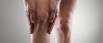
To understand the causes of pain behind the knee when walking, it is necessary to understand the anatomical features of this area.
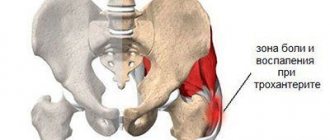
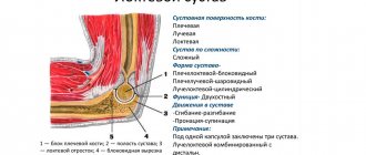
The knee joint is an articulation of the heads of the femur and tibia bones, covered with a layer of cartilage tissue and enclosed in a synovial membrane (bursa), which contains synovial fluid. The spacer between the heads of the bones is a ring-shaped cartilage - the meniscus. The joint is strengthened by several ligaments. In the front part is the kneecap, held in place by ligaments and tendons. In the popliteal region there is adipose tissue through which large blood and lymphatic vessels and nerve bundles pass. Lymph nodes, ligaments of the knee joint are also located here, tendons and muscles are located above and below. Pathology of any of these components can cause pain in the legs behind the knees.
Arthrosis
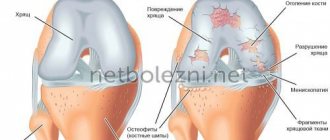
Arthrosis is a degenerative-deforming disease of the joint, during the development of which the composition of the synovial fluid changes, cartilage tissue and other articular elements are destroyed.
The early stage of the disease is precisely characterized by such a symptom as “stiffness” of the joint. The patient notes that it hurts to start moving and his leg feels like wood at first. For normal movement you need to diverge a little. Then the pain subsides. In addition, there may be a crunch in the knee. The second stage of arthrosis is characterized by additional symptoms such as:
- swelling of the knee area;
- local increase in temperature;
- possible redness of the skin;
- pronounced crunching sound when walking;
- limitation of mobility of the articular area. The patient cannot straighten his leg freely. In addition, the leg does not bend completely;
- pain accompanies not only at the beginning of movement, but also during walking and other loads.
At the third stage, the knee stops bending and straightening altogether. The patient suffers from constant sharp, sometimes shooting, pains, which often worsen at night. With arthrosis of the last degree, cartilage tissue is completely destroyed, in addition, bones are susceptible to destructive processes. Usually the third stage ends with the destruction of articular elements and the assignment of disability.
Does it hurt when your knee bends or straightens? In this case, the possibility of developing arthrosis cannot be ruled out.
Causes of pain
The most common cause of pain behind the knee when bending is injuries and diseases of the knee joint, but other diseases can often cause it. To establish an accurate diagnosis, you need to contact a clinic specializing in the treatment of the spine and joints. You should not delay seeing a doctor, as some conditions with pain in the leg below the knee require immediate surgical intervention.
Pain can be caused by:
- Gonarthrosis is the gradual destruction of the articular cartilage of the knee joint due to metabolic disorders in the cartilage tissue. Arthrosis develops after injury or during the aging process. Depending on the location of the source of destruction, aching pain may be felt under the knee at the back, which intensifies after exercise and subsides after rest.
- Arthritis is an inflammation of the knee joint that is most often caused by an infection or an autoimmune reaction when immune cells attack the body's own tissues. Arthritis is characterized by increased temperature in the joint area and severe pain under the knee in the back, front and sides, which does not go away with rest.
- Bursitis is an inflammation of the joint capsule, characterized by the appearance of an elastic round swelling, redness and swelling. Bursitis is usually accompanied by an increase in body temperature.
- Baker's cyst. If arthrosis, arthritis and bursitis can have different localizations, then Baker's cyst is a disease that develops only in the popliteal fossa. It is a fluid-filled cavity made of connective tissue that can be felt under the knee. Its danger is that it can compress the vein, causing thrombosis and varicose veins.
- Knee injuries: bruises, dislocations, sprains and torn ligaments. Pain behind the knee during extension usually appears immediately after a traumatic impact. Urgent diagnosis and assistance from a traumatologist are required.
- Meniscus tear resulting from injury. Immediately after the injury, sharp pain appears under the knee when squatting and any attempts to bend and rotate the knee joint.
- Meniscus cyst. Most often, this disease develops during high physical activity in athletes. It is characterized by gradually increasing pain; in the later stages, protrusion appears on the side of the knee joint and a sharp limitation of mobility.
- Tendinitis is inflammation of the tendons. It begins with the appearance of pain under the knee at the back of a pulling nature after exercise, over time it intensifies, becomes paroxysmal in nature and does not go away even at rest. An increase in body temperature, local redness and swelling, and the appearance of nodules may be observed.
- Deep vein thrombosis in the popliteal region. This dangerous condition can be fatal when a blood clot breaks off and is carried by the bloodstream into the pulmonary artery. Requires immediate diagnosis and treatment. It manifests itself as pain under the knee at rest, pain when pressed, swelling and blue discoloration of the leg.
- Inflammation of the popliteal lymph nodes (lymphadenitis), a complication of which can be an abscess. It is accompanied by pain behind the knee when walking, enlarged lymph nodes that can be felt like balls, swelling, redness and increased temperature of the popliteal region.
- Pinched nerve in the popliteal area. It manifests itself as sharp pain, numbness and weakness of the leg.
If one of the above conditions occurs, under no circumstances should you use folk remedies or heat the affected area. The only thing you need to do if you have pain behind your knee is to go to a medical facility.
Causes of the disease
All diseases and conditions that cause sciatica are divided into two groups:
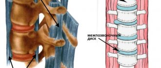
- vertebrogenic, i.e. associated with injuries and diseases of the spine - this includes osteochondrosis, injuries and deformations of the vertebrae or intervertebral discs, spondylosis, stenosis of the lumbosacral region;
- non-vertebral, i.e. not associated with the spine, are infectious inflammation of the genitourinary system or muscle tissue, hypothermia, excessive physical exertion on the muscular corset of the lower back and pelvic region, a sedentary lifestyle, excess weight, pregnancy, tumor development, etc.
Any compression of the sciatic nerve leads to the development of the disease. The most common cause is a herniated intervertebral disc, a ring of fibrous tissue that separates the vertebrae.
Diagnostics
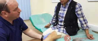
Due to the large number of diseases manifested by pain in the popliteal region, diagnosis of this condition is difficult and should be carried out in well-equipped modern clinics. First, the doctor examines the patient, asks about lifestyle, injuries, occupational and genetic factors.
To detect the inflammatory process in the body, blood and urine tests are prescribed. Then hardware diagnostics are carried out. Depending on the expected diagnosis, the patient is prescribed:
- Doppler testing for suspected deep vein thrombosis;
- radiography with contrast to detect arthritis and gonarthrosis;
- Ultrasound of the knee joint;
- magnetic resonance imaging, which gives a picture of the condition of the meniscus;
- arthroscopy, in which the joint cavity is examined by inserting an endoscope;
- joint puncture to collect synovial fluid.
Depending on the clinical picture, other specialists may be involved in the diagnosis: a rheumatologist (if rheumatoid arthritis is suspected), a neurologist (if a nerve is pinched in the popliteal region).
Ligament damage
Ligament damage is a fairly common disease, especially among athletes.
Ligament rupture can be partial, incomplete (tear) or complete.
The external collateral ligament is damaged less frequently than the internal one. Most often, damage to the external ligaments occurs when the tibia is excessively deviated inward. The rupture may be combined with a separation of part of the head of the fibula. The internal collateral ligament is injured more often, however, its damage is usually incomplete. The injury occurs when the tibia deviates excessively outward. This ligament damage is often combined with a rupture of the internal meniscus and damage to the joint capsule.
Symptoms of ligament damage include:
- acute pain in the damaged area;
- restriction of movement;
- swelling of the joint area;
- with a complete rupture of the ligaments, increased mobility in the articular area is observed;
- muscle spasms.
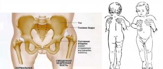
Meniscus tears in children most often heal on their own
Treatment of pain under the knee
The doctor determines how to treat pain under the knee after making a diagnosis. If the problems are caused by infectious or systemic inflammation (lymphadenitis, rheumatoid arthritis), then treatment of the underlying disease is prescribed. In some cases, surgery is required. The operation is performed when:
- some types of injuries;
- thrombosis;
- abscesses;
- gonarthrosis grade 3–4 (knee replacement);
- tumors.
In the postoperative period, the patient is prescribed anti-inflammatory and painkillers and wearing fixing bandages.
For inflammatory diseases, non-steroidal anti-inflammatory drugs are prescribed. In case of injury, a plaster cast or splint is applied to the damaged area. After removing the fixator, therapeutic massage, gymnastics, and physiotherapeutic procedures are used to restore mobility of the knee joint. Non-advanced stages of gonarthrosis are treated similarly, for which chondroprotectors are also prescribed that can slow down the degeneration of cartilage tissue. Folk remedies can only be used as additional treatment if they are approved by a doctor.
Meniscal injuries
Menisci are cartilage fibers that are located between the femur and tibia joint area. They act as shock absorbers, serve to distribute loads, stabilize the joint, and protect articular cartilage from overstrain due to loads.
The following types of meniscus damage are distinguished:
- separation of cartilage tissue from the attachment site;
- stretching;
- inflammation;
- body rupture;
- rupture of the anterior or posterior horns.
If the patient experiences pain during extension or an inability to fully straighten the knee, most likely there is a sprain or tear of the body or anterior horn of the meniscus. When the dorsal horn is affected, the limb usually cannot bend completely.
Diagnosis of meniscus damage is carried out by a doctor using MRI, ultrasound or arthroscopy.
As a rule, a small tear of the meniscal body can heal on its own, especially if it occurs in a child. If the horns rupture, surgery is necessary because there are no blood vessels in this area and the cartilage tissue here does not regenerate. An interesting fact remains that in a child, the cartilage fibers of the menisci are densely penetrated by blood vessels, so in the child the process of fusion and healing of the menisci is much better and faster. If the horn of the meniscus is slightly damaged in a child, it may be possible to do without surgery.
Preventing knee pain
Risk factors are excess weight, physical inactivity or excessive stress on the knee joints, frequent injuries, and a hereditary predisposition to joint diseases. Following a healthy lifestyle will help keep your knee joints healthy. This concept includes:
- maintaining normal weight;
- optimal level of physical activity, including strength and aerobic training;
- balanced diet;
- good sleep;
- hardening;
- avoiding traumatic situations and overwork;
- increasing stress resistance;
- periodic monitoring of blood composition and taking measures to eliminate identified deficiencies of essential vitamins and minerals.
To prevent loss of ability to work and the development of dangerous life-threatening complications, you must consult a doctor at the first sign of discomfort or pain in the popliteal area.
First aid measures
Of course, if you feel restricted in movement, you should consult a doctor.
If you do not have the opportunity to visit a specialist in the near future, and the pain is already tormented, you can take a painkiller. The most effective drugs include non-steroidal anti-inflammatory drugs, including:
- ibuprofen;
- indomethacin;
- ketanov;
- diclofenac;
- arthrothec;
- flubiprofen.
Among the folk remedies, compresses made from fresh cabbage leaves, raw potato pulp, and nettle decoction are very effective in relieving pain.
Non-steroidal anti-inflammatory drugs will help you relieve pain
Human legs are designed in such a way that they can support the weight of the entire body. It is to their structure that we owe upright posture. However, the price to pay for this is various joint diseases. A large load is constantly placed on the lumbar spine, as well as on the knee joint, which often leads to serious problems. If the knee does not bend completely, then in most cases the problem indicates the presence of some pathological process. Therefore, if such a symptom is detected, it is necessary to urgently visit a doctor. After all, any pathology associated with this part of the musculoskeletal system prevents a person from leading a full life and can lead to the fact that the patient simply cannot walk.
Limited mobility
Symptoms
Since there can be quite a lot of reasons for this condition, it is sometimes very difficult to determine what exactly is the matter. Therefore, it is very important to pay attention to other accompanying symptoms. For example, you need to try to determine whether difficulties with bending are caused by the inability to perform this movement without severe pain, or by the fact that the joint has actually lost mobility.
List of possible symptoms:
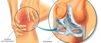
- The appearance of signs of inflammation. The knee swells, increases significantly in size, and redness of the skin appears.
- There may also be a feeling that the cartilage is rubbing against each other due to a lack of synovial fluid. A crunching sound may occur.
- Periodically, sudden, sharp pain occurs. In a calm state, such a symptom may not be acute, but nagging. Sometimes joint diseases can be painless for a long time.
These symptoms most often accompany limited mobility. They are especially acute in advanced stages of disease. In this case, even any attempt to stand up or ordinary walking will bring acute pain.
Symptoms