Diseases of the musculoskeletal system appear at any age. In children, this is a congenital pathology associated with heredity. In adults – consequences of injuries, inflammatory diseases. In older people, dystrophic and degenerative processes predominate due to age-related deterioration of blood supply. Their bone tissue is more fragile, so long-term recovery is necessary after fractures. Muscles must be constantly dosed to maintain normal tone.
Causes of joint diseases
With the development of civilization, the number of factors that negatively affect human health has increased. In particular, on his musculoskeletal system. At the same time, the age threshold for joint diseases is reduced. Obvious reasons for their appearance:
- insufficient physical activity (physical inactivity) leads to excess weight and muscle sagging;
- poor environmental conditions and poor nutrition provoke allergies, which, in turn, cause inflammation and degenerative changes in the joints;
- a careless attitude towards the first pain in the joints (it will go away on its own) often makes a visit to the doctor too late;
- Age matters – the risk of getting sick increases with age.
It should be remembered that joint diseases cause damage to internal organs. Therefore, treatment cannot be delayed.
Symptoms of infectious arthritis
Infectious arthritis is characterized by suddenness: pain appears unexpectedly, as if out of nowhere, and extremely intensely signals the onset of the disease. Almost immediately, stiffness sets in in movements - both active (using only muscles) and passive (produced with the help of hands or other aids). Typically, the first symptoms of infectious arthritis are observed in only one joint, with the possibility of spreading to others. But with blood diseases, viruses, gonococcal infections, polyarthritis is possible, in which symptoms are observed in several joints at the same time.
The main signs of infectious arthritis are:
- redness of the skin over the joint, local or general increase in body temperature;
- pain when touching the joint;
- accumulation of fluid in adjacent tissues caused by an inflammatory process;
- a sharp deterioration in mobility (the joint moves tightly, even if you bend it with your hand);
- fever, chills.
Specific symptoms and treatment for infectious arthritis vary depending on the pathogen. Gonococcal arthritis may be accompanied by ulcerative rashes on the skin and mucous membranes, in the mouth or in the genital area. If infection occurs through a bite, the lymph nodes usually become enlarged. Fungal infections of the joints can occur without fever or other systemic manifestations.
The disease is usually localized in the shoulder, elbow, wrist and interphalangeal joints, as well as in the knee and hip joints. Certain diseases can “select” specific joints - for example, borelliosis most often affects the knees.
Joint diseases: causes, types
Diseases of the musculoskeletal system are divided into groups: dystrophic (arthrosis), inflammatory, infectious (arthritis).
Arthrosis. Dystrophic group
- The disease affects all parts of the joints: ligaments, cartilage, surrounding muscles. Occurs under the influence of the following factors:
- injuries or previous operations;
- low estrogen levels (in women during menopause);
- excess weight;
- heredity.
Arthrosis affects mainly people over 40-45 years of age, as well as the elderly. The disease comes in different types:
- Gonarthrosis. Destruction and deformation of cartilage in the knee. It is difficult for a person to walk and lameness appears.
- Coxarthrosis. Impaired mobility in the pelvic area and the appearance of skin spurs. Surgery is recommended.
- Periarthrosis. Stiffening of the feet, as well as the shoulder - scapula, elbow and wrist areas.
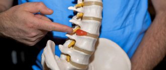
- Osteochondrosis. Damage to intervertebral discs and nerve roots. May lead to intervertebral hernia.
- Ankylosing spondylitis. Affects intervertebral units. In the worst case scenario, it immobilizes the spine.
Arthritis. Inflammatory and infectious group
This disease can occur in different forms: acute or chronic. In all cases, it is characterized by severe pain. Arthritis occurs against the background of injuries, allergic reactions, diseases of the nervous system, metabolism and infection.
Symptoms of the disease:
- acute pain when walking;
- stiffness of movements;
- red spots on the affected area;
- elevated temperature.
At a doctor’s appointment, the patient needs to share his observations. This will speed up the diagnosis.
There are 4 types of arthritis:
- Bursitis. As a result of injury, the periarticular bursa fills with inflammatory fluid. The disease is fraught with complete incapacity of the infected limb.
- Hoffa's disease (lipoarthritis). Inflammation of the fatty tissues of the knee due to increased stress. The leg does not straighten. Most often professional athletes get sick.
- Schlatter's disease. The knee joint is poorly supplied with blood, causing the core of the bone to collapse.
- Perters disease. Affects the hip joint and causes tissue necrosis. Most often, the disease affects children from 2 to 14 years old.
Arthropathy - symptoms and treatment
The diagnosis of arthropathy is made when all possible joint diseases are excluded.
Analysis of complaints and medical history collection
Diagnosis of arthropathy begins with an analysis of complaints and anamnesis. The doctor’s task at this stage:
1. To clarify the nature of joint pain:
- is there a connection with the time of day, with the load and its nature, with the position of the joint;
- Is the pain accompanied by symptoms of inflammation: swelling of the joints and surrounding tissues, a change in shape due to the accumulation of exudate (fluid in the joint cavity), a change in skin color, an increase in the temperature of the joint.
2. Find out the number of joints involved in the process, the presence of deformities, and limitations of mobility [1][3][9].
3. To clarify the nature of the debut (onset) of the disease and its subsequent course. In rheumatoid arthritis, for example, joint damage occurs in childhood or adolescence against the background of streptococcal tonsillitis. In this case, the inflammation moves from one joint to another, but subsequently disappears without a trace [9]. With gout, arthritis of the first toe may recur periodically. Then exacerbations occur more often, and other joints become involved in the inflammation, and the arthritis no longer goes away, but becomes wave-like [4]. With rheumatoid arthritis, the process steadily progresses, affecting more and more new joints and destroying previously affected ones. The inflammatory process worsens.
Risk factors for debut:
- infections: tonsillitis, enterocolitis, genital and acute respiratory infections (ARI);
- cooling, stress, climate change;
- pregnancy, lactation and the postpartum period [7][9].
Physical examination
During an objective physical examination, the doctor examines the patient in standing, sitting, and lying positions on a couch. Changes in posture, gait, joint shape and position sometimes indicate arthropathy.
When examining the skin, you need to pay attention to the presence of rashes. The rash can be a sign of psoriasis, which in 70% of cases is accompanied by psoriatic arthritis. In rheumatoid arthritis, rheumatoid nodules appear.
Examination of the scalp is necessary to identify alopecia. Hair loss characteristic of diffuse connective tissue diseases, such as systemic lupus erythematosus.
To diagnose damage to the tendon-ligamentous apparatus, joints are examined. It includes inspection, palpation, study of the volume of active, passive and resistive movements:
- the patient performs active movements himself, for example, squats, bending his arms at the elbow, raising his arms above his head, etc.;
- passive ones are performed by the doctor when the patient’s muscles are completely relaxed;
- active resistive movements are performed against resistance, i.e. the doctor tries to make a movement in the joint, and the patient resists this movement by tensing the corresponding muscles.
Changes in the shape and volume of the joints may be associated with exudate (liquid) in the joint cavity. In the knee joints, for example, effusion can be seen as a horseshoe sign over the patella [9][11].
Instrumental and laboratory diagnostics
Instrumental research methods have different information content and have their own indications.
Ultrasound of joints helps to identify degenerative changes in tendons, ligaments and articular cartilage, and the presence of fluid in the joint cavity.
X-ray examination allows you to visualize bone structures and identify erosions of articular surfaces or osteophytes.
Using MRI of joints, it is possible to evaluate the internal environment of the joints, soft tissue and bone structures. The study must be carried out in case of injuries and for the purpose of differential diagnosis. MRI makes it possible to differentiate arthritis in the early stages, especially of deep-lying joints (sacroileal joints, for example), internal tears of the meniscus and ligaments. Due to the high cost, it is not always prescribed.
Laboratory diagnostics help identify signs of systemic rheumatic diseases. To do this, rheumatoid tests are performed: the level of rheumatoid factor, antinuclear antibodies, antistreptolysin O and C-reactive protein in the blood is determined. They also do a general blood test with a detailed leukocyte formula, an analysis for total protein, albumin, circulating immune complexes and uric acid.
Antinuclear antibodies are specific for various systemic diseases: rheumatoid arthritis, systemic lupus erythematosus, systemic scleroderma, ankylosing spondylitis, Sjogren's disease, etc.).
In the diagnosis of infectious arthropathy, microbiological studies (cultures) and PCR are needed to diagnose chlamydia, ureaplasmosis and other infections. Studying the level of calcium, vitamin D and markers of osteoporosis can detect additional risk factors for arthropathy [5][9].
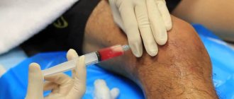
Invasive methods are used in both diagnosis and treatment. Using joint puncture, it is possible to examine the synovial fluid and inject drugs into the joint cavity.
Arthroscopy is a surgical procedure that uses optical equipment to examine the joint cavity and perform interventions on articular cartilage and ligaments.
Differential diagnosis
The differential diagnosis of arthropathy is aimed at clarifying the nature of joint damage, location and cause.
Traditional methods of treating sore joints
In medical practice, as a rule, an integrated approach is used to treat patients suffering from musculoskeletal disorders. Let's name a few popular methods:
- Drug therapy. Medications relieve pain and inflammation and improve the patient’s general condition. Intended for internal (tablets, injections) and external (gels, ointments) use.
- Blockade. Direct injection of the drug into the lesion to relieve pain.
- Physiotherapy. Warming and ultrasound procedures that massage and relax damaged tissues.
- Physiotherapy. Full life of joints is possible only in constant movement. The therapeutic set of exercises must be selected individually, in consultation with a doctor.
- Manual method. Effectively affects the restoration of damaged periarticular tissues. Has no side effects.
- Surgical intervention. An emergency measure used in the most difficult and dangerous cases for the patient’s health.
Timely consultation with a doctor will ensure your joints have a long and fulfilling life. Medical specialists will make the correct diagnosis and prescribe adequate treatment. A high level of service and complete confidentiality are guaranteed.
Infectious arthritis in children
Infectious arthritis occurs in every 5-10 children, girls are especially vulnerable to the disease. In patients under 16 years of age, the disease is often accompanied by loss of appetite, increased irritability, temperature from 37.1 to 38 ° C (sometimes higher), and there may be complaints that the limb is as if paralyzed. Sometimes the disease is hidden and makes itself felt only by minor pain, lameness, and allergic rashes, for which there are other explanations. If a child is atypically lethargic, losing weight, gets tired quickly, or “pulls” a limb, this is a reason for an immediate examination!
Infectious arthritis can result from congenital infections (eg, staphylococcal infections). Children under one year old become inactive, are not interested in food, and avoid putting stress on the affected joints. They behave more cautiously or, on the contrary, restlessly, and begin to whine when touching the sore joint.
More than half of children suffering from infectious arthritis are under 3 years of age—particularly vulnerable to bacterial, viral, and fungal infections.
In no case should you neglect vaccinating children against influenza (Pfeiffer bacillus) and pneumococcus - it helps reduce the risk of infectious arthritis several times. Young children are especially susceptible to staphylococci, streptococci and gram-negative bacteria.
Please note: infectious arthritis in children can develop due to hypothermia, high physical activity, taking glucocorticosteroids and other drugs, as well as post-vaccination complications.
If left untreated, infectious arthritis can leave a minor with lifelong disabilities, especially if there are genetic disorders of the immune system.
Are there tests to determine inflammation?
There are general tests that look for signs of inflammation. They can help to suspect joint diseases and monitor their dynamics and the effectiveness of their therapy. The erythrocyte sedimentation rate (ESR) reflects how severe the inflammatory process is in the body. In healthy people, ESR is usually low and increases in the presence of inflammation. ESR does not indicate any specific disease, but characterizes the severity of any inflammatory reaction in the body. C-reactive protein (CRP) levels also indicate the degree of inflammation present. A high CRP (CRP) suggests that there is significant inflammation or damage in the body. This indicator may indicate, among other things, an exacerbation of joint diseases.
CRP and ESR levels are used to monitor the activity of chronic joint diseases and evaluate the effectiveness of treatment.
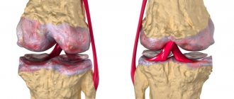
Knee
Knee joint injury is the most common and varied in its manifestations. Everything can happen: from a simple bruise to intra-articular fractures, dislocations and meniscus tears. The joint is a large one, bears the weight of the entire body, and experiences constant stress. Three bones connect here: the femur, tibia and patella. There are five synovial bursae and three types of ligaments: lateral, posterior and intra-articular, the largest of which are cruciate.
The most severe cases are ruptures of the cruciate ligaments, fractures of the condyles or spherical ends, and damage to the meniscus.
The knee has a unique structure, and traumatologists always try to preserve tissue as much as possible. Thus, torn ligaments are replaced with synthetic tape, only small fragments are removed, and large fragments are fixed with metal, the meniscus is only partially removed.
Orthopedics and traumatology services at CELT
The administration of CELT JSC regularly updates the price list posted on the clinic’s website. However, in order to avoid possible misunderstandings, we ask you to clarify the cost of services by phone: +7
| Service name | Price in rubles |
| X-ray of bones and joints of the limbs | 2 200 |
| MSCT of the ankle joint | 7 500 |
| MRI of knee joints (2 joints) | 10 000 |
| Diagnostic arthroscopy | 55 000 |
All services
Make an appointment through the application or by calling +7 +7 We work every day:
- Monday—Friday: 8.00—20.00
- Saturday: 8.00–18.00
- Sunday is a day off
The nearest metro and MCC stations to the clinic:
- Highway of Enthusiasts or Perovo
- Partisan
- Enthusiast Highway
Driving directions
Types of joint diseases in older people by location
An experienced doctor immediately diagnoses the disease, without waiting for test results and x-rays. The fact is that joint diseases in older people usually have a persistent localization.
- Thus, glenohumeral periarthritis and osteochondrosis of the cervical spine, which are accompanied by constant pain in the shoulder girdle, are detected in elderly or mature people who have been engaged in heavy physical labor for a long time.
- Diseases of former athletes include epicondylitis, deforming osteoarthritis and osteochondritis. The pain is usually localized in the elbow joint, which makes movement much more difficult.
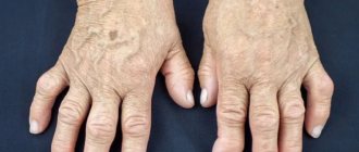
- If in medical practice a doctor encounters diseases such as rheumatoid arthritis, then most likely he is dealing with a former musician, typist, engraver, jeweler, i.e. with those whose professional activity is associated with great tension in the joints of the hands. Inflamed joints, usually of both hands, do not allow you to straighten your fingers in the morning. You need to knead them for a long time and carefully to restore at least some mobility.
- Coxarthrosis, a disease of the hip joint, is considered a real disaster among joint diseases in older people. Softening of the structure of the femur (osteoporosis) threatens a fracture of its neck that is difficult to heal.
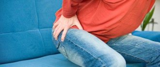
- Unfortunately, people of all ages are familiar with diseases of the knee joint. In older people, gonarthrosis is more common, affecting the destruction of the knee joint.
- Diseases of obese elderly people with a sedentary lifestyle can be called ankle arthrosis, coxarthrosis and gonarthrosis. The diseases accompany walking with severe pain, which entails uncertainty in gait and fear of falling.
How is the joint structured?
A joint is a movable joint between two bones. It is needed in order to redistribute the load on the limbs. The ends of the bones or epiphyses are congruent to each other: where there is a bulge on one bone, there is a depression on the other. The epiphyses are covered with hyaline cartilage. The joint is surrounded by a dense fibrous membrane. The cavity is filled with synovial viscous fluid having high viscosity. The joint receives nutrition from this fluid. There are ligaments around the joint that strengthen it on all sides. The movements performed in the joint are strictly defined - flexion-extension, adduction-abduction, rotation and rotation.
For joint health, their correct geometry is incredibly important. At the slightest violation - a ligament tear, a displacement of the articular surfaces invisible to the eye - the remaining parts of the joint receive chronic overload, and their wear accelerates many times over. The reasons for this are manifold: microtraumas, monotonous daily loads, athletic jerks or excessive physical activity. If the joint is left untreated, the biomechanics and structure of the intra-articular cartilage are irreversibly altered. To stabilize the joint, the body grows osteophytes, or bony spurs, that change the joint so much that movement may become impossible.
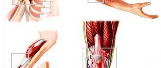
Common Elbow Injuries
Elbow joint injury is one of the most difficult. A neurovascular bundle runs near the joint, which is almost always damaged. Associated neuritis significantly complicates the course of the injury - this is additional pain, impaired trophism, developing contractures or immobility. In addition, the elbow joint does not respond well to physical therapy and this method is not used, which prolongs the recovery time.
The olecranon often breaks off and cannot be put back into place without surgery. Such a fracture can be successfully treated only with the help of metal osteosynthesis, when the fragment is attached to the ulna with a metal structure. The prognosis in this case is favorable, movements return in full after recovery.
The prognosis for complete recovery is less guaranteed for comminuted fractures, especially intra-articular ones. An open access operation is performed when those fragments that are unlikely to grow are removed. The prognosis depends on how much bone tissue had to be removed. The use of alloplants is sometimes possible, sometimes not - it all depends on the clinical picture and the general level of health of the patient.
Traumatologists consider elbow injuries to be the most “fastidious”; complications can arise at any stage.