(average: 4.50 out of 5)
The structure of the joints of the human body allows you to perform many complex movements necessary in the process of life. The possible level of load is indicated by the structure itself, which divides joints into simple and complex. However, some activities involve a significant increase in loads, which can lead to the development of hygroma, affecting the synovial tendon sheaths and joint capsules.
Causes
The pathology is especially unpleasant because it can develop without any obvious reasons, but getting rid of it is quite problematic. Today, experts cannot answer with absolute accuracy what exactly causes hygroma of the hand and other parts of the extremities, but there are a number of prerequisites leading to its formation:
- Constant loads.
- The presence of injuries that were not treated in a timely manner.
- Genetic predisposition.
- Hygroma of the wrist joint of the hand can develop against the background of inflammatory processes.
- Monotonous movements when performing certain types of work.
As statistics show, for programmers, typesetters, and office workers, hygroma on the wrist is a fairly common occurrence. It is for this reason that when constantly performing monotonous processes, it is recommended not to forget about hand exercises and periodic rest.
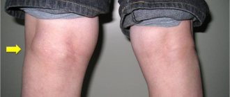
Symptoms of hygroma, photo
A small and uncomplicated hygroma (see photo) does not cause a person any discomfort other than aesthetic. It can be so small that the person himself will not notice it for some time. It does not need to be treated, as it does not cause any inconvenience. If the growth of the hand hygroma continues, then moderate pain in the wrist area may occur. In this case, you should immediately consult a doctor for effective treatment (puncture or surgery).
Symptoms of progressive wrist hygroma:
- dull pain at the site of the cyst;
- a dense oval or round formation appears on the hand in the joint area;
- the skin on the arm at the site of the cyst may change slightly in color;
- loss of sensation in the hand.
In some cases, a hygroma of the wrist may open on its own (usually due to injury). Then an open wound forms on the surface of the hand, which oozes for a certain time - this is exudate flowing out of the cyst. If the hygroma has opened, you need to take special care, as there is a risk of infection. Infection with bacteria can provoke the development of a purulent process and lead to a severe form of the disease.
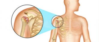
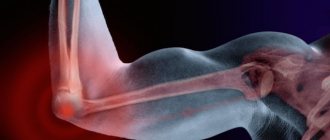
The mechanism of occurrence of hand hygroma
The joint cavity is normally filled with fluid, which moves inside the joint capsule during physical activity and movement. If a defect in the capsule is formed, then a protrusion occurs in this place - a hygroma. It is formed when, as a result of load, this formation is disconnected from the capsule and a closed space is formed. Since the inner surface of the cyst is covered with articular endothelium, the production of synovial fluid continues, and the hygroma gradually increases in size.
Conservative therapy
If hygroma of the wrist, hand, or fingers was detected at the initial stage of formation, the attending physician prescribes a set of physiotherapeutic procedures along with immobilization of the joint.
How to treat hygroma on the wrist, hand and fingers conservatively:
- A course of electrophoresis with iodine.
- Blockades with a glucocorticoid agent (Diprospan, Hydrocortisone).
- Applications with mud and paraffin.
- Soda and salt baths.
- UHF with deep heating of tissues and improvement of local blood circulation.
- Ultrasound therapy.
- Ultraviolet irradiation.
To ensure immobility and rest in the joint, the patient is given an immobilization bandage for a week: a plaster splint or tight bandage. If conservative therapy does not produce results, surgical excision of the tumor is indicated.
Diagnosis and main methods of treatment of hygroma
Diagnosis of a neoplasm at the initial stages of development consists of examining the affected area and palpation. The presence of soft contents detected in this way makes it possible to diagnose hygroma. In addition to examination and palpation, especially in advanced stages of the disease, ultrasound, X-ray, computed tomography, and sampling of material by puncture are also used for subsequent histological examination.
If, as a result of the diagnosis, the development of a tumor was confirmed, then treatment of hygroma on the wrist must be started as quickly as possible. Such efficiency is more than justified, since this disease is prone to becoming chronic and gradually infecting the entire body with the appearance of tumors throughout the body.
In addition, there is a danger of getting damaged by a hygroma. This situation will not bring anything good. If a hygroma on the wrist bursts, for example, due to injury or a deliberate attempt to get rid of a cyst, a wound of impressive size may form, which will bother the patient for a long time with the leakage of contents and pain.
The risk of infection also remains high. Therefore, the question of whether it is necessary to remove the hygroma of the wrist does not even arise. It’s a different matter when it comes to the method of treatment at different stages of the development of the disease. Most often, experts recommend surgery to remove a benign tumor.
Top
Puncture
This method is also used in cases where there is a non-advanced hygroma of the hand; treatment in this case comes down to suctioning out the contents of the tumor capsule using a syringe with a long needle. Puncture is also used to make an accurate diagnosis and exclude the presence of a malignant tumor instead of a hygroma.
Today, this treatment method is used less and less, as it often causes relapses of the disease. This is explained by the fact that after puncture the capsule shell remains in place and over time may again begin to secrete pathological serous fluid. To prevent recurrence of the disease, the patient is recommended to use elastic bandages or bandages for the wrist joint, and, if possible, limit physical activity on the affected joint of the hand.
Diagnosis of hygroma
To make a diagnosis, as well as to differentiate various types of periarticular pathology, use:
- radiography, which allows you to distinguish hygroma from osteosarcoma, hematoma, lipoma, tuberculosis lesion, osteomyelitis;
- computed tomography, which makes it possible to determine not only the structure of the tumor, but also to identify its exact location and connection with the articular structures;
- puncture biopsy, with the help of which the contents of the cyst are obtained for analysis for the presence of atypical cells - this makes it possible to distinguish a benign process from a malignant one. If pus, caseous masses, or blood are detected in the punctate, the diagnosis of hygroma is considered doubtful.
Folk remedies
People who have the disease in its early stages often ask about how to treat hygroma on the wrist with folk remedies. Or expensive operations are impossible. Therefore, the question of how to cure hygroma at home is by no means idle. Traditional medicine offers many methods. Let's look at the most effective of them.
- Dissolve sea salt (2 teaspoons) in half a glass of warm water. Then mix the saline solution with a glass of red clay. Apply the resulting composition to the hygroma and apply a bandage. Keep for at least a day, periodically wetting.
- Treatment of hygroma with dimexide. Dimexide, used as a solution for compresses, effectively removes hygroma. Take dimexide (5 ml) with the addition of dexamethasone or prednisolone (2 ml). Novocaine 2% (2 ml) and aloe (1 ml) are also included in the composition.
- Fresh wormwood must be chopped and thoroughly crushed. Apply the resulting slurry to a piece of cloth. Apply to the site of hygroma formation and fix. Be sure to insulate and keep for more than two hours.
- Various ointments for hygroma also help. The list includes both modern drugs and ointments used for many decades.
- Experts recommend an ointment for hygroma such as chondroxide. The treatment method involves applying chondroxide to the skin and fixing the wrist with an elastic bandage. Usually a week is enough to see noticeable results.
- Hygroma is well cured with Vishnevsky ointment. Vishnevsky's liniment is applied to fabric that repeats the size of the hygroma of the wrist. A tissue with ointment is applied to the tumor, covered with cellophane and secured with an elastic bandage overnight. In the morning the procedure is repeated. Treatment is carried out until the hygroma completely disappears.
- You can also use Flexen gel against hygroma. The method of treatment does not differ from the method of treatment with chondroxide.
Another answer to the question of how to get rid of hygroma without surgery comes from the experience of patients who used ordinary iodine. The method is effective and at the same time simple. It is enough to lubricate the hygroma with iodine at night until it disappears. At the same time, carefully monitor the condition of the skin to avoid burns. Similar negative results were noted when using conventional alcohol compresses, if the alcohol was used in its pure form.
How does melanoma manifest?
The symptoms of melanoma are very varied. It can have different sizes, shapes, colors and surfaces. Dimensions vary from 1–2 mm to several centimeters, and the shape can be very different. As for color, there are options here too. Melanoma is most often black, but can be brown, purple, or even pink. There are often cases when several colors are combined in one neoplasm.
The surface of the melanoma may be unchanged, but over time it may ulcerate, bleed, or become weepy. At the initial stage, the symptom of patent leather is characteristic, when there is no skin pattern on the tumor.
From the point of view of clinical manifestations, the following forms of melanoma are distinguished:
- Superficial spreading melanoma. At first it looks like a brown or black spot or plaque; if it rises above the surface of the skin, it is only slightly. She can remain in this state for up to 7 years. As it progresses, it thickens and turns into a node, and its color may change.
- Nodular melanoma. It has the shape of a node, polyp or mushroom, blue-red or black in color. Gradually its surface ulcerates and begins to bleed.
- Lentigo melanoma. It is the result of malignant degeneration of Durrey's melanosis. For a long time (up to 20 years) it exists in the form of a spot or plaque, and then its vertical growth begins. At the same time, the lesion takes on an irregular shape and uneven coloration.
- Acral melanoma. It develops on the fingers or toes in the nail bed and appears as a dark spot under the nail.
When is surgery indicated?
You should know that conservative treatment, as well as puncture, does not always give the desired result, but only temporarily eliminates the development of the tumor. After all, puncture eliminates only the quantitative indicator of fluid in the cavity; its shell itself remains in the same place and without any special changes. It is advisable to perform the puncture several times. However, without surgery, it is impossible to completely eliminate the problem.
- The risk of relapse after puncture can be reduced only by eliminating the original source that led to the development of hygroma. It is especially important to ensure that the wrist is not exposed to physical stress, but physical therapy must be carried out, and, of course, a timely examination by a doctor.
- As medical practice shows, more than half of people, if there are no acute symptoms, do not pay any attention to hygroma, quietly living like this all their lives. But this is fundamentally wrong, since timely treatment is much better than dealing with the associated consequences. It is especially important not to self-medicate, and also not to completely trust alternative medicine. Untimely, uncontrolled treatment or lack of treatment always leads to complications that cannot be eliminated without surgical intervention.
- Surgery to eliminate a cyst on the wrist completely eliminates recurrence. Indications for surgery include a sharp increase in the volume of the cyst, acute pain, cellularity of the cyst, as well as limited joint movement. Moreover, the longer the operation is delayed, the worse the consequences can develop. In particular, ligaments of blood vessels and periarticular tissues may be damaged.
When performing surgery, an incision is made in the capsule, which allows the joint fluid to be brought out. When making an incision, the doctor separates the tissue around the cyst and removes the neoplasm itself. Before the procedure, the patient receives local anesthesia. Afterwards, the joint is immobilized, which prevents further growth of the hygroma. The finale is suturing the wound and applying a bandage. To improve the state of calm, a cast may be applied for 20 days. Antibiotic therapy is required after surgery.
Treatment of melanoma
The choice of treatment tactics is determined by the stage of the disease. For stages 1–3, the main method is surgery, which, if indicated, can be supplemented with chemotherapy. For 4 common stages, immunotherapy is indicated taking into account the molecular profile of the tumor. If such treatment is not possible, chemotherapy is performed.
Treatment of stages 1–2
The main point in the treatment of stage 1–2 melanoma is radical surgical removal of the tumor. It must be removed within healthy tissues, and the amount of indentation is determined by the data of a morphological study:
- For melanoma in situ, 0.5 cm recede.
- If, according to histological examination, the tumor is more than 2 mm, it is recommended to retreat from its edge by at least 1 cm.
- If the melanoma thickness is more than 2 mm, the indentation should be at least 2 cm.
It is allowed to reduce the indentation when removing melanoma on the fingers in order to maintain their functionality. If an excisional biopsy was used and the diagnosis was confirmed, the scar is excised with the above indentations within 4–8 weeks.
Routine prophylactic removal of regional lymph nodes or their irradiation is not indicated. Instead, a sentinel node biopsy is performed. Only if its results are positive, lymphadenectomy is performed.
Treatment of common forms of the disease
Treatment of patients with advanced forms of melanoma will depend on the possibility of surgical removal of the tumor.
If melanoma is in a resectable state, its radical removal with regional lymphadenectomy is recommended. If metastases were detected during a biopsy of the sentinel node, complete removal of the lymph nodes of the axillary region is carried out with the most complete removal of the fatty tissue in which these nodes were located.
After surgery, the patient is offered immunotherapy and radiation therapy to the area of regional metastasis. RT reduces the likelihood of regional relapse in patients with high-risk factors:
- Damage to 4 or more lymph nodes.
- If the metastasis has gone beyond the lymph node capsule.
Irradiation is carried out in fractionation mode at a total focal dose of 48 Gy.
Patients with high and intermediate risks of melanoma progression are offered adjuvant (postoperative) immunotherapy. The risk group includes patients with the following characteristics of melanoma:
- The thickness of the tumor is from 2 to 4 mm with an ulcerated surface of the tumor.
- The thickness of the tumor is more than 4 mm with any surface.
As part of adjuvant immunotherapy, recombinant interferon preparations and CTLA4 receptor blockers (ipilimumab) are used. This treatment increases the median disease-free survival and overall life expectancy. Adjuvant immunotherapy begins no later than 9 weeks after surgery and continues for up to 12 months.
Treatment of unresectable and metastatic melanoma
Treatment of unresectable and metastatic forms of melanoma is determined by the molecular genetic characteristics of the tumor. The presence of a mutation in the BRAF gene is of primary importance. If the test result is negative (no mutation), a test is performed for a mutation in the CKIT gene.
Treatment of melanoma with a BRAF mutation
First-line therapy uses one of the following treatments:
- Monotherapy with inhibitors
- Monotherapy anti-
- Combined use of BRAF+ inhibitors anti-PD
- Combined use of BRAF+ MEK inhibitors (for patients with rapid progression or large volume of tumor mass).
Treatment is carried out for a long time, until progression or development of severe complications requiring discontinuation of drugs. If the molecular genetic status of the tumor is not specified, these drugs are not prescribed, since there is a possibility of paradoxical acceleration and progression of melanoma growth
These drugs can also cause the development of skin cancer. Therefore, it is necessary to regularly examine the skin for the presence of suspicious growths. If there are any, they are subjected to surgical treatment. Canceling treatment or reducing the dose of drugs is not carried out.
Treatment for the presence of a CKIT mutation
First-line treatment includes anti-PD1 therapy or imatinib (a CKIT inhibitor). Therapy is used until progression or development of complications requiring discontinuation of the drug.
As the disease progresses and BRAF and CKIT mutations are present, nivolumab or pembrolizumab is used as second-line therapy. For slow progression, the CTLA4 inhibitor ipilimumab is used. If immunotherapy is not possible, cytostatics are used.
It should be noted that chemotherapy is less effective in treating melanoma and causes more complications. Therefore, it is not recommended for first-line therapy and is used only when all resources have been exhausted or other treatment is not available.
Treatment in the absence of BRAF and CKIT mutations
In the absence of these mutations, PD1 inhibitors are indicated. If the tumor turns out to be insensitive to them, or progression occurs, they switch to CTLA4 inhibitors (ipilimumab). As part of 3rd line therapy, cytostatics are used.
Hyperthermic perfusion with melphalan can be used to treat unresectable arm melanoma. This method is used as part of palliative therapy for limb preservation in patients who have not responded to immunotherapy and cytotoxic therapy.
Prevention
If people cannot always prevent the occurrence of many diseases, then the likelihood of developing hygroma can be significantly reduced. For this purpose, you need to adhere to certain rules:
- You cannot ignore injuries and consult a doctor in a timely manner.
- When performing monotonous movements with the hand, it is necessary to distribute the load.
- When performing physical exercises, you need to fix the joint with a bandage or elastic bandage.
Even if wrist hygroma is not accompanied by painful symptoms, it is still better not to self-medicate and not to advance the disease. Indeed, in some cases, pathology can lead to complications.
Stages
- Stage 0, or melanoma in situ - there is dysplasia or non-invasive damage by malignant cells.
- Stage 1 – the thickness of the melanoma is no more than 1 mm, or up to 2 mm but without ulceration of the surface.
- The second stage is assigned if the thickness of the ulcerated tumor is more than 2 mm, or less than 2 mm in the absence of ulceration of its surface.
- Stage 3 is assigned if melanoma metastasizes to regional lymph nodes.
- Stage 4 – any melanoma with metastases to internal organs.