Paresis of the left or right foot is a symptom of many diseases of the nervous system. The Yusupov Hospital has created the necessary conditions for the treatment of patients with sagging feet:
- modern methods are used to determine the cause of foot paresis;
- individual approach to choosing a treatment regimen;
- the use of modern medicines that are effective and have a minimal range of side effects;
- innovative methods of physical rehabilitation.
The team of rehabilitation clinic specialists (physical therapy instructors, physiotherapists, massage therapists, reflexologists) works harmoniously and coordinates their actions. Professors and doctors of the highest category at a meeting of the expert council discuss severe cases of the disease and collectively make decisions regarding further tactics for managing patients with foot paresis. Psychologists, using the latest psychological techniques, restore the patient’s mental balance, help to gain confidence in recovery and actively participate in the treatment process.
What is foot paresis
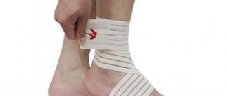
Foot paresis is a complete or partial loss of muscle strength in the foot. The leg seems to drag along the ground, since the patient is unable to lift the front part of the foot. Such disorders are provoked by neurological diseases. As a result, the patient cannot walk normally. If we examine the lower arch, there is a strong bend that does not allow the patient to lower his leg normally to the surface.
One or both limbs are affected by pathology. The disease can develop at any age. This condition is called “foot paralysis.”
Main types of operations
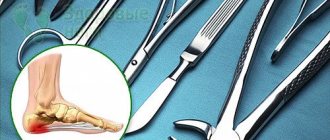
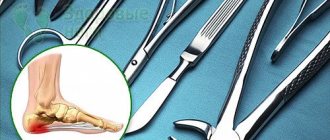
Modern doctors use different surgical techniques during the treatment of spinal diseases. During the intervention, different types of access are used to the affected vertebra. Previously, this could only be done through open access. Now, depending on the location of the operated area, the following types of access are used:
- Posterior – the skin is cut from the back.
- Lateral - used only during treatment of the cervical spine. The surgeon makes an incision on the right or left side of the neck.
- Anterior - the affected area is reached through the abdominal space. This type of access is used in the treatment of the lumbar region.
The decision to choose the type of access is made by the surgeon, taking into account the location of the damaged vertebrae, the severity of the pathology, and the individual characteristics of the patient’s body.
Most often, surgical treatment of pathologies of the spinal column is carried out using the following techniques:
- A discectomy is a surgical procedure in which a doctor removes a portion of the intervertebral disc that extends beyond the spinal column. It is prescribed for disc protrusion or herniation. After the operation, it is possible to release the nerve bundles that are compressed by the cartilage, eliminate the inflammatory process, swelling, back pain, numbness of the legs, and restore their mobility.
- Laminectomy is a surgical procedure to remove the lamina of the vertebra that is located above the spinal cord. The procedure helps relieve compression of the damaged nerve bundle, as a result, blood flow to it improves, swelling disappears, and pain is relieved.
- Spinal fusion is a stabilizing operation aimed at immobilizing adjacent vertebrae and connecting them. Due to fixation with special structures, the affected area is stabilized and the risk of spinal cord injuries is reduced. The operation is prescribed for injuries, degenerative and dystrophic changes in the vertebrae, their discs, deformation, and narrowing of the spinal canal.
- Vertebroplasty is a minimally invasive procedure that stabilizes specific components of the spine. During the intervention, the doctor injects polymethyl methacrylate (bone cement) into the vertebral body or small bones. The injection is made through the skin with a special needle, so the operation is minimally invasive. Vertebroplasty is indicated for compression injuries, osteoporosis, and tumors.
During the operations described above, general anesthesia is used, so the patient will not feel pain during the manipulations. Vertebroplasty is a minimally invasive procedure and is therefore performed under local anesthesia.
To receive high-quality surgical treatment, contact large medical centers with a good reputation. Pay attention to patient reviews, level of service, qualifications of doctors, availability of modern equipment.
Other possible symptoms of foot drop:
- tingling, numbness and mild pain in the foot due to damage to the sciatic nerve;
- disturbances in the flexion of the foot and its toes;
- difficulty climbing stairs;
- loss of sensitivity on the sole and in the area of the outer edge of the foot;
- atrophy of the leg muscles due to intervertebral hernia or spinal cord injury.
Foot paresis requires immediate treatment. Otherwise, the person will lose the ability to move independently and an irreversible foot deformity will develop.
Indications for spinal surgery
Without treatment, pathologies of the spinal column provoke constant back pain, reduce the quality of life, and cause serious consequences, including disability. To avoid this, treatment should be started as early as possible.
Spinal surgery is necessary in the following cases:
- Pinching of the spinal cord or nerve bundles by the vertebrae, the presence of neurological disorders (numbness of the limbs, impaired mobility) or an increased risk of such conditions. Typically, these symptoms include narrowing of the spinal canal and intervertebral hernia.
- Scoliosis, in which the angle of deformation exceeds 40°.
- Rapidly progressive curvature of the spinal column, which disrupts the functioning of internal organs.
- Deformations that worsen a person’s appearance (if desired by the patient), for example, a hump.
- A tumor or cyst of the spine, the presence of neoplasms in the spinal cord, its membranes, their growth into the vessels, nerves, surrounding tissues.
- Severe spinal column injuries, such as compression fractures.
- Increased mobility of the vertebrae in a certain area, disruption of the normal relationship between them.
- Severe back pain that cannot be relieved with conservative methods.
- Conservative treatment does not have an effect within six months.
- The functionality of the pelvic organs is impaired.
- Cauda equina syndrome (back and leg pain, groin numbness, loss of control over urination, defecation, etc.).
- Disc herniation with sequestration (complete loss of the disc core, pinching the spinal cord and nerve bundles).
The decision to choose a method of surgical intervention is made by the doctor, taking into account the capabilities of the clinic, the indications and the wishes of the patient.
Complications of foot paresis
Moving in this way causes great inconvenience to a person, increasing the load on the hips. If the disease is not treated, degenerative changes will only increase. Due to incorrect positioning of the legs, foot deformation develops. At the initial stage, it will not be difficult to return it to an anatomically correct state. But a protracted process leads to irreversible changes.
At the first signs of the disease, you should seek help from a neurologist. He will be able to accurately identify the cause of “foot paralysis.” Elimination of the provoking factor will lead to the cure of foot paresis (intervertebral hernia, consequences of injury), since this disease is not independent.
In severe cases of the disease, paralysis develops and the person loses the ability to move independently.
Spine operations
Modern people lead a predominantly passive lifestyle, this is due to the nature of their work. Therefore, most of them suffer from spinal diseases. The most common pathologies include osteochondrosis, intervertebral hernias, scoliosis, spinal column injuries, etc. However, not all patients rush to the doctor when the first symptoms of the disease appear. They choose to endure the pain, thereby making the problem worse. Many of them go so far as to say that the disease can only be cured with surgery.
There are a large number of surgical techniques that help get rid of even severe diseases. Spinal surgeries are considered quite complex and risky, so it is important to contact a qualified specialist and follow his recommendations after the procedure in order to speed up recovery and avoid dangerous complications. In addition, there are innovative minimally invasive surgeries that help solve the problem with minimal risk to the patient.
Diagnosis of foot paralysis
It is impossible to make a diagnosis on your own, much less identify the cause of foot paresis. The specialists of the Noosphere clinic will help with this. Only a comprehensive diagnosis will be able to accurately identify the provoking factor of “cauda equina.” It is important, before visiting a specialist, to remember how long ago the foot stopped moving normally, the front part of the foot, which was in anticipation of this problem. The following methods are used to carry out diagnostics:
- MRI. Magnetic resonance imaging
- Ultrasound examination (ultrasound)
- Electrocardiogram (ECG)
- Laboratory research
Minimally invasive techniques
Minimally invasive surgeries are safer for patients. Such procedures are performed through a small incision; soft tissues are not as injured as with standard surgery. Blood loss is reduced and recovery time is reduced.
During the treatment of spinal diseases, the following modern techniques are used:
- Laser treatment (vaporization) is used for protrusions and disc herniation at the initial stage. During the procedure, a needle is inserted into the disc through which a laser beam passes. Under the influence of radiation, the inner part of the disk seems to evaporate. As a result, the protrusion decreases and the pressure on the nerve bundles of the spinal cord is relieved. The operation is performed through a small puncture, it lasts no more than 60 minutes, the likelihood of complications is minimal, and the patient recovers faster.
- Nucleoplasty - a conductor is inserted into the disc between the vertebrae, which destroys its internal section. The procedure is indicated in the presence of protrusions or small hernial protrusions. Cold plasma, an electrode or chymopapain (a substance with enzymatic properties) is used as a conductor. Under their influence, the inner part of the nucleus pulposus of the disc is destroyed and the protrusion is retracted back. This is a short-term procedure during which local anesthesia is used. However, after nucleoplasty there is a possibility of relapses.
- Percutaneous discectomy - this operation is performed through a small incision into which a special instrument is inserted to remove the excised disc tissue.
If doctors cannot understand why the patient’s spine hurts, then he is prescribed an epiduroscopy. The therapeutic and diagnostic procedure is also performed when pain occurs after open surgery. It allows you to examine the spinal canal. During an epiduroscopy, the doctor makes a small incision through which the endoscope is inserted. The procedure is carried out under X-ray control. And a contrast image appears on the monitor, which allows you to carefully examine the spinal canal.
With the help of epiduroscopy, adhesions, dead areas, inflammatory processes, fibrosis and stenosis can be identified. This procedure is minimally invasive and is therefore performed under local anesthesia. The advantage is that after diagnosis you can immediately begin treatment. For example, administer anti-inflammatory drugs, stop bleeding, cut connective tissue adhesions with forceps or a cold laser, etc.
Separately, I would like to highlight endoscopic operations, which are increasingly used in the treatment of the spine. During the procedure, special endoscopic equipment is used. Manipulations are performed through 3 small holes in the skin (up to 1 cm), into which instruments are inserted. The operation is carried out under X-ray control, so the surgeon controls the movements of the instruments.
Most often, endoscopic operations are performed for disc herniation and other pathologies that are accompanied by destruction of intervertebral discs.
Pros of endoscopic surgery:
- There is no extensive soft tissue trauma as with standard open surgery.
- The patient recovers in 2–4 days.
- You need to stay in the hospital for no longer than 3 days.
In addition, after endoscopy, the risk of anesthetic and postoperative complications is reduced.
Treatment of foot paresis
Foot paresis can be treated in two ways - surgery and conservative therapy. In many ways, the choice of technique is influenced by the reasons that caused the disease and the degree of deformation of the foot. The Noosphere clinic uses only conservative treatment methods.
An individual course of therapy is selected for each patient, based on the examination results and the patient’s condition. A full course of recovery takes up to 1.5 months. The procedure schedule is 2-3 times a week. The general treatment regimen includes the following therapeutic procedures:
- Resonance wave UHF therapy
Resonance wave therapy is a method of therapeutic effects on the aquatic environment of the body with low-intensity, high-frequency electromagnetic waves.
- Fermatron injections
Fermatron intra-articular injections are an effective method of treating various diseases of the musculoskeletal system by introducing a drug (chondroprotector) into the affected joint.
- Rehabilitation on the Thera-Band exercise machine
Treatment of the spine and joints using the Thera-Band simulator will restore limb mobility in a short period of time without expensive treatment in specialized sanatoriums.
- Block of joints and spine
Joint blockade is a type of drug treatment of the spine and joints aimed at relieving acute pain, inflammation and muscle spasms.
- Drug treatment
Drug treatment of joints and spine at the Noosphere clinic is used in a wide range and in combination with physiotherapy. Intra-articular injections, blockades and droppers.
Treatment of foot paresis at the Noosphere clinic (St. Petersburg) involves the return of normal muscle tone, restoration of the anatomically correct position of the leg and general improvement of the body. As a result, metabolic processes and blood circulation improve.
To consolidate the positive results of treatment, the patient is recommended to undergo a course of rehabilitation exercises. Within a year after completing the course, the patient can visit the clinic’s specialists free of charge. To get rid of horse foot forever, you need to strictly follow all the recommendations of the doctors at the Noosphere clinic.
Customer Reviews
We strive to provide quality services with a high level of service. We are grateful to our patients for their trust and positive feedback on our work together.
He was admitted with severe pain in the cervical and shoulder joints. The examination revealed a cervical hernia. The examination and operation were carried out by doctor Sergey Borisovich Mulin. After the operation the pain went away. Thanks to the doctor and all the hospital staff for the treatment provided.
I was admitted to the clinic with a problem of varicose veins of the lower extremities.
The doctor, V. G. Khudashev, carefully and competently examined my problem and performed an ultrasound of the lower extremities.
Surgery was prescribed for both limbs, laser surgery.
The postoperative period went well, without any complications. I would like to express my gratitude to V. G. Khudashev for the skillfully performed operations, as well as to the anesthesiologist E. N. Bessonova.
