Age-related hyperkyphosis affects 20–40% of older adults and can be described as an excessive frontal curvature of the thoracic spine that occurs during the aging process.
- The frontal curvature of the thoracic spine is natural (kyphosis), it is caused by the shape of the vertebral bodies and intervertebral discs.
- Kyphosis can turn into hyperkyphosis if its angle exceeds 40°.
The main causes of age-related hyperkyphosis are poor posture, dehydrated intervertebral discs and weak extensor muscles. Age-related hyperkyphosis may:
- lead to decreased mobility of the chest (it is connected to the thoracic spine), which can cause difficulty in the functioning of the lungs;
- increase the biomechanical load on the spine, which can lead to compression fractures;
- increase the risk of falls and fractures due to difficulty walking;
- reduce the quality of life by limiting the functionality of the body;
- be a risk factor for premature death. As the angle of kyphosis increases, the likelihood of death increases. Some studies have linked this growth to impaired lung function.
Clinically Relevant Anatomy
Clinically Relevant Anatomy
The spine consists of vertebrae and intervertebral discs, has a curved shape and is divided into several sections.
Friends, on June 3, Dmitry Gorkovsky’s webinar “Strategies for treatment and training of adolescents and adults with scoliosis” will take place. Find out more...
The important elements of the thoracic spine are bones, joints, nerves, ligaments, intervertebral discs and muscles. The thoracic region is characterized by relatively high rigidity compared to other regions due to its attachment to the rib cage, ligaments and thin, inactive vertebral discs. It is the thoracic region that is most often susceptible to hyperkyphosis.
Epidemiology/etiology
- The kyphosis angle varies from 20° to 29°.
- Hyperkyphosis occurs in 20–40% of people, regardless of gender.
- After 40 years, the angle of kyphosis in women usually increases faster than in men.
- The angle of hyperkyphosis varies from 43° to 52° in women aged 55–60 years and reaches 52° in women aged 76–80 years.
Excessive curvature of the thoracic spine can be associated with many physiological factors and reasons:
- Osteoporosis and vertebral fractures. However, only 1/3 of patients with hyperkyphosis have radiographically confirmed fractures.
- Depression, feelings of insecurity, sadness and anxiety.
- Gradual changes in the structure and mechanics of connective tissue, over time leading to loss of elasticity and mobility, causing an inability to resist the force of gravity pulling the body forward.
- Muscle weakness (most often due to weakness of the back extensor muscles).
- Disturbances in the functioning of the somatosensory, visual and vestibular systems, leading to weakened control over posture.
- Loss of proprioceptive and vibration sensitivity of the joints of the lower extremities, making it difficult to maintain a straight vertical body position.
Characteristics/clinical picture
Clinical picture
Thoracic hyperkyphosis is characterized by the following main symptoms:
- Increased frontal curvature of the thoracic spine. It can develop over a long period of time and is most often noticed not by the patient himself, but by his relatives (sudden curvature of the spine can be a sign of not only hyperkyphosis, but also other diseases).
- Difficulty getting up from a chair without using your hands.
- Loss of balance (the patient feels like he might fall).
- Reduced walking pace.
- Reduced walking speed on stairs.
- The need to use walking aids and maintain body position to avoid falling.
- In severe cases of hyperkyphosis, difficulty breathing occurs due to decreased lung volume.
Reasons for the development of stage 2 kyphosis
The main cause of grade 2 kyphosis is the lack of treatment for mild curvature at the initial stage. Pathology can progress quickly. Provoking factors are: lack of control of correct posture, weakness of the muscular corset, injuries and heavy loads on the spinal column.
Pathological biomechanics
The presence of an imbalance of muscle mass and changes in the geometry of the spinal column lead to the progression of curvature. The spine develops incorrectly, the curve worsens.
Injuries
The pathology manifests itself against the background of strong mechanical or physical impact - blows to the back, jumping or falling from a height. Compression of the vertebrae leads to pathological curvature of the spine.
Excessive loads
Lack of muscle support causes improper distribution of the load on the skeleton during the period of active growth of the spine. If muscle formation does not keep pace with the growth of the spinal column, the load becomes excessive, the spine cannot cope and bends.
Diagnostics
X-ray of the spine in lateral projection
There are several generally accepted methods for diagnosing hyperkyphosis. One of these methods is radiography of the spine in a lateral projection in a standing position. It is a kind of gold standard for the quantitative assessment of thoracic kyphosis by measuring the Cobb angle.
Cobb angle
In addition to radiography, other non-invasive methods for clinically measuring the Cobb angle are used, in particular, the Debrunner kyphometer, Flexicurve and Spinal Mouse® devices are used.
- A kyphometer measures the angle of kyphosis. The ends of the kyphometer are located on the upper and lower extremities of the thoracic spine (from above - on the 1st thoracic vertebra, from below - on the 11th/12th vertebra). By applying the appropriate mathematical formula, it is possible to estimate the Cobb angle, which matches the X-ray data quite accurately.
- Flexicurve is a ruler made from a strip of flexible metal coated with plastic. It is applied tightly to the back along the spine (from the 7th cervical vertebra to the lumbosacral joint) to take a shape that matches its curvature. The kyphosis index is calculated as the width of the ruler divided by the length of the thoracic curve and multiplied by 100. Hyperkyphosis corresponds to an index value greater than 13.
- The Spinal Mouse® device is an electronic device that, together with an appropriate computer program, allows you to evaluate the curvature of the spine without the use of x-rays.
Note: There is a significant correlation between the above methods for measuring kyphosis angle and radiological studies (intraclass correlation coefficient is 0.68).
Outcome assessment
- Scoliosis Research Society Outcomes Instrument (SRS-22). The SRS-22 assesses a patient's pain, function, self-perception, mental health, and satisfaction with treatment. There is evidence that hyperkyphosis is associated with increased pain, decreased self-perception, and decreased overall function and activity of the body. The values determined by this questionnaire for kyphosis (0.40–0.66) are significantly higher than for scoliosis (0.16–0.26). Thus, the SRS questionnaire is a very effective method for assessing hyperkyphosis.
- Occiput to Wall Distance test. The patient leans his back against the wall, then the doctor measures the distance from the back of the patient's head to the wall.
- Tragus to wall test. Similar to the previous one, the distance is measured from the tragus of the ear to the wall.
- Neck Pain and Disability Scale.
- “Get up and go” test (the patient gets up from the chair, walks 3 meters and returns to his place; the test is carried out for a time).
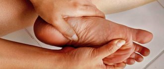
Radiculopathy (radicular syndrome)
The compression of the nerve root described above can be caused not only by a hernia. Pinching is the direct cause of back pain caused by:
- spondylosis;
- ankylosing spondylitis;
- injury;
- neoplasm;
- syphilis, tuberculosis, meningitis, osteomyelitis (inflammatory nerve damage).
Nerve damage causes a burning, stabbing pain. The affected area gets very hot and shoots, as if it had been struck by lightning.
Clinical assessment
Clinical assessment
Hyperkyphosis can be qualitatively assessed by visual examination of the patient. The inspection can be carried out in the following ways:
- Assessment of the frontal curvature of the spine. It is worth keeping in mind that the curvature of the thoracic region can be affected by changes in other areas of the spine (for example, in the lumbar region);
- Test “from the tragus to the wall” or “from the back of the head to the wall”;
- Determining the number of 1.5 cm blocks to support the head.
There are currently no studies comparing the above measures with radiography.
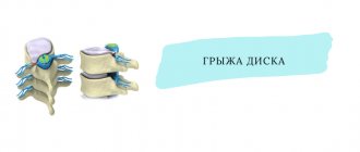
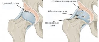
Herniated disc
Hernial protrusion is caused by the destruction of the fibrous ring of the disc, which, under the weight of the overlying vertebrae, is flattened and extends beyond its anatomical boundaries. Hernias are detected during examination for osteochondrosis and are its complication. Hernias, like protrusions, often develop asymptomatically and may not manifest themselves in any way. Otherwise, patients complain of dull pain at the location of the hernia. It increases with walking and standing, and goes away after resting in a lying position. If a nerve root near the disc is pinched, the pain is difficult to endure because it resembles an electric shock and is sharply shooting, piercing in nature. In the area of innervation of the pinched nerve, numbness and muscle weakness are possible.
Treatment
Pharmacological treatment primarily prescribes anti-resorptive or bone-regenerating drugs, since many patients with age-related hyperkyphosis have low bone density or vertebral fractures.
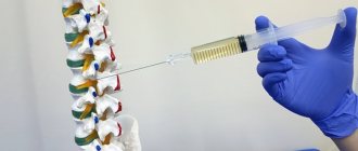
As part of surgical treatment, there are two options: vertebroplasty or kyphoplasty (mainly to relieve pain, reduce the angle of kyphosis and eliminate body dysfunction).
- Kyphoplasty helps relieve pain and deformity in patients with spinal fractures.
- Vertebroplasty involves injecting acrylic bone cement directly into the vertebral fracture space. The surgery has the same risks as kyphoplasty, including the possibility of damaging the spinal cord and leaking cerebrospinal fluid.
Physical therapy
In a systematic review of the effects of exercise in the treatment of hyperkyphosis (Bansal et al., 2014), the authors report that there are currently only a small number of robust studies documenting a beneficial effect of exercise in treating hyperkyphosis (this effect, however, , is very modest: the kyphosis angle decreases by 1.67°–3.74°). Initially, the patient should exercise under the supervision of a physical therapist or certified athletic trainer for the greatest benefit and safety. The load and frequency of these exercises are selected depending on the individual characteristics of the patient.
Taping for hyperkyphosis
The main objectives of physiotherapeutic care are:
- Increased strength of the back extensor muscles.
- Increased mobility of the spine.
- Strengthening postural control.
- Preventing compression fractures.
Exercises for hyperkyphosis
Physical therapy care strategies include:
- Correcting posture through stretching and strengthening exercises to reduce hyperkyphotic curvature and prevent its development.
- Breathing exercises to adapt to physical activity by increasing lung capacity (for example, diaphragmatic breathing exercises).
- Relieving pain using heat, cold, and/or electrical stimulation (eg, transcutaneous electrical nerve stimulation).
- Manual therapy (including soft tissue mobilization techniques) to increase spinal flexibility.
- The use of specialized orthopedic products or therapeutic taping to reduce the kyphotic angle.
- Informing the patient about the need to maintain correct posture and daily physical activity to maintain functional activity.
- Balance and walking exercises to maintain overall fitness and reduce the risk of sudden falls.
Manual therapy
The following exercises are recommended for the treatment and prevention of hyperkyphosis (the effect of exercises was studied in randomized controlled trials, level of evidence - 1B):
- Hatha yoga adapted for people with hyperkyphosis. Emphasis should be placed on breathing techniques, stretching tight muscle groups, and strengthening weak muscle groups (such as the rhomboids).
- Manual mobilization techniques. Mobilization should be carried out in accordance with the Maitland Joint Mobilization Grading Scale.
- Active mobilization/stretching of the pectoral muscles, shoulder girdle and latissimus dorsi.
- Strengthening muscles: paravertebral muscles, abdominal muscles, scapula muscles, other muscles of the upper extremities.
- Maintaining correct posture (standing, sitting) using visual (images, mirrors), auditory (therapist's advice) or tactile (taping, therapist's help) posture control measures.
To train correct posture you should:
- Explain to the patient what postures can lead to hyperkyphosis.
- Demonstrate the correct posture with an explanation of each necessary movement: knees slightly bent;
- the shoulders are pulled back (the shoulder blades are retracted), you can also perform external rotation of the shoulders at the same time;
- chest arched forward;
- the chin is slightly lowered.
Prevention and diet
For the purpose of preventive measures, from childhood you should monitor the child’s position at the table during lessons, sleep on a fairly solid base, and engage in regular physical exercise.
When working sedentarily, you should take breaks every hour, with exercise if possible. Avoid heavy weights. You should always control your back position.
Pay attention to what your posture should be
Obesity can lead to spinal deformation, so you should eat right, always including vitamins, water and fruits in your daily diet.