Frontal hyperostosis is a rare pathology, but sometimes it is dangerous for the patient. Due to its low prevalence, it has not been studied enough, so there are not many serious studies in this area. In ICD-10, the disease is defined as No. M85 along with other disorders of bone density.
The very concept of “hyperostosis” means bone growth of a pathological nature. It is often accompanied by tumor processes and serious hormonal disorders. Hyperostosis also occurs as a consequence of malignant neoplasms.
Causes of diffuse ankylosing hyperostosis
Diffuse hyperostosis, according to many doctors, is a hereditary disease. This form is distinguished by simultaneous damage to several tubular bones of the upper or lower extremities, skull or spine. For timely diagnosis, you should pay attention to family history. If one of the baby’s parents or other close relatives on the father’s and mother’s side is sick with a similar disease, then a diagnosis should be made during the first year of life and the possibility of developing the process of bone tissue proliferation should be excluded.
Ankylosing hyperostosis can be caused by the following reasons:
- increased physical activity under conditions of insufficient development of the muscular frame of the body;
- infectious and aseptic inflammatory processes in the area of the bone heads (including necrotic changes that occur after compression or serious injury);
- fractures and cracks of bones, formation of hematomas due to ruptures and stretching of soft tissues (ligaments, tendons, fascia and muscles);
- chronic intoxication with salts of heavy metals, which provoke disruption of the process of differentiation of bone tissue;
- osteomalacia against the background of hematogenous and lymphogenous spread of infection of tuberculosis, syphilis or osteomyelitis;
- the effect of radiation exposure on bone marrow structures and areas of the periosteum;
- disruption of hematopoietic processes with long-term use of non-steroidal anti-inflammatory drugs (they are often prescribed for the treatment of orthopedic diseases);
- hormonal disorders, in particular thyroid dysfunction, increased levels of pituitary hormones in the blood;
- diabetes mellitus and diabetic angiopathy developing against it;
- disruption of the blood supply to certain areas of the body, for example, with obliterating endarteritis, extensive hyperostosis of the bones of the toes is observed;
- improper prevention of rickets in children of the first year of life.
The idiopathic form of hyperostosis develops against the background of a combination of several pathological factors. Usually, with an unspecified etiology, doctors talk about the hereditary nature of the occurrence of this disease. It is more difficult to treat such forms, since it is not possible to eliminate the potential cause of impaired differentiation of bone tissue.
Varieties and classification
Hyperostosis is considered a rather rare disease of bone tissue.
No serious statistical studies have been conducted on the prevalence of this pathology. In the International Classification of Diseases (ICD-10), hyperostosis is included in the class of diseases of bone and connective tissue with code M 85 “other disorders of bone density and structure.”
Until now, there is no clear answer to the question “Why does hyperostosis occur?” scientists cannot. But the main factors of the disease leading to impaired bone tissue growth have been sufficiently studied.
These include:
- pathology of the endocrine system with acromegaly (disease of the anterior pituitary gland), increased function of the parathyroid glands, decreased activity of thyroid hormones;
- consequence of chronic poisoning with toxic substances containing compounds of phosphorus, arsenic, fluorine, lead, bismuth;
- hypervitaminosis of vitamins A, D;
- excessive physical activity;
- exposure to penetrating radiation or excessive radiation dose;
- response to chronic inflammatory processes, long-term infectious diseases, acute viral infection;
- kidney pathology, which complicates the process of removing excess phosphates through the tubules;
- blood diseases (myeloma, lymphogranulomatosis, leukemia, sickle cell anemia);
- cancerous tumors;
- Paget's disease at the last stage of sclerosis;
- manifestation of neurofibromatosis;
- consequences of diseases (echinococcosis, cirrhosis of the liver).
- local, local forms - when one bone is affected, for example, hyperostosis of the frontal bone develops in women during menopause, in men with obesity;
- general or generalized process - found in children in infancy, when the balance of sex hormones is disturbed, as a result of genetic mutations.
Restriction of hyperostosis to only tubular bones is called periostosis. The disease affects the legs, forearms, and deformation of the fingers is detected.
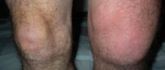
Local hyperostosis of the knee joint (tibia and femur)
In most cases, local hyperostosis develops against the background of pathologies of the lumbosacral spine and pathologies associated with impaired blood supply and innervation of one of the lower extremities. There is a gradual decrease in muscle mass and dystrophy occurs. The affected limb cannot fully cope with the loads. They are transferred to the healthy leg. In this case, hyperostosis of the knee joint begins to develop, which for a long time manifests itself only by a slight change in gait.
When walking, the patient tries to transfer most of the load to the healthy leg, which is why the effect of lameness occurs. The first pain in the development of tibial hyperostosis occurs after prolonged physical exertion.
Symptoms of femoral (and tibial) hyperostosis may then include:
- local swelling, the presence of compaction in the area of bone tissue growth;
- increased muscle tone in the lower leg and thigh area;
- regular development of attacks of clonic convulsions in the femoral and calf areas;
- increase in joint volume;
- violation of the process of flexion and extension of the leg;
- the appearance of the first signs of contracture, which soon lead to complete immobility of the limb in the knee joint;
- the skin is pale, tense, and the process of capillary blood supply is disrupted.
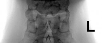
To make a correct diagnosis, it is necessary to take an x-ray. Differential diagnosis is carried out to exclude deforming osteoarthritis of the knee joint (gonarthrosis) and oncological neoplasm.
Causes of pathology
The main reason for the appearance is heredity or the body’s response to acute osteomyelitis and damage in the radiation zone. Additional reasons are:
- injuries resulting in the development of inflammatory processes;
- exposure to arsenic, lead;
- oncology;
- syphilis;
- echinococcosis;
- leukemia;
- can develop when the body reaches the stage of oterosclerotization;
- kidney and liver diseases that develop into chronic ones. Also lack of the required amount of vitamin A, D.
Often the disease is diagnosed in athletes or people who have a constant load on one of the bones of the skeleton. For example, if one leg is in constant motion and the other is at rest, then development is guaranteed. Therefore, the load should be uniform and the same for the body as a whole.
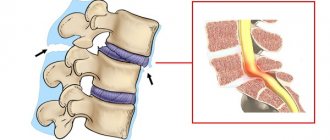
Fixing hyperostosis of the thoracic spine
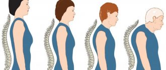
Hyperostosis of the thoracic spine is a consequence of degeneration and dystrophy of the cartilage tissue of the intervertebral discs. Less commonly, it develops after a back injury. Primary changes begin with wedge-shaped deformation, which occurs with a significant decrease in the height of the fibrous ring of the intervertebral disc. Then the process of thickening the vertebral bodies begins in order to replace the function of the intervertebral disc.
In infants, fixing hyperostosis of the thoracic spine is almost always triggered by falling onto the back. It is very difficult to recognize such pathologies in a baby. It often manifests itself as nervousness, tearfulness and crying of the child when trying to put him on his back. When you turn onto your stomach, all this goes away. If your child cannot lie quietly on his back, then you need to see an orthopedist. It is likely that he develops fixation hyperostosis, which causes pain when lying on his back.
Fixing hyperostosis in an adult can give the following clinical symptoms:
- feeling of lack of air and inability to take a deep breath;
- pain when trying to take a sharp deep breath;
- frequent relapses of intercostal neuralgia;
- smoothing the physiological curvature of the spinal column in the thoracic region;
- feeling of a straight back;
- difficulties when trying to bend the body or make a turn to the left and right;
- pain in the heart area, etc.
With a unilateral process of bone tissue proliferation, secondary scoliosis and deformation of the chest can form. Diagnosis is made using an x-ray. It shows foci of pathological changes in bone density.
Tests and diagnostics
Despite the level of development of modern medicine, it is quite difficult to recognize hyperostosis after studying the clinical picture. X-ray examination becomes the most revealing, and tomography is used to accurately determine the localization of the pathological process within the bone structure. In some cases, additional radionuclide testing may be required.
Cervical spine with Forestier hyperostosis
Forestier's disease on an x-ray is characterized by the presence of thickening of the anterior longitudinal ligament and its fusion with the ventral surfaces of the vertebral bodies, but the height of the intervertebral spaces is not reduced. Laboratory values sometimes reveal hyperglycemia .
When making a differential diagnosis with other pathologies that deform the spine, take into account the absence of laboratory signs, manifestations of inflammatory activity, sacroiliitis , ankylosis of intervertebral joints , and narrowing of intervertebral discs.
Cortical reactive hyperostosis of the lumbar region
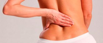
Cortical hyperostosis is characterized by a long asymptomatic course and very severe consequences. Reactive hyperostosis often occurs, provoked by inflammatory reactions in the area of bone tissue deformation. Most often it is diagnosed in the cervical and lumbar spine, which bear the maximum physical load.
Hyperostosis of the lumbar region leads to the inability to bend over and perform a number of other movements. The process of secondary compression of the radicular nerves quickly begins. This is accompanied by constant exacerbations of osteochondrosis. The doctor prescribes typical anti-inflammatory treatment, but once you take an x-ray, it becomes clear that compression of the radicular nerves is a consequence of bone tissue proliferation. In such situations, emergency initiation of treatment is indicated.
Hyperostosis of the lumbar spine is characterized by the following symptoms:
- constant pain in the lumbar and sacral areas;
- numbness of the lower extremities (more often the process is bilateral);
- dysfunction of the internal organs of the abdominal cavity and pelvis;
- inability to lean forward and in different directions;
- increased fatigue of the back muscles;
- constant hypertonicity of the muscular frame of the back;
- palpation of bone growths on the bodies and spinous processes of the vertebrae through the skin.
An x-ray is often sufficient for diagnosis. In some cases, to exclude an oncological process, the doctor will ask for a puncture and histological blinding of the resulting tissue.
Approach to therapy
It is impossible to make a diagnosis based only on the patient’s external symptoms and complaints.
The following methods are used for final diagnosis:
- radiography,
- CT scan,
- Radionuclide testing is rarely carried out.
If the pathology is a complication of another disease, then the underlying disease must be treated.
The result comes with the use of anti-inflammatory drugs, hormonal agents that eliminate the effect of somatotropin, and antibiotics for chronic pneumonia.
In the treatment of the primary disease the following is used:
- high doses of corticosteroid hormones;
- restorative treatment is carried out;
- A diet with sufficient amounts of protein and vitamins is recommended;
- joints are developed using massage and physiotherapy.
In most cases of early diagnosis, good results can be achieved after several months of treatment. The prognosis for hyperostosis is favorable.
Treatment of Forestier disease requires a special approach:
- to eliminate pain - non-steroidal anti-inflammatory drugs, it is possible to use spinal blockades;
- a group of chondroprotectors is prescribed to strengthen the intervertebral discs;
- pentoxifylline is used to improve blood circulation in the spinal cord and spine;
- It is recommended to wear special orthopedic bandages that support the spinal column.
Treatment of hyperostosis
Currently, conservative treatment methods are mainly used to treat bone hyperostosis. Corticosteroids (hormones) are prescribed, which are designed to suppress bone growth. But in most cases, the effects of manual therapy are sufficient to stop the pathological process and reverse it.
In our manual therapy clinic, the following techniques are used to treat hyperostosis:
- diet therapy – the patient receives comprehensive individual recommendations on how to properly build their diet and diet;
- osteopathy and massage - accelerate metabolic processes, enhance microcirculation of blood and lymphatic fluid in the lesions;
- therapeutic exercises and kinesiotherapy prevent the formation of contractures and maintain full body mobility;
- reflexology restores metabolic processes and triggers the regeneration of damaged tissues;
- physiotherapy allows you to create suitable conditions for the restoration of bone tissue.
In some cases, shock wave therapy, laser treatment and electrical myostimulation are used.
If you need treatment for hyperostosis, we recommend making a free appointment with an osteopath or orthopedist at our manual therapy clinic. During the initial appointment, the doctor will make an accurate diagnosis and talk about the prospects for conservative treatment in your individual case.
Depending on the stage of the disease, we choose one or more treatment methods:
Osteopathy
Soft technique of working with the spine, joints, muscles, ligaments, internal organs. Eliminates pain and starts the self-healing process.
Therapeutic massage, osteopathy, manual therapy
Helps bones and joints take the correct physiological position, relieves pain and spasms, relaxes muscles.
Acupuncture
Work on biologically active points. It affects the affected area and the body as a whole. Eliminates the cause of the disease and removes the symptoms.
In addition, according to indications, the following are used: taping, pharmacopuncture, FormTotix insoles, exercise therapy with an instructor and other methods. The choice of procedures depends on the current condition; taken together, they act faster and give a more lasting result.
how to find a good osteopath in Moscow
Structure and anatomy
The frontal bone is a part of the skull and its base, which consists of four sections:
- Two orbital.
- Arched nasal.
- Frontal scales. Bone lobules located vertically. They are the ones we are interested in.
The frontal scales consist of:
- The outer smooth surface, which has an elevation in the lower part, is the remains of a frontal suture. As a child, he divided the bone into two halves.
- Two temporal ones.
- The inner surface has a concave shape along the midline of the upper part.
It is this internal part, to which the falciform process of the meninges is attached, that will be discussed below. But first, it is important to understand what hyperostosis is in general and how it manifests itself on the inner surface of the frontal bone.