Instability of the spine is a displacement of its articular elements - the vertebrae - with excessive activity. Normal mobility does not cause pain, but instability causes severe pain.
In this case, the vertebrae arbitrarily move forward or backward, even at rest, pinching the nerve roots during their movement.
In medical practice, 5 types of spinal instability are classified:
- pathological;
- dysplastic;
- degenerative;
- spondylolysis;
- traumatic.
The causes of this anomaly may be previous injuries, the consequences of oncological pathologies and inflammatory processes. In addition, the following factors can provoke spinal instability:
- disturbed structure of the vertebral joints;
- ligament weakness;
- degenerative changes;
- osteochondrosis;
- consequences of surgical procedures.
This disease occurs against the background of excessive physical activity, as a result of intense training.
As the disease develops, the intervertebral disc wears out, the intervertebral gap decreases, and the ligaments relax and do not properly support the affected part of the spine. Against this background, pathological instability of the vertebrae occurs in a specific part of the spinal column.
What it is?
Spinal instability is a pathological condition of the spine in which the spinal column is unable to maintain its physiological position either in motion or at rest..
The vertebrae can move laterally and anteroposteriorly. In this case, pain occurs, the source of which is irritation of the capsule of the facet joints or deformation of the spinal canal and compression of the nerve roots.
Spinal instability can be a consequence of degenerative changes in the spine , dysplasia, previous surgeries and injuries. The pathology occurs in patients of different ages and, in the absence of therapy, leads to disability of the patient.
Clinical picture
Instability of the spinal column is accompanied by constant mechanical irritation of pain-sensitive structures of the spinal motion segment (muscles, ligaments, fibrous ring, capsule of facet joints).
The result is the formation of chronic pain syndrome:
- Radicular pain in the lower extremities (one or two). Occurs when the nerve roots of the spinal cord are compressed.
- Backache. Appears or intensifies with prolonged walking, as well as in an extension-flexion position. It can be reflected, often spreading to the thighs and buttocks.
Pain due to spinal instability occurs due to radicular syndrome
Classification
The classification of spinal instability is based on the causes of this disease.
Spinal instability may be:
- Post-traumatic . It occurs against the background of fractures, subluxations and dislocations of the vertebrae caused by sports injuries, road traffic accidents, etc.
- Dysplastic . The prerequisite for its appearance is the pathology of the connective tissue located in the vertebral bodies, vertebral joints and intervertebral ligaments.
- Postoperative . It occurs as a result of laminectomy and an incorrect rehabilitation period (due to early excessive load on the spinal column).
- Degenerative . Develops in people with degenerative processes in the spine (usually against the background of osteochondrosis).
Prevalence and significance
Fortunately, spinal instability is not the most common phenomenon today . It is much less common than other musculoskeletal diseases.
In most cases, spinal instability is found in newborns and infants who have immature ligaments and who have suffered birth injuries to the spinal column.
Video: “What is spinal instability?”
Clinical picture
Instability of the spinal column constantly irritates pain-sensitive muscles, ligaments, the fibrous ring and the capsule of the facet joints, forming a chronic pain syndrome.
Pain is located in different locations and has its own characteristics:
- radicular pain: localized in the legs, occurs due to compression of the spinal roots;
- back pain: felt during long walks and flexion-extension, for the same reasons it can intensify. It is often reflected and spreads to the buttocks and thighs.
Risk factors, causes in adults and children
The causes of spinal instability include:
- various spinal injuries (car accident, heavy lifting, fall, etc.);
- osteochondrosis;
- age-related changes;
- increased sports loads;
- underdevelopment or congenital weakness of the articular-ligamentous apparatus;
- surgeries on the spinal cord and spine, etc.
In children, spinal instability is usually caused by injuries (including those received during childbirth), sports activities and birth defects.
Instability does not appear by itself. Its occurrence is due to a number of predisposing factors.
These include:
- metabolic disorders;
- sedentary lifestyle;
- deficiency of essential vitamins in the body;
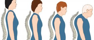
- curvature of the spine at any stage;
- atherosclerosis, which is accompanied by deterioration of blood supply;
- dysfunction of the endocrine system;
- failure to follow the recommendations of the attending physician regarding recovery after surgery.
Thus, spinal instability occurs more often in old age and childhood . In the first case, it can be caused by age-related changes occurring in the body, such as muscle sagging and ligament weakness. In the second case, instability appears against the background of rapid development and growth of all tissues of the body. Most often, children experience abnormal mobility of the cervical vertebrae, which can lead to the development of infantile torticollis.
Why is spinal instability dangerous?
Without proper treatment, spinal instability can lead to serious complications. The most common diseases that can develop against the background of vertebral instability are:
- Spinal cord compression
- Cervicalgia
- Muscular-tonic syndrome
- Radiculopathy
- Periarthritis
With timely diagnosis and treatment of the disease in a specialized clinic, it is possible to completely eliminate the pain syndrome and return normal mobility to the spine.
vertebrology spinal diseases spinal instability
Consequences
If the vertebrae are unstable, osteophytes may grow. Pathological mobility of the vertebrae is fraught with accelerated development of osteochondrosis and the appearance of arthrosis of the intervertebral joints. Spinal instability increases the load on muscles and ligaments, causes impaired muscle tone, and contributes to pain. Painful sensations can be triggered by prolonged sitting or performing simple movements.
Spinal instability can limit movement , cause spasms and various neurological disorders (epicondylosis, cardiac syndrome and others). If excessively mobile vertebrae are located in the neck, headaches with a general feeling of weakness and nausea may occur.
Among the most dangerous consequences of the disease is spondylosis . This pathology causes changes in the tissues of the discs, which leads to the formation of spiky bone growths on the sides of the vertebrae.
Video: “Instability of the cervical spine”
Diagnostics
Diagnosis for spinal instability includes:
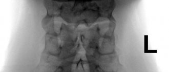
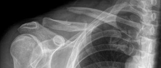
- functional spondylography: an x-ray of the entire spine in a straightened and flexed position. The study allows you to detect spondylolisthesis and exclude mobility of other vertebrae;
- Magnetic resonance imaging: this is a less informative study for vertebral mobility, since displacement is not always visible in the supine position. But an MRI can show pathologies in the tissues surrounding the spine.
Symptoms and diagnostic methods
Typically, the following symptoms indicate the presence of spinal instability::
- back pain (in different parts of the spinal column), which may intensify after exercise;
- limited mobility when rotating and bending the body;
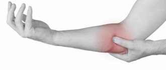
- leg pain;
- lower back pain, especially when lifting heavy objects;
- a feeling of discomfort in the lower back, neck or other segment (where the vertebrae are unstable);
- dizziness, headaches (with displacement in the neck area).
The source of pain in spinal instability is compression of the nerve roots and compression of the spinal canal . In the area of the damaged area, the back becomes as if “petrified” due to constant muscle tension. As for the remaining muscle groups, they become weak and flabby.
A person tries to take a position in which he does not feel pain, which is why muscle tone is disturbed. The tissue is unable to provide support for the abnormally mobile vertebra, and it constantly changes its position. Sometimes the displacement of a vertebra is accompanied by a crunching or clicking sound when tilted.
If any symptoms from the list presented above appear, you should consult a doctor for an examination and choose a treatment regimen.
Diagnosis of spinal instability includes the following measures::
- Functional spondylography . It is an x-ray performed in the position of maximum lumbar extension and flexion (the doctor’s task is to minimize movements in the hip joints). This study allows us to identify spondylolisthesis and additional pathological displacement of the overlying vertebrae.
- MRI . Used to visualize spinal structures. Please note: MRI images taken in the supine position do not always show the presence of pathological displacement.
The easiest way to see an unstable spine is on MRI images in a standing position
More about spinal instability in articles like this?
- What causes instability of the cervical spine?
- You can learn about the symptoms and treatment of lumbosacral spondylolisthesis here
- More information about what to do with cervical spondylolisthesis is described on the page
Symptoms of vertebral instability
- Loss of mobility in the affected part of the spine
- Acute and sharp pain during movements, nagging pain when at rest
- Pain when bending or making sharp turns
- Feeling of heaviness in the affected part of the spine
- Dizziness
- Pain in the neck
A characteristic feature of spinal instability is radiating pain, that is, sensations appear not only in the affected part of the back, but are also reflected in other parts of the spine or in the legs.
Treatment
Today, there are several ways to treat spinal instability. The task of a neurologist is to examine the patient and choose a method of therapy based on the nature of the pathology, its stage, and the individual characteristics of the patient.
Drugs
Did you know that...
Next fact
Drug treatment of spinal instability is usually carried out when severe pain occurs. In some cases, painkillers are taken in parallel with non-steroidal anti-inflammatory drugs.
The most effective medications include drugs from the Diclofenac group : Diclobene, Ortofen ointment, Voltaren, Diclak and others.
Medicines from the Ibuprofen group (Nurofen, Dolgit) and Ketoprofen (Bystrumgel, Ketonal, Fastum) are also prescribed. The latter are prescribed in cases where it is necessary to relieve inflammation. The new generation of drugs used for spinal instability include Nimesil and Nise.
Only a doctor can decide what kind of therapy a patient needs. He must prescribe the dosage of the drug, choose the frequency and duration of use of certain drugs.
Surgery
The indication for surgical intervention may be:
- lack of results in the treatment of pain syndrome for 1.5 months;
- the presence of subluxation due to spinal instability;
- the presence of contraindications to conservative treatment methods;
- lack of results from physiotherapy and other types of conservative therapy.
The goal of surgery is to re-stabilize the spinal segments . During the operation, the surgeon uses modern stabilizing implants, designed taking into account the physiological structure of the spine.
The installed implants help restore and maintain the natural mobility of all joints and parts of the spine. The tactics of the operation, the option of surgical access, stabilizing implants - all this is selected on an individual basis, based on the patient’s constitution and the nature of spinal instability.
Surgery is performed under general anesthesia. After the operation, the patient remains in the hospital for some time and then goes home. Subsequently, he has to visit the clinic again to have the stitches removed.
To avoid complications, you should not strain your back muscles for a week after installing the implants, and you should not lift heavy objects for two months . If you follow the surgeon’s recommendations, recovery will be as quick and painless as possible.
Video: “What to do in case of spinal instability?”
Exercises, exercise therapy, massage
For minor forms of spinal instability, exercises can be very effective . A doctor should select a set of exercises, and an instructor should monitor their implementation.
Sometimes the most effective exercises are those designed to strengthen the back muscles . Strong, properly developed muscles support the spinal column in the desired position and speed up the healing process. They prevent displacement of the vertebrae and provide the necessary level of mobility.
One of the most effective exercises to strengthen your back
Often the treatment of spinal instability is complex, i.e. combines several techniques (gymnastics, massage, various types of physiotherapy). It is important to remember that the selection of a treatment regimen should be carried out by a doctor.
Massage for spinal instability stimulates blood circulation, relieves muscle tension, and restores mobility in those areas of the spine that are immobile due to muscle stiffness. Massage procedures should be carried out by professionals, because... inept actions can only aggravate the situation.
Treatment at home
There is no home treatment for spinal instability. Spinal diseases should be treated by a doctor .
Prevention
To avoid spinal instability, you should pay attention to the following recommendations::
- strengthen the muscular frame of the back through physical exercises;
- eat rationally and balanced;
- do not attempt self-medication;
- avoid overloading the spine;
- promptly treat concomitant diseases of the spine.
Science and clinical practice
Spine development
Normally, the development of the spine continues until 20–22 years of age. Osteogenesis of different parts of the spine occurs in the following order: upper cervical, midthoracic, cervical, lower thoracic, lumbar and sacral. The apophyses of the vertebrae become ossified from 8 to 15–16 years of age. Ossification of the C2 vertebra occurs at the age of 4–6 years. Physiological curves of the spine are visible between the ages of 2 and 4 years and become distinct at 6 years of age. The magnitude of cervical lordosis decreases up to 9 years. With age, there is a change in the orientation of the facets of the intervertebral joints. In early childhood they have a relatively horizontal arrangement. The increase in the angle of inclination until the facets assume a horizontal position continues up to 10 years, after which they are able to limit the movement of the vertebrae.
Anatomical and functional features of the upper cervical spine
The cervical spine has characteristic structural and functional features.
The first cervical vertebra, the atlas (C1), and the second, the axis, or epistropheus (C2), connect the spine to the skull and form the atlantoaxial-occipital complex. Vertebra C 1 does not have a body, but there are anterior and posterior arches that limit the lumen of the spinal canal. The superior surface of the C1 vertebra has slightly concave articular processes that are connected to the condyles of the occipital bone. Vertebra C 2 has a body that passes into the odontoid process. It protrudes upward, articulates with the inner surface of the anterior arch of the atlas and reaches the level of the foramen magnum. Vertebra C 1 connects to the condyles of the occipital bone. There are three joints between vertebrae C1 and C2: two paired joints between C1 and C2 and one between the odontoid process of C2 and the arch of the C1 vertebra.
Functionally, these joints are combined into a combined joint, in which rotational movements of the head together with the C1 vertebra are possible. Approximately half of all neck movements occur at the atlantooccipital and atlantoaxial joints.
In the spine, axial and rotational loads are placed on the vertebral bodies and intervertebral discs. Combined into a single structure, they ensure a vertical position of the body, withstand axial pressure, absorb and distribute shock loads. Intervertebral discs connect the vertebrae to each other and provide the stabilizing function of the spine. Fixation of the intervertebral disc to the vertebral body is carried out by the fibers of the fibrous ring. The nucleus pulposus distributes the load applied to the spine. Intervertebral joints, reinforced by articular capsules, do not bear axial load. They determine the direction of movement of the vertebrae. Spinal ligaments fix the vertebrae and intervertebral discs together and influence the range of motion of the spine. The anterior longitudinal ligament prevents the extension of the spine, the posterior longitudinal, supraspinous, interspinous ligaments, as well as the nucleus pulposus limit the flexion of the spine, the intertransverse ligaments limit lateral bending. Of all the ligaments of the spine, the anterior longitudinal one is the strongest. With age, their strength decreases.
The extensibility of the ligaments is most pronounced in places of maximum physiological kyphosis and lordosis, where vertical loads on the spine are absorbed. The greatest extensibility is found in the posterior longitudinal ligament in the cervical spine, which results in greater mobility. The ability of the vertebrae to move is related to their location and the direction of the shear force. The displacement at the level of the apex of lordosis and kyphosis occurs in the direction of the convexity of the curvature. The mobility of the entire spine is the sum of movements of individual segments, which on average fluctuates within 4°
The spine moves around three axes:
1) flexion and extension around the transverse;
2) lateral tilts around the sagittal;
3) rotational movements around the longitudinal.
Circular movements are possible along all three axes, as well as movements along the vertical one.
Mobility and stability of the spine
- The spine combines the properties of mobility and stability. Mobility depends on the structural features of the vertebrae, the size of the intervertebral disc, and the mechanical strength of the structures that provide stability in this section. The most mobile part of the spine is its cervical region. In it, one half of all movements are carried out in the atlantoaxial and atlantooccipital joints, and the other half in the lower cervical region.
- Spinal stability is the ability to maintain such relationships between the vertebrae that protect it from deformation and pain under physiological load. The main stabilizing elements of the spine are the fibrous ring and the nucleus pulposus of the intervertebral disc, the spinal ligaments and the capsule of the intervertebral joints. The stability of the entire spine is ensured by the stability of its individual segments. A vertebral segment consists of two adjacent vertebrae connected by an intervertebral disc. The segment contains several supporting complexes that perform a stabilizing function. According to Holdsworth, the spine has two supporting complexes
Spinal instability
Instability is pathological mobility in the spinal segment. This can be either an increase in the amplitude of normal movements, or the emergence of new degrees of freedom of movement that are uncharacteristic for the norm. An indicator of spinal instability is the displacement of the vertebrae. Vertebral displacement is a radiographic finding, while spinal instability is a clinical concept. Displacement of the vertebrae can occur without pain, but instability is characterized by pain.
Instability has characteristic signs:
1. Violation of the load-bearing capacity of the spine occurs when exposed to external loads, both physiological and excessive. The spine loses its ability to maintain certain relationships between the vertebrae.
2. Instability indicates the failure of the supporting complexes, which protect the spine from deformation, and the spinal cord and its structures protect from irritation.
3. The disorder manifests itself in the form of deformation, pathological movement of the vertebrae or destruction of the elements of the spine. Instability causes pain, neurological damage, muscle tension and limited movement.
There are factors that predispose to excessive mobility of spinal segments. Normally, for the cervical spine, excess mobility is determined by two factors: age and vertebral location. The range of mobility of the spine in children is greater than in adults. Increased mobility of the C2–C3 segment is observed until the age of 8 years. In children, excessive mobility is observed in the upper cervical spine in 65% of cases, which is associated with the absence of an intervertebral disc at the C1–C2 level. In children, the most mobile segment is C2–C3. Disorders at this level are diagnosed in 52% of cases of spinal instability.
The main symptom of instability is pain or discomfort in the neck. In the cervical spine in patients with instability in the atlanto-occipital joint, irritating pain can be periodic and intensify after physical activity. Pain is the cause of chronic reflex tension of the neck muscles. In children, instability is the cause of the development of acute torticollis. At the onset of the disease, there is increased tone of the paravertebral muscles, which leads to their overwork. Microcirculation disorders occur in the muscles, the development of malnutrition and decreased tone. There is a feeling of uncertainty when moving in the neck. The ability to withstand normal load is impaired. There is a need for means of additional immobilization of the neck, including supporting the head with the hands. In the clinic, cervical spine instability in adults is measured using a clinical scoring system.
Post-traumatic instability
Develops as a result of trauma, which includes fracture, fracture-dislocation and dislocation of the vertebrae. When the spine is damaged, flexion, flexion-rotation, extension and compression mechanisms of injury operate. The first two mechanisms operate in road and sports injuries and account for 70% of the total number of spinal injuries. The extensor mechanism occurs when the head is thrown back sharply and is found in whiplash-like motor vehicle injuries. The compression mechanism is associated with a sharp load along the axis of the spine and acts when falling from a height. Post-traumatic instability complicates about 10% of cases of vertebral fractures and fracture-dislocations. After an injury, the segment of the spine in which damage to either the anterior or posterior supporting complexes occurs is unstable. Post-traumatic instability produces spinal or radicular symptoms. In case of traumatic injury, displacement of the vertebrae by more than 3.5 mm and an increase in the angle between the endplates of adjacent vertebral bodies by more than 11° are interpreted as the result of gross damage to the ligamentous apparatus. Damage to the posterior supporting complex with displacement of the vertebral body up to 2 mm and articular processes up to 1/3 can be considered a favorable variant of the course of instability. At the same time, complete destruction of the anterior supporting complex with displacement of the vertebral bodies of 2 mm or more is considered unfavorable in terms of prognosis. In such patients there is reason for the progression of instability.
Post-traumatic instability occurs in all age groups. In children, instability develops both as a consequence of intranatal trauma to the cervical spine during obstetric care, during which damage to the ligaments of the cervical spine occurs, and as a result of postnatal compression fractures of the vertebral bodies with unstable damage to the intervertebral discs and ligaments. In adults, after a spinal injury, a decrease in the height of the intervertebral discs, pathological mobility and displacement of the vertebrae caused by disc damage and ligament rupture are found in the damaged area. In isolated vertebral fractures, which are not accompanied by ligament rupture and vertebral displacement, there is residual stability due to the preservation of the supporting complexes of the spine.
Degenerative instability
Degeneration of intervertebral discs occurs with spinal osteochondrosis. Degenerative-dystrophic changes consist in the disintegration of the fibrous ring and fragmentation of the disc tissue, which leads to a decrease in its fixation ability. Degeneration of the intervertebral disc can be either primary (based on a violation of cartilage metabolism) or secondary, caused by a violation of the statics of the spine. When a load is placed on a segment of the spine with a degenerated defective disc, pathological mobility and displacement of the vertebrae occurs, which is called degenerative spondylolisthesis or pseudospondylolisthesis. Displacement of the vertebra causes overload in the posterior supporting complex with the development of degenerative spondyloarthrosis. When the vertebrae are displaced, extensive changes develop in both supporting complexes, from where pain impulses come. In 85% of cases, instability with severe disc degeneration is observed at the C3–C4, C4–C5 and C5–C6 levels.
Postoperative instability
Postoperative spinal instability is associated with disruption of the integrity of supporting complexes during surgery. In practice, postoperative instability is most often observed after laminectomy, the extent of which has a significant impact on spinal stability. Unilateral laminectomy is a relatively gentle operation, while bilateral laminectomy with resection of the articular facets significantly worsens the support ability of the spine. A decrease in the load-bearing capacity of the posterior supporting complex leads to a redistribution of the load in the spine, resulting in an increased load on the vertebral bodies and intervertebral discs, which contributes to the further progression of their degeneration after a significant period of time after the intervention. The development and progression of postoperative spinal instability is associated with the influence of several factors:
1) inadequate load on the spine in the postoperative period;
2) ongoing disc degeneration and recurrent hernia;
3) errors and shortcomings in surgical technique in the form of an unreasonably large volume of resection of bone and ligamentous structures and lack of fixation of the spine;
4) development of instability at a level adjacent to the level of surgery. Spinal fusion after laminectomy can lead to overload of segments ranging from 60 to 180%, which are located above and below the level of spinal fixation.
To eliminate postoperative instability, repeated operations are required, which are not inferior in complexity to the primary intervention.
Dysplastic instability
Dysplastic instability of the spine develops due to dysplastic syndrome. Signs of dysplasia are found in the vertebral body, intervertebral disc, intervertebral joints and spinal ligaments.
Instability at the lower cervical level is associated with congenital inferiority of the intervertebral disc. A manifestation of dysplasia is the eccentric position of the nucleus pulposus, narrowing of the intervertebral disc, disruption of the integrity and parallelism of the endplates, as well as wedge-shaped vertebral bodies. In dysplasia, a change in the structure of collagen fibers leads to disruption of the hydration process of the tissue of the nucleus pulposus, which causes a violation of the mechanical properties of the intervertebral disc, a decrease in the rigidity of fixation of the vertebrae, and a violation of the relationship between the nucleus pulposus and the fibrous ring. These changes lead to the development of spinal instability at the C1 to C7 level.
Dysplastic changes can affect any element of the spine. Congenital underdevelopment of the bursa-ligamentous apparatus forms the syndrome of the posterior support complex, described by A.V. Demchenko in adolescents. Dysplastic changes lead to the early development of a degenerative process at a young age with impaired spinal stability. Congenital asymmetry of intervertebral joints, or tropism, manifests itself in changes in the size and position of the articular facets. Changes in the plane of the intervertebral joints cause changes in the rigidity of the posterior supporting complex, redistribution of the load on both supporting complexes, and early degeneration of the spinal segment, which is complicated by its instability. Underdevelopment of the articular processes leads to overstretching of the joint capsule, displacement of the articular surfaces, narrowing of the intervertebral foramina, looseness of the intervertebral joints, a decrease in the rigidity of the posterior supporting complex and an increase in the load on the anterior supporting complex. Disproportionately large articular processes lead to increased rigidity of the posterior supporting complex, which contributes to an increase in the load on this part of the spine.
In adulthood, dysplasia of the intervertebral joints is the cause of the development of dysplastic osteochondrosis, which can lead to both spinal instability and spondyloarthrosis.
Conservative treatment
- Conservative treatment is based on the controlled process of fibrosis of the intervertebral disc in an unstable segment of the spine. Wearing a head support promotes the development of disc fibrosis and stops the progressive displacement of the vertebrae. In an adult patient, the development of fibrosis can lead to gradual relief from pain. As a rule, treatment of spinal instability begins with the use of conservative methods. Conservative treatment methods are indicated in patients with instability of minor severity, which is not accompanied by severe pain and spinal symptoms. Conservative treatment methods include the following:
1) adherence to a gentle regimen;
2) wearing a soft or hard head holder;
3) taking non-steroidal anti-inflammatory drugs (NSAIDs);
4) novocaine blockades for exacerbation of pain syndrome;
5) massage and physical therapy of the back muscles;
6) physiotherapy (electrophoresis, ultrasound).
Surgical treatment
The main principles of surgical treatment of cervical spine instability are spinal stabilization and decompression of neural structures. The operation eliminates compression of the nerves and creates conditions for ankylosis of the spine. Surgical treatment has the following indications:
1) unsuccessful treatment of pain syndrome for 1–1.5 months;
2) persistent radicular and spinal symptoms caused by compression of nerve structures by exostoses, disc herniation, hypertrophied ligamentum flavum;
3) subluxation due to instability;
4) intolerance to certain types of conservative treatment (NSAIDs, physiotherapy, etc.);
5) frequent exacerbations of pain syndrome with short remission.
The choice of surgical treatment method depends on the type of instability. In severe post-traumatic instability with vertebral subluxation, the most reliable stabilization of the spine is achieved with a combination of intervention by anterior and posterior approaches. A combined intervention provides the opportunity to take advantage of the benefits of each approach. A wide laminectomy is performed using a posterior approach to decompress all nerve structures. An anterior approach is used to perform spinal fusion, which achieves spinal stabilization.
To summarize what has been written, I would like to urge my colleagues not to make a diagnosis
Instability of the cervical spine in children - and adults - without any specific clinical reasons for this (based only on radiological signs, which may be a feature of the child’s body). The diagnosis frightens parents and relatives terribly and leads to the prescription of incorrect treatment, which not only does not help, but often worsens the clinical situation.
Health to you and your FAMILY!