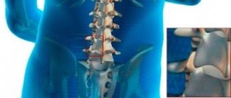
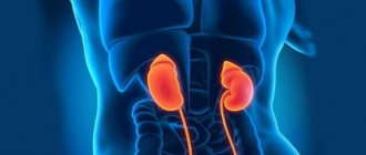
Spondyloarthrosis is a chronically progressive degenerative disease that affects the facet joints of the spine. All structures are involved in the pathological process - bone and cartilage tissue, joint capsule, musculo-ligamentous apparatus.
Spondyloarthrosis is a type of arthrosis. It develops against the background of degenerative-dystrophic changes, when the natural aging process is aggravated by negative factors. Systematic overload of the spinal column associated with hard work, intense sports training, and congenital and acquired vertebral defects is of key importance in the onset of pathology. The cervical region and lower back are most often affected.
Symptoms of spondyloarthrosis
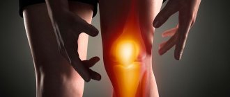
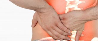
Pain is the main sign of damage to the articular processes of the vertebrae. In the early stages, it is periodic, wave-like in nature and worries mainly when moving. At rest, pain decreases or disappears completely. The pain syndrome is felt only in the damaged area and does not tend to spread. It does not radiate to the arms and legs, as with osteochondrosis and intervertebral hernias. There is no numbness or sensory disturbances. Spondyloarthrosis of the 2nd degree is accompanied by stiffness and limited mobility in the morning. It lasts about half an hour and gradually passes. If left untreated, symptoms become extremely severe. A pronounced stoop appears due to displacement of the vertebrae, pain and motor discomfort intensify.
Complications include compression of the nerve roots, which is manifested by sharp, burning pain that radiates - radiates - to different parts of the back and limbs. A consequence of the chronicity of the process may also be spinal canal stenosis.
Medicines for osteochondrosis
Osteochondrosis is a dangerous disease of the spine, which manifests itself as a result of degenerative pathological processes in the intervertebral discs. The soft tissue around the damaged disc gradually becomes inflamed and puts pressure on the nerve endings, resulting in pain. Since osteochondrosis is a systemic disease, its treatment should be carried out comprehensively. What medications for osteochondrosis effectively fight the pathology?
Principles of treatment of osteochondrosis
Medicines used in the treatment of pathology must perform a number of tasks:
- eliminate pain,
- stop inflammatory processes,
- stimulate blood circulation in damaged tissues,
- restore cartilage tissue,
- restore natural joint mobility,
- improve the patient's emotional state.
Medicines treating osteochondrosis are divided into several main groups.
Nonsteroidal anti-inflammatory drugs (NSAIDs)
NSAIDs eliminate pain, relieve swelling and stop the inflammatory process in tissues. Similar medications for osteochondrosis are available both in the form of ointments and rubbing gels, and in the form of injections and tablets:
1. Diclofenac is a popular and effective drug for the treatment of osteochondrosis; it is the active ingredient of many medicines:
- Voltaren,
- Diklak,
- Diclobene,
- Dikloberl,
- Dicloran plus,
- Diklo-F,
- Ortofen.
2. Ibuprofen has an anti-inflammatory and analgesic effect. Analogues:
- Dolgit,
- Ibuprom,
- Ibupron,
- Nurofen,
- Reumafen.
3. Indomethacin has an anti-inflammatory and analgesic effect directly on damaged tissue. Analogues:
- Arthrocid,
- Indobene,
- Indovazin,
- Indomethacin,
- Indocide,
- Inteban.
4. Ketoprofen (Fastum gel, Ketonal, Flexen, Febrofid).
5. Nimesulide is a new generation drug. Analogues:
- Nise,
- Nimesil,
- Nimika,
- Nimulid.
Vasodilators
Pain due to osteochondrosis provokes constant muscle tension, which results in vasoconstriction. This is a dangerous condition, as oxygen deficiency develops in the tissues and the functions of internal organs are disrupted. To prevent such consequences, vasodilators are used:
- Actovegin,
- Berlition,
- Xanthinol nicotinate,
- Pentoxifylline,
- Trental,
- Eufillin.
Drugs that restore cartilage tissue
In the process of complex treatment of osteochondrosis, it is important to restore the cartilage tissue responsible for joint mobility. For these purposes, patients are prescribed chondroprotectors - effective medications for osteochondrosis of the cervical spine, which have an anti-inflammatory and restorative effect:
- Collagen Ultra,
- Oseoartisi,
- Teraflex,
- Chondroxide.
For these purposes, special vitamin complexes containing vitamins A, B, C, D, E, as well as calcium and phosphorus are also used. B vitamins eliminate pain and restore the sensitivity of damaged nerve endings. B vitamins are included in the following vitamin complexes:
- Milgama,
- Neurorubin,
- Neurobion,
- Neuroplex.
Muscle relaxants
Treatment of osteochondrosis of the thoracic region involves the use of muscle relaxants, which promote muscle relaxation and also have a calming effect. As a result, natural blood circulation is restored, pain goes away, and active regeneration of damaged tissue occurs. These are the following medicines:
- Baclofen,
- Mydocalm,
- Sirdalud,
- Cyclobenzaprine.
Sedatives
Prolonged pain can cause depression and depression in the patient, so doctors often prescribe sedatives. It could be valerian, motherwort tincture. For severe depressive conditions, antidepressants are prescribed:
- Gidazepam,
- Donormil,
- Eglonil.
Related products View all products
Aponil, tab.
100 mg No. 20 210.00 RUR
More details
Arketal Rompharm, solution d/inf. and IM input. 50 mg/ml No. 10 ampoules
174,00 ₽
More details
← Previous article Medicines for asthma
Next article → Medicines for bloating
What is included in the examination
The diagnosis is made based on physical examination and X-ray, MRI and/or CT data. To detect inflammation of the facet joints, a radioisotope scan of the spine is performed. In order to exclude the development of vertebral artery syndrome in cervical spondyloarthrosis, an MRI or CT scan of the brain vessels is performed, as well as a duplex ultrasound scan of the arteries of the head and neck. In some cases, diagnostic blockades with Novocaine and steroids are performed. Reduction or disappearance of pain after the procedure clearly indicates the presence of spondyloarthrosis.
Types of treatment for spondyloarthrosis
The choice of treatment method depends on the stage of the disease, but they always begin with the removal of acute symptoms. The main goals of therapy are pain relief, unloading of the spine and stopping further progress of the disease.
Pharmacological
Nonsteroidal anti-inflammatory drugs (NSAIDs) are prescribed for the shortest possible course to eliminate pain and inflammation. The drugs of choice are Diclofenac, Indomethacin, Ibuprofen, Ketoprofen and their derivatives. Important! If pain persists, they switch to selective NSAIDs that are less toxic to the body - Meloxicam, Celecoxib, Nimesil.
Persistent pain is relieved with steroid medications. If NSAIDs do not help, then they are replaced with one of the following drugs:
- Diprospan;
- Hydrocortisone;
- Dexamethasone.
They are most effective in the form of intramuscular injections.
In especially severe cases, cytostatics (Methotrexate) and immunosuppressants (Leflunomide), Sulfasalazine are prescribed. If pronounced muscle spasms are observed, muscle relaxants are used - Sirdalud, Mydocalm, Baclofen.
Non-drug
After the severe pain caused by spondyloarthrosis has been relieved, treatment continues with non-drug methods. It includes physiotherapy, manual therapy sessions and therapeutic exercises. The most effective physiotherapy procedures are:
- phonophoresis with Hydrocortisone;
- ionogalvanization with local anesthetics - Lidocaine, Novocaine;
- magnet;
- SMT therapy (sinusoidally modulated currents).
With the help of manual techniques, the range of motion of the spine increases by relaxing spastic muscles and optimizing the load. Manual techniques of manual therapy can minimize the negative consequences of pathology - pain, stiffness in the body. After a course visit to the procedures, posture improves significantly./p>
Therapeutic exercises help correct the motor pattern and strengthen the back muscles. Thanks to regular exercise, the overall tone of the body increases.
Surgical
Surgery is performed if conservative methods do not produce results. The essence of the operation is to install a special structure between the spinous processes of the damaged vertebrae. After implantation, the spinal motion segment experiences less load, the spinal ligaments and posterior sections of the intervertebral disc are stretched. As a result, the foraminal openings for the exit of the nerve roots and the spinal canal itself expand.
Comparative effectiveness of non-steroidal anti-inflammatory drugs for back pain
IN
Currently, back pain (BP) is widespread, and in developed countries, according to WHO experts, it is comparable to a non-infectious epidemic, which in most cases is associated with increasing stress on a person [4]. The high level of disability among people of working age due to lesions of the musculoskeletal system raises the problem of treating BS to the rank of urgent. Population studies have revealed an association of low back pain (LBP) with factors such as gender and age, posture, muscle strength, and spinal mobility. A recent population-based study of spinal pain among people aged 35–45 years in Sweden found that the incidence of pain in the past year was 66.3%, and the rate was slightly higher among women than among men. 25% of respondents had significant problems with their ability to work and the degree of impairment of their functional status. In the UK, 90 million working days were lost due to back pain in 1992, second only to respiratory and circulatory diseases. Moreover, 75% of the patients were patients from 30 to 59 years old, that is, during the period of maximum working capacity.
All cases of back pain are divided into primary and secondary.
Primary BS syndrome
is a pain syndrome in the back caused by dystrophic and functional changes in the tissues of the musculoskeletal system (facet joints, fascia, muscles, tendons, ligaments) with possible involvement of adjacent structures.
Secondary BS syndrome
may be associated with congenital anomalies, traumatic lesions of the spine, tumor and infectious processes, osteoporosis, and diseases of internal organs.
BS most often develops between the ages of 20 and 50 years (the peak incidence is between the ages of 35 and 45 years). It is in this age group that primary (mechanical) BS syndrome is usually diagnosed, while in patients under 20 years of age and over 50 years of age, secondary BS syndrome predominates (Table 1).
Diagnosis of BS requires the participation of doctors of different specialties: therapist, rheumatologist, neurologist, orthopedist and surgeon, whose task, in particular, is to exclude the secondary nature of pain in the lower back.
Provoking factors for the acute course of PS can be trauma, lifting an excessive load, unprepared movements, prolonged stay in a non-physiological position, and hypothermia. Chronic pain can occur both after regression of acute pain and independently of it.
Treatment is carried out taking into account the form of the disease and its course using conservative and surgical approaches.
Currently, the indications for surgical treatment have been narrowed, since evidence has accumulated that even with a very good method of surgical decompression and stabilization, the disease tends to recur.
rest was previously recommended
within a few weeks.
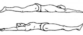
However, all studies in recent years emphasize that not bed rest, but early activation of patients
should be the main component of the treatment program, which helps to improve the nutrition of the intervertebral disc, and therefore bed rest in acute cases is limited to several days.
Nonsteroidal anti-inflammatory drugs (NSAIDs)
should be included in the treatment program as early as possible - on the 1st-2nd day of the onset of the disease. These data are based on the results of studies on the treatment of low back pain from the perspective of evidence-based medicine [1].
Thus, the recommendation to the patient to “continue daily activities or resume them as soon as possible” has a level of evidence of category A, “for acute pain in the lumbar region, active physical exercises during the first 2 weeks of the disease are ineffective” - category B. There is no reliable evidence of the effectiveness of wearing a support corset exists (level of evidence category C). Regarding the use of medications for BS, only NSAIDs have been proven effective - level of evidence category A.
The degenerative process in the intervertebral disc activates nociceptors along the periphery of the fibrous ring, in tendons, fascia, and muscles, with subsequent transmission of impulses to the spinal cord. In this case, immune and biochemical reactions are triggered, culminating in the formation of aseptic neurogenic inflammation. In this regard, the advisability of prescribing NSAIDs is justified (Scheme 1) [3].
Scheme 1. Mechanisms of aseptic neurogenic inflammation in spondyloarthrosis (Borenstein D., 2000).
The main mechanism of the anti-inflammatory effect of NSAIDs is
the suppression of the activity of the enzyme cyclooxygenase (COX) and prostaglandin biosynthesis
. The anti-inflammatory effect of NSAIDs is due to their ability to inhibit COX-2, a pro-inflammatory isoenzyme. The development of adverse reactions inherent in most NSAIDs is associated with the simultaneous suppression of the physiological enzyme - COX-1. Although at the population level all NSAIDs at equivalent doses have similar efficacy and toxicity, clinical experience suggests significant differences in response to NSAIDs in individual patients. For some, one NSAID is significantly more effective at suppressing pain and inflammation or, conversely, is more likely to cause toxic reactions than another. The reasons for this phenomenon are not completely clear. The importance of individual characteristics of absorption, distribution and metabolism of drugs, the relative predominance of mechanisms of action dependent and independent of COX inhibition are discussed. Since NSAIDs are racemic mixtures, differences in effect may depend on the ratio of levo- and dextrorotatory enantiomers, one of which has more pronounced anti-inflammatory and analgesic activity, and the other has toxic activity [2].
Currently, it is possible to use an NSAID such as ketoprofen (Ketonal)
for osteoarthritis, as well as for the treatment of back pain. Ketoprofen is a derivative of propionic acid. A feature of its anti-inflammatory effect is not only the inhibition of prostaglandin synthesis at the level of cyclooxygenase; it also inhibits lipoxygenase, has antibradykinin activity, and stabilizes lysosomal membranes. When taken orally, the drug is quickly and completely absorbed from the gastrointestinal tract. The maximum concentration in the blood is reached after 1-2 hours. Ketonal penetrates well into synovial fluid and connective tissue. Significant concentration levels are achieved within 15 minutes after a single intramuscular injection of 100 mg of ketoprofen. An important advantage of the drug is that it is available in various dosage forms: capsules, forte and retard tablets, solution for intramuscular injection, cream and suppositories.
Ketonal has a “balanced” activity in relation to the inhibition of COX-1 and COX-2, which makes it possible to optimally combine high efficiency with good tolerability, comparable, according to some authors, with the tolerability of selective NSAIDs [3,5]. Researchers associate the powerful analgesic effect of Ketonal with its proven central effect, which is realized at the level of the dorsal horns of the spinal cord, as well as through a direct effect on the thalamic centers of pain sensitivity, which is apparently associated with inhibition of prostaglandin synthesis in the central nervous system [4,5 ].
In 1st City Clinical Hospital named after. N.I. Pirogov at the Department of Faculty Therapy named after. acad. A.I. Nesterov Russian State Medical University conducted a comparative analysis of the effectiveness and safety of ketoprofen (Ketonal, Lek) 300 mg per day and diclofenac 150 mg per day in patients with primary BS. The study group included 60 people aged from 35 to 70 years (average age was 42.7+12.4 years). There were 36 men, 24 women (Table 2). The duration of spondyloarthritis among those examined ranged from 1 to 7 years, averaging 4.3 years. Either Ketonal or diclofenac was assigned by random sampling.
To determine the severity of pain and limitation of movements in the back, the following parameters were used: pain index, according to a 5-point system; morning stiffness in the lumbar spine in min; assessment of pain severity using a visual analogue scale (VAS) in cm; McGill pain index, points; The effectiveness and tolerability of the drugs were assessed using a 4-point system by the patient and the doctor.
The study included outpatients of both sexes with primary LBP syndrome (low back pain), severe pain syndrome - pain on movement of 3.0 cm or more according to VAS, requiring NSAIDs, radiographically with stages I-III of spinal osteochondrosis according to Kellgren.
Exclusion criteria were the presence of secondary LBP syndrome, mild pain syndrome - less than 3.0 cm according to VAS.
The first group of patients (30 people) took Ketonal forte at a dose of 100 mg 3 times a day to relieve BS, the second group (30 people) took diclofenac at a dose of 50 mg 3 times a day for 10 days. Clinical examination of patients was carried out before treatment and on the 10th day of treatment.
When analyzing pain indicators according to VAS 10 days after the start of therapy, a significant decrease in its severity was revealed from 8.1 cm to 2.4 cm, as well as the duration of morning stiffness from 25.6 to 3.9 minutes, the pain index from 4.2 to 3.9 minutes. 1.4 points, McGill pain index from 7.3 to 2.5 points when taking Ketonal forte. There was a significant decrease in pain severity from 7.3 cm to 2.8 cm according to VAS, pain index from 4.1 to 1.9 points, McGill pain index from 7.1 to 3.2 points, and a decrease in the duration of morning stiffness from 24.4 to 4.6 minutes during diclofenac therapy (Fig. 1,2).
Rice. 1. Dynamics of clinical parameters in patients with back pain before and after treatment with Ketonal forte
Rice. 2. Dynamics of clinical parameters in patients with back pain before and after treatment with diclofenac
Assessments of the effectiveness of treatment by the patient and the doctor practically did not differ from each other and indicated the pronounced clinical effectiveness of the drugs. High efficiency was noted in 93.3% of patients taking Ketonal forte, in 73.3% of patients taking diclofenac.
Tolerability of the drug “good” or “very good” was noted in 80% of patients taking Ketonal, and in 64% of those taking diclofenac. The main side effects when taking both diclofenac and Ketonal were heartburn and discomfort in the epigastric region. Discontinuation of treatment was required only in 1 case, when the patient developed urticaria while taking diclofenac, which was regarded as a side effect of moderate severity.
A study of Ketonal showed its high effectiveness in patients with back pain with a low number of adverse reactions
. In acute cases of BS, Ketonal Forte is recommended to be prescribed 3 times a day at a dose of 100 mg for 7-10 days, followed by a dose reduction. It is possible to switch to other dosage forms of the drug (retard tablets, rectal suppositories, cream), as well as their combinations. The minimal risk of adverse reactions is an advantage of Ketonal, which is especially important in the treatment of elderly patients suffering from chronic LBP and in most cases having concomitant diseases (arterial hypertension, diabetes mellitus, gastric and duodenal ulcers).
Thus, at present, the treatment of back pain is optimized by the use of drugs from the NSAID group, in particular, Ketonal, due to its high efficiency, which is manifested by clearly expressed positive dynamics with regression of pain manifestations in patients with BS.
Literature:
1. Clinical recommendations for practitioners, based on evidence-based medicine. Ed. house "GEOTAR-MED", 2001, p. 606-611.
2. Nasonov E.L. Non-steroidal anti-inflammatory drugs (prospects for use in medicine). M., 2000, 262.
3. Nasonov E.L., Chichasova N.V., Shmidt E.I. Prospects for the use of non-selective non-steroidal anti-inflammatory drugs (using the example of ketoprofen) and selective COX-2 inhibitors in clinical practice. Russian Medical Journal, volume 10, no. 22, 2002, pp. 1014-1017.
4. Vetshev P.S., Vetsheva M.S. Principles of analgesia in the early postoperative period. Surgery, 2002, No. 12, p. 49-52.
5. Chichasova N.V., Nasonov E.L., Imametdinova G.R. The use of ketoprofen (ketonal) in medical practice. Pharmateka, 2003, No. 5, p. 30-32.
6. Borenstein D. “Epidemiology, etiology, diagnostic evaluation and treatment of low back pain.” Intl. Medical Journal, 2000, No. 35, p. 36-42.
7. WHO. Department of noncomunicable disease menagement. Low back pain initiation. Geneve.1999.
Prevention
There are no preventive measures that would guarantee 100% prevention of the disease. The risk of developing spondyloarthrosis can be minimized by strengthening the muscular frame of the back and eliminating physical overload. It is also recommended to follow a diet to maintain normal weight, consume sufficient amounts of vitamins and microelements, and exercise therapy. Treatment of spinal spondyloarthrosis is successfully carried out in our center. Here you can undergo all the necessary procedures in accordance with your diagnosis. Consultations are carried out by high-level specialists, so during the visit patients feel as comfortable as possible. Modern equipment is at your service, allowing you to perform even complex manipulations.