ICD-10 code: M25.0 (Hemarthrosis)
Hemarthrosis is not an independent disease, but is a secondary sign of articular pathology.
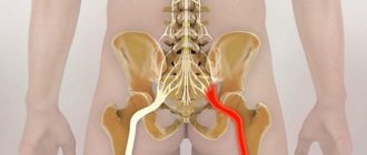
This diagnosis is made when blood accumulates inside the joint cavity due to rupture of blood vessels. Hemarthrosis of the knee joint is most often diagnosed, less often - ankle, shoulder, hip and elbow.
Hemarthrosis of the knee joint
This disease has varying symptoms and severity, occurs in adults and children, requires treatment, has serious complications and can cause irreversible processes.
Flow
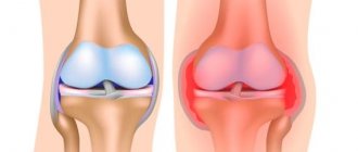
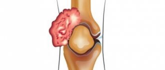
With hemarthrosis, the vessels supplying intra-articular tissues rupture and, as a result, hemorrhage into the joint.
This causes disruption of blood circulation, its accumulation in the cavity, the formation of clots and an increase in intra-articular pressure on surrounding tissues. The joint swells, becomes spherical, increases in volume, pain and a feeling of fullness appear, loss of function occurs and range of motion is limited.
Blood in the knee joint
If the disease is left without proper treatment, complications may develop: a chronic form of the disease, secondary infection, arthritis.
Causes and symptoms of the disease
A description of hemarthrosis is included in the reference book on the International Classification of Diseases, which contains diagnostic information.
In ICD 10, the disease is coded M25.0. Codes are used for the convenience of processing and storing information data. Clinical and diagnostic terms used in the Russian version of the ICD 10 reference book correspond to domestic practice. Hemarthrosis of the knee or other joint can be caused by various types of injuries.
- Bleeding is possible when bones that have articulation in the joint are fractured or dislocated.
- Hemarthrosis can occur as a result of sprained or torn ligaments.
- Damage to the capsule or meniscus can also cause this disease.
- Injury to blood vessels due to soft tissue bruises can cause bleeding and accumulation of blood in the joint capsule.
Symptoms manifest themselves more quickly in children, so after a bruise or injury, the child must be immediately shown to a specialist.
Hemarthrosis, which appears as a result of injury, is characterized by the presence of a certain clinical picture.
- Due to the sudden rupture of blood vessels, blood begins to penetrate into the cavity of the injured joint, which leads to its rapid swelling.
- Pain in the knee joint and decreased mobility are caused by an increase in the size of the capsule, which begins to put pressure on surrounding tissues.
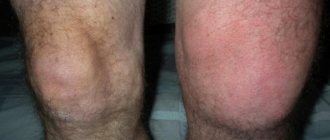
- After bleeding starts, the knee quickly swells and swelling appears on its sides.
- If the blood continues to flow, then swelling appears on the front of the joint, its contours are smoothed out.
- The rupture of a large vessel is manifested by cyanosis of the skin; with the destruction of small and medium-sized vessels, the skin acquires a purple tint, and hyperthermia of the integument is present.
- There is a feeling of weakness in the joint, and the pain intensifies during palpation. Post-traumatic hemarthrosis manifests itself quickly, often its symptoms develop within 1.5-2 hours.
Causes
Hemorrhage in the joint cavity can be caused by two reasons: various injuries and somatic diseases.
Traumatic hemarthrosis in 100% of cases develops with intra-articular fractures and can accompany milder injuries, for example, bruise of the elbow or dislocation of the ankle joint. Non-traumatic causes include a group of diseases associated with blood clotting disorders: hemophilia, hemorrhagic diathesis. In these cases, even minimal trauma can cause bleeding.
The most common causes of hemarthrosis
Hemorrhage and accumulation of blood in the cavity can affect any large joint. Hemarthrosis of the knee, ankle, shoulder, elbow and hip joints occurs. For treatment, you need to contact a surgeon or traumatologist, the doctor will conduct an examination and collect an anamnesis.
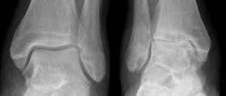
If there was an injury, then an x-ray of the affected joint will be prescribed to rule out bone damage. If hemarthrosis is suspected of being non-traumatic, the patient will need to consult a hematologist.
Diagnostics
The diagnosis of hemarthrosis is made based on clinical data and medical history. In order to exclude bone damage, all patients with suspected hemarthrosis are referred for an X-ray of the knee joint. If necessary (for example, if a ligament rupture or cartilage damage is suspected), other additional studies may be prescribed: CT of the knee joint or MRI of the knee joint, arthroscopy. Suspicion of non-traumatic hemarthrosis is an indication for consultation with a hematologist.
Symptoms and degrees of hemarthrosis
Hemarthrosis has generally similar symptoms, however, depending on the location of the affected joint, some features of the course of the disease can be traced. For example, hemarthrosis of the knee joint is a common consequence of various injuries, due to the fact that a person’s legs are often subject to shocks and bruises.
Injuries to the knee joint limit the victim's ability to move - they require rest for the injured limb and the use of crutches.
In turn, the elbow joint, being a very complex anatomical and functional node with special blood circulation and innervation, is characterized by high reactivity.
Even after a minor injury or bruise of the elbow joint, local pain, swelling, partial dysfunction, and hemorrhage appear.
Hemarthrosis of other joints
When moving, the ankle joint takes on a large load and provides rotation of the foot, stability and support for the entire body. Having a complex structure, the ankle joint is extremely vulnerable to injury; sprains and ruptures of ligaments are especially common, which can be accompanied by hemarthrosis.
This condition is manifested by high pain in the ankle joint, swelling and swelling of not only the joint itself, but also the foot.
Symptoms of hemarthrosis
- swelling and enlargement of the joint;
- spherical shape;
- smoothing the contours of the joint;
- local or arching pain;
- limitation of range of motion;
- soft tissue tension;
- bluish skin color.
Degrees of hemarthrosis
3 stages (degrees) of the disease
- In grade I, a small volume of blood enters the cavity, which does not exceed 15 ml. The symptoms of the main injury, local pain, and slight swelling dominate.
- In grade II, the blood volume in the joint reaches 100 ml, the joint increases in volume, takes on a spherical shape and smoothed contours, and bursting pain appears in the joint.
- With continued bleeding, when more than 100 ml of blood accumulates in the cavity, stage III is diagnosed. It is characterized by the previous symptoms, to which are added soft tissue tension and burgundy-bluish skin color.
Hemarthrosis of the joints
Hemarthrosis of the knee joint (ICD 10 code - M25.0)
Photo
Causes
Bruises and sprains of the ligaments of the knee joint are quite common, which should be attributed to the anatomical features of the knee joint, which is easily damaged by a fall or blunt force. When movement in a joint exceeds normal movement, overstretching of the ligaments occurs, and sometimes partial or even complete rupture. Damage to the ligamentous apparatus and capsule is accompanied by hemorrhage, and blood enters the intra-articular space, forming hemarthrosis of the knee joint (ICD 10 code - M25.0).
Clinic
The knee joint affected by hemarthrosis swells, its contours are smoothed. The leg is half bent at the knee joint, movements in it are sharply painful.
Diagnostics
With hemarthrosis of the knee joint, fluctuation (oscillation) in the joint area and protrusion of the patella are determined.
The method for determining the patella balloting is simple: with the palm and fingers of both hands, lightly press on the lateral surfaces of the joint, and with the thumbs, push the anterior surface of the patella; at the same time there is a feeling of “floating” of the patella.
Through X-ray examination, it is sometimes possible to establish a widened joint space.
Treatment
Treatment of hemarthrosis of the knee joint is carried out using conservative methods or puncture, which will be discussed below.
Hemarthrosis of the ankle joint
Photo
Causes
Hemarthrosis of the ankle joint occurs:
- when struck by blunt objects,
- with uncoordinated movements in the joint,
- when walking on uneven surfaces, when going down stairs,
- In a cavalryman, when a foot gets stuck in the stirrup and falls, a sharp movement of the entire body occurs with a fixed foot.
Clinic
The victim immediately feels severe pain and is often unable to walk. Upon examination, the joint appears to be very swollen, its configuration is smoothed, and palpation is painful. Movement in the joint is severely limited and causes pain.
Hemarthrosis of the shoulder joint
Photo
Very common injuries include closed injuries of the shoulder joint with the occurrence of hemarthrosis.
Causes
The causes of hemarthrosis of the shoulder joint can be:
- direct impact of injury, in particular when falling on the shoulder joint from a height or on the side with the arm abducted,
- blow to the joint with a blunt object,
- indirect injury transmitted to the shoulder joint, for example, along the axis of the limb.
The latter type of injury causes sprain of the ligamentous apparatus.
Clinic
Immediately after the injury, the first symptom occurs - pain, which is initially felt even in a quiet position. Active and passive movements in the shoulder joint are severely limited due to pain. Within a few hours, swelling of the joint area is noted, which is most pronounced 24 hours after the injury.
Hemarthrosis of the elbow joint
Photo
Causes
Hemarthrosis of the elbow joint occurs for the following reasons:
- direct blow to the elbow,
- excessive flexion of the elbow joint (wrestling, playing tennis, etc.),
- falling on hands.
Clinic
Immediately after the injury, the victim complains of pain. There is swelling in the joint area, its contours are smoothed, the arm is bent at the elbow, movements are sharply painful.
Hemarthrosis of the wrist joint
Photo
Causes
Hemarthrosis of the wrist joint occurs due to:
- impact with blunt objects,
- heavy parts falling on your hand when working in industrial enterprises.
Stretching of the ligamentous apparatus of the joint usually occurs when the hand is twisted during a fall, less often when it is excessively bent in the event of an attempt to hold on during a fall.
Clinic
Clinically, with hemarthrosis of the wrist joint the following are observed:
- smoothing the contours of the joint,
- limitation of hand movements,
- sharp pain on palpation of the joint area.
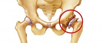
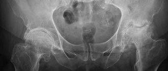
Hemarthrosis of the hip joint
Photo
The deep location of the hip joint and the powerful muscles covering it pose a significant difficulty in recognizing hemarthrosis of the hip joint.
Clinic
Clinically, hemarthrosis of the hip joint manifests itself:
- painful sensations,
- restriction of movements,
- swelling of the joint area.
Diagnostics
In some cases, to make an accurate diagnosis it is necessary to resort to joint puncture. When examining a patient with hemorrhage in a joint, it is necessary to pay attention to the mechanism of injury, the patient’s complaints, local changes in the joint and its functional state. An intra-articular fracture can be identified by severe local pain on palpation, as well as using an x-ray.
Treatment
If a person has the above symptoms of hemarthrosis, timely treatment will alleviate the condition and prevent the development of complications. At the pre-medical stage, the injured joint should be kept at rest and, if possible, given an elevated position.
With grade I hemarthrosis, the accumulation of blood is insignificant, it does not require special manipulations and will resolve on its own. A fixing bandage is applied to the joint, it is recommended to apply cold for several days, ensure rest and limit the load.
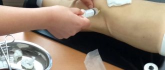
If the blood volume is more than 30 ml, a puncture is used: blood suction and washing of the joint cavity. At the end of the procedure, medications are injected into the cavity and immobilization is performed, the duration of which depends on the underlying injury. If necessary, the puncture is repeated. Hemarthrosis of the third degree, as a rule, appears as a result of severe injuries and requires combined treatment in a hospital for the main injury and hemorrhage in the joint. Punctures are performed as blood accumulates in the cavity. Hemarthrosis of the knee joint that is difficult to treat and recurrent is an indication for an additional, more detailed examination, since it is necessary to establish the cause of repeated bleeding.
Treatment of hemarthrosis in hemophilia is carried out in the hematology department.
Possible complications
If you do not pay attention to a severe bruise and do not visit a doctor for prolonged pain, you may miss the development of serious consequences. These include damage to the meniscus, sprain, rupture of ligaments, damage to the Achilles tendon, synovitis, and the development of the inflammatory process.
The first complications are hemarthrosis and synovitis. They are sure signs that the joint is damaged inside. Most often they are expressed as follows:
- The victim has a feeling that the joint is bursting and its volume is increasing.
- If you press on the kneecap, it seems to float, i.e. movement occurs in a liquid.
The consequences of a knee injury are very varied.
- An abrasion on the knee and a bruise are the easiest consequences.
- Inflammation of the meniscus occurs without timely treatment after a bruise of the knee joint. This pathology can become chronic with the development of cysts on damaged connective tissue.
- Fluid accumulation in the knee. Swelling of various sizes and pain indicate the development of synovitis.
- If the injury is severe, a fracture of the kneecap may occur. And as a result, loss of basic functions - motor and support.
- Bruises of the knee joint may be accompanied by stretching or tearing of the connective tissue. When a complete rupture occurs, the joint ceases to function (extends, but does not bend). When sprained, stiffness occurs.
- Habitual dislocations of the patella are accompanied by pain, damage to the connective tissue of the knee apparatus, and muscle atrophy.
- Prepatellar bursitis as a result of trauma. Inflammation of the bursa is accompanied by swelling, hyperthermia, and severe pain in the area of injury. If the injury is open, inflammation may begin due to infection.
In case of a knee injury, regardless of the severity of the injury, consultation with a traumatologist is necessary to avoid all sorts of negative consequences.
Trauma in the ankle and foot area causes disruption of blood flow, provokes infection with pathogenic microbes (in the presence of open wounds), which subsequently leads to serious disorders of the musculoskeletal system, inflammation, synovitis (accumulations of synovial fluid inside the joint capsule), decreased performance, loss of ability to performing movements, sepsis may develop, which will lead to amputation of the limb.
Consequences and complications
For example, if there was an injury to the left hand and hemorrhage occurred, but no treatment was carried out, then clots will form from the blood that has entered the joint, which will subsequently undergo fermentation (destruction of blood cells). Fermentation processes disrupt the integrity of cartilage tissue and can provoke the development of degenerative diseases of the elbow joint.
Possible consequences of the disease
In case of relapse with repeated hemorrhages into the cavity of the left elbow joint, the fermentation process always occurs much more intensely and causes more harm.
Accumulations of blood impair the blood circulation of tissues in the joint cavity and can cause dystrophic changes, often found in recurrent hemarthrosis.
Often an unpleasant consequence of fermentation is inflammation of the synovial membrane - synovitis, which, in the case of the addition of pathogenic flora (through blood, lymph, or if sterility is not observed during puncture), turns from aseptic into infectious and can cause purulent arthritis.
Providing first aid for a knee injury
For any injuries, you must call an ambulance and, if possible, provide first aid to the victim. General first aid rules are the same for all types of injuries.
Help for a knee injury is as follows:
- Lay the victim down, placing a cushion over the bruised knee. The leg should be higher than the body. This will ensure minimal blood flow to the injury site.
- It is necessary to ensure immobility of the injured limb. Secure with a bandage (elastic bandage, scarf, scarf, etc.) The bandage should not be too tight.
- If your knee is swollen, apply cold to the bruised area. The duration of the cold compress should not exceed 15 minutes. The procedure can be repeated every ten minutes.
- If the pain is very severe, you can give an analgesic, but be sure to consult your doctor before doing so.
If the knee is severely bruised, severe swelling may develop, and the joint may be larger than the width of the hip. The reason for this swelling is the accumulation of fluid and blood in the joint cavity, internal damage to connective and bone tissue.
If the knee is severely bruised, transport the patient to the nearest hospital independently to provide professional medical care to the victim.
A knee bruise is a fairly serious injury and timely professional treatment will allow you to avoid complications that arise in advanced cases.
What to do if you are injured
Children regularly experience injuries such as knee bruises. Often such an injury in this age category is accompanied by damage to the integrity of the skin (wounds, abrasions, etc.). What to do if a child has a bruised knee?
httpv://www.youtube.com/watch?v=embed/MsmYQcU9aOU
First aid for a knee injury at home for adults:
- Apply cold to the joint;
- Lay the victim down, the leg should be slightly higher than the level of the body;
- bandage with an elastic bandage.
At home, you can apply various painkillers and anti-inflammatory creams and gels and at first try to limit the load on the injured leg.