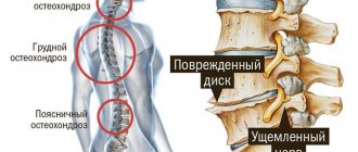
Cervical osteochondrosis is a common phenomenon, since the cervical spine is very mobile and its cartilages are subject to intensive use with a large range of motion. Cervical osteochondrosis, the symptoms and treatment of which are discussed in this article, is a pathology that is characterized by the development of dystrophic disorders in the intervertebral discs of the cervical spine. The latter are “responsible” for its flexibility and mobility. If you do not seek medical help in a timely manner, the process will develop further and will lead to the appearance of a vertebral hernia, as well as changes in the vertebrae themselves.
A course of treatment for cervical osteochondrosis can be completed at the CELT clinic. We employ highly qualified doctors from the Pain Clinic department: neurologists and orthopedic traumatologists who have extensive experience in this area. They will develop an individual treatment plan that is sure to be successful.
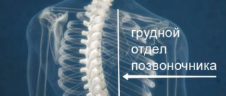
What is thoracic osteochondrosis?
Osteochondrosis of the thoracic spine is a regenerative (destructive) process localized in the interval from 8 to 19 vertebrae, which forms the human thoracic spine.
Unlike other types of the disease, symptoms of thoracic osteochondrosis can occur in both young and elderly people.
A feature of the disease is the difficulty of its early diagnosis, which is associated with low mobility of the spine and the difficulty of identifying the main symptoms. For this reason, it is important to promptly consult a specialist when detecting the first signs of thoracic osteochondrosis in order to prevent irreversible processes.
Treatment of cervicothoracic osteochondrosis and symptoms of the disease
The need to consult a doctor specializing in the treatment of cervicothoracic osteochondrosis occurs when local and referred pain appears in the back, shoulder girdle or chest. The following should also be serious causes for concern:
- curvature of the spine and limitation of its mobility in the problem segment;
- neuroosteofibrosis, neuromyofibrosis, dystrophic and vascular changes in soft tissues in the affected area;
- sensory disturbances in the back area;
- paresis and muscle paralysis;
- Difficulty raising your arms and bending over.
Osteochondrosis, supplemented by damage to the spinal cord, is often accompanied by disturbances in the processes of defecation and urination.
Causes
The main cause of osteochondrosis of the thoracic spine is the occurrence of dystrophic processes (disorders of cellular metabolism) that transform tissues and provoke complications of metabolic actions, which is caused by an insufficiently balanced diet and improper load on the intervertebral discs.
In addition, the list of factors that increase the risk of developing thoracic osteochondrosis includes:
- the presence of intervertebral hernias;
- impaired blood supply to the spinal cord;
- disturbance of mineral balance in the body;
- regularly increased loads on the spine;
- predominantly sedentary lifestyle;
- consequences of previous injuries;
- engaging in active sports.
The main symptoms of osteochondrosis of the thoracic region
Chest pain with osteochondrosis is the main symptom of the clinical picture. However, considering the general symptoms of osteochondrosis of the thoracic region, we can also highlight:
- difficulty breathing, chest tightness;
- difficulty in bending;
- periodic occurrence of a feeling of freezing of the extremities due to circulatory failure;
- brittle nails and hair;
- nausea, digestive disorders;
- pain that is easily confused with that that accompanies cardiovascular diseases;
- soreness of the mammary glands in women;
- discomfort and difficulty swallowing, cough.
The manifestation of several symptoms of thoracic osteochondrosis requires immediate consultation and a full examination by a specialist in order to make an accurate diagnosis and determine a treatment plan for thoracic osteochondrosis.
Stages of disease development
Experts distinguish three main stages of development of thoracic osteochondrosis:
Stage 1 – the beginning of the development of the disease. Characterized by the absence of clinical manifestations.
Signs of stage 1 thoracic osteochondrosis:
- barely perceptible, slight painful lumbago, accompanied by a nagging pain in the chest. Unpleasant sensations intensify after strenuous work or heavy lifting;
- muscle spasms, constant tone of the muscle frame for no apparent reason;
- discomfort in the heart area.
Seeing a doctor at the initial stage of the disease guarantees a complete cure of the disease.
Stage 2 – the progression of the disease leads to the formation of microcracks in the intervertebral discs, which causes limited mobility and the appearance of severe pain.
Clinical manifestations include:
- visually noticeable deformation of the spinal column;
- decrease in pressure;
- accompaniment of an attempt to place the hand behind the head with palpable pain in the sternum;
- chronic feeling of fatigue;
- discomfort in the area of the heart and spinal column of the thoracic region.
If the disease is detected at this stage, the process of restoring health may require a fairly long period of time.
Stage 3 is an advanced process that has a negative impact on the entire body as a whole.
Dangerous manifestations such as:
- limited mobility of the spine;
- sharp pain;
- the appearance of intervertebral hernias;
- pinching of blood vessels and nerve roots;
- diseases of the biliary tract.
Refusal to treat thoracic osteochondrosis at this stage can lead to disability.
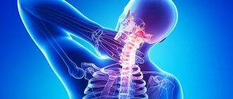
Features of complaints, symptoms and treatment of headaches with cervical osteochondrosis
A characteristic feature of pain in this disease is the unexpectedness of its appearance, intensity and strength. The pain often makes the patient freeze; during an attack it becomes difficult for him to breathe and even blink his eyes. This specificity of manifestations must be taken into account when diagnosing and subsequently prescribing a treatment method for headaches with chondrosis (osteochondrosis). The pain can radiate to any part of the head, but most often the back of the head and parietal region suffer. The spread of painful sensations extends to the shoulder joints, chest, and arms. During an attack, dizziness, nausea may occur, extraneous sounds appear in the ears, and darkness appears in the eyes. The patient's sleep, appetite, and mood are disturbed. Therefore, treatment for cervical headaches is required immediately. It must be carried out under the supervision of a doctor.
How to treat thoracic osteochondrosis?
Before determining how to treat thoracic osteochondrosis, it is important to visit a specialist. Diagnosis of the disease should be carried out only by a specialist. Treatment of osteochondrosis of the thoracic spine is determined by a neurologist after a high-quality examination.
The key rule for starting treatment for thoracic osteochondrosis is getting rid of pain. Today, there are several optimal treatment options. Let's look at each of them in more detail.
Physiotherapy
Physiotherapeutic treatment of thoracic osteochondrosis is used in medical practice quite often, both at the acute stage and after it, in order to achieve long-term remission of the disease.
For osteochondrosis of the thoracic spine, physiotherapy methods are used such as:
- medicinal electrophoresis - the combined effect of direct current and drugs;
- UHF therapy – exposure to high-frequency electromagnetic fields;
- Magnetic therapy is an alternative medicine that involves the use of a constant or alternating magnetic field;
- phonophoresis of drugs - administration of drugs under the influence of ultrasound;
- detensor therapy – relaxation of the muscular frame of the spine using a specialized mattress/mat.
It is important to note that when prescribing physiotherapeutic methods for treating a degenerative disease, the stage of its development is taken into account. Inappropriate use of physiotherapeutic treatment can aggravate the situation and exclude the possibility of long-term remission.
Massage for osteochondrosis of the thoracic region
Massage is one of the most effective methods of non-drug treatment of thoracic osteochondrosis.
Providing a reflex massage effect provides a positive effect on the affected area of the spinal column, which provokes a positive dynamics of treatment.
Unlike other parts of the spine, the thoracic spine requires a special approach when massaged. The muscle fibers of this zone have a special threshold of action.
The positive effects of massage procedures are manifested in:
- reducing the level of pain;
- improving blood circulation at the location of the disease;
- eliminating muscle spasms;
- increasing arm muscle strength;
- increasing the mobility of the spinal column and launching regenerative processes of cartilage tissue.
Among the basic principles of thoracic massage it is worth highlighting:
- the beginning of the session should be carried out with light strokes, which will ensure blood flow and reduce pain;
- during the massage it is recommended to use squeezing movements, rubbing and patting;
- each movement must be repeated 4-5 times;
- movements should be carried out along a single trajectory: from the shoulder blades to the neck, upward;
- each side of the thoracic region should be treated separately.
Exercise therapy for thoracic osteochondrosis
Physical therapy (therapeutic physical education) classes provide the opportunity to provide high-quality support for the optimal tone of the muscular frame of the back and help relieve soreness of the paravertebral muscles.
Exercise therapy for osteochondrosis of the thoracic region is most effective in the initial stages of the development of the disease.
When doing gymnastics, it is important to consider and adhere to the basic rules for performing exercises for osteochondrosis of the thoracic region, including:
- systematic repetition over several months;
- the duration of the lesson should not exceed half an hour;
- gymnastics for thoracic osteochondrosis should be performed without the use of additional equipment;
- You should only exercise in comfortable clothes and shoes;
- taking into account existing contraindications.
Absolute contraindications include:
- acute cardiac pathologies;
- increased body temperature (more than 37.6°C);
- oncological diseases;
- mental instability;
- dermatological diseases;
- high blood pressure.
Consultation with a neurologist in combination with a reasonable, competent approach to exercise ensures the successful achievement of remission at various stages of the disease.
Treatment of cervical osteochondrosis
Treatment of osteochondrosis of the cervical spine is aimed at eliminating pain and slowing the progression of the pathological process. It is carried out in two directions: limiting the impact of unfavorable factors and suppressing the mechanisms of disease development.
Therapeutic and preventive measures to minimize the influence of causative agents include:
- rational selection of work furniture
- use of orthopedic mattresses and pillows
- correction of hearing, vision and posture
- wearing special fixing devices
- restriction of work activities associated with long-term forced conditions
- adequate physical activity
- proper nutrition
There are many different methods of therapeutic correction designed to slow down the development of the degenerative process.
Massage for cervical osteochondrosis
Massage procedures aimed at relieving inflammation and eliminating pain are included in the complex of mandatory therapeutic measures. The most effective types of neck massage:
- classical
- therapeutic (manual)
- acupressure (acupuncture)
- vacuum (can)
- hardware
Thanks to massage techniques, local blood and lymph circulation is enhanced, tissue trophism is accelerated, muscle tension is eliminated, tension in the neck is relieved, muscle tone is improved and muscle elasticity is increased.
Orthopedic collars
To fix the cervical spine in the correct position, special orthopedic devices (Schanz collars) are used. Removable structures of various sizes, shapes and degrees of rigidity limit the usual pathological position of the head, control movement in the neck, reduce pressure on the spinal segments, warm and relax tense muscles and prevent further progression of the disease.
The cervical collar for osteochondrosis is available in several modifications:
Soft splints made of medical foam rubber
or other porous hypoallergenic materials have a recess for the chin and lower surfaces of the neck, and retainers. They are used to correct minor disorders in the upper spine, maintain the vertebrae in an anatomically correct position and relax the muscles of the shoulder girdle.
Pneumatic (inflatable) collars
designed to prevent pain, smooth traction and eliminate compression of the vertebral artery.
Semi-rigid bandages
equipped with metal inserts, reliably stabilize the intervertebral segments. They significantly limit the range of movements and contribute to the expansion of the spaces between the vertebral bodies.
Rigid corsets made of durable plastic
designed to completely immobilize the cervical spine in a neutral position. Prescribed in the later stages of the disease, accompanied by compression disorders.
The collar for osteochondrosis of the cervical spine is selected by the doctor, taking into account age, anatomical features and the stage of the degenerative process.
Manual therapy
Manual therapy treatment is aimed at identifying and eliminating blockades in the motor segments. A local dosed effect on the vertebral joints helps to normalize blood flow and blood supply to the brain, eliminate compression (pinching) and restore the normal functioning of nerve fibers. Specific manipulations by a chiropractor allow you to achieve maximum relaxation, eliminate muscle spasms, cervicogenic headaches arising from damage to the anatomical structures of the neck, and tension headaches.
Acupuncture
Acupuncture, which involves installing acupuncture needles into bioactive points in the neck and shoulder blades, is aimed at restoring the disturbed energy balance. By stimulating rapid contractions of sensory nerve fibers and the release of endorphins and neurotransmitters, acupuncture for cervical osteochondrosis has a powerful anti-inflammatory and analgesic effect. Thanks to this technique, numbness in the hands, dizziness, tinnitus disappears, blood flow improves and mobility is optimized.
Physiotherapy
Physiotherapy for degenerative pathologies of the spine is aimed at relieving pain and stimulating recovery processes. The greatest therapeutic effect is provided by:
- Ural Federal District
- ultrasound treatment
Medications
Drug treatment of osteochondrosis of the thoracic spine is carried out by taking tablets, a course of injections, suppositories or using ointments/gels.
Treatment with medications is carried out over several months. In the absence of positive changes, the disease is eliminated through surgery.
Anti-inflammatory nonsteroidal drugs (PVNS)
They are used to reduce pain and relieve inflammatory processes occurring in the nerve roots. Among the most effective drugs are:
- Artradol;
- Nimesulide;
- Diclofenac.
Self-administration of PVNS is extremely dangerous to health and can cause irreparable harm. Specialist consultation is required.
Glucocorticosteroids
This category of drugs for osteochondrosis of the thoracic region includes Dexamethasone and Prednisol.
Due to the fact that the drugs are hormonal, their use must be agreed upon with the attending physician. Otherwise, irreparable harm may be caused to the body.
Diuretics
Diuretics that relieve swelling in the presence of pinched nerve roots. Drugs of this type include Furosemide and Diacarb.
Vitamin complexes
Taking vitamins improves the metabolic processes occurring in the nervous tissue.
In order to restore high-quality metabolism, patients are prescribed B vitamins, as well as Pentoxifylline and Thioctic acid.
Chondroprotectors
Medicines, the use of which ensures the restoration of elasticity and shock-absorbing functions of the intervertebral discs.
One of the most effective remedies is Artracam.
The intensity and dosage of taking medications for thoracic osteochondrosis is determined taking into account the stage of development of the disease.
Antispasmodics
They help relax the muscle frame and help get rid of the problem of constant spasms, starting the recovery process of thoracic osteochondrosis.
The most effective include:
- Mydocalm;
- Buscopan;
- Eufillin.
Muscle spasm during back pain - pathogenesis, diagnosis and treatment
B
Back pain or dorsalgia is one of the most common complaints in general medical practice. Many patients experience an increase in the tone of individual muscles of the back and limbs (muscle spasms), which can lead to spinal deformation (scoliosis, changes in physiological curves) and limitation of its mobility. Most often, muscle spasm during dorsalgia is a manifestation of reflex or compression complications of spinal osteochondrosis or myofascial pain [1,5,6].
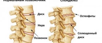
The development of spinal osteochondrosis - degenerative damage to the cartilage of the intervertebral disc and reactive changes in the adjacent vertebral bodies - is associated with repeated injuries (heavy lifting, excessive static and dynamic load, falls, etc.) and age-related degenerative changes. The nucleus pulposus, the central part of the disc, dries out and partially loses its shock-absorbing function. The fibrous ring, located along the periphery of the disc, becomes thinner, cracks form in it, towards which the nucleus pulposus moves, forming a protrusion (prolapse), and if the fibrous ring ruptures, a hernia. In the affected spinal segment, relative instability of the spine occurs, osteophytes of the vertebral bodies develop (spondylosis), and ligaments and intervertebral joints are damaged (spondyloarthrosis). Disc herniations in the posterior and posterolateral direction can cause compression of the spinal root (radiculopathy), spinal cord (myelopathy at the cervical level) or their vessels. Pain reflex syndromes, which are caused by irritation of pain receptors in the discs, ligaments and joints of the spine, are observed much more often than compression ones.
The development of myofascial pain is associated with muscle overstrain, leading to dysfunction of myofascial tissue. For each muscle there is an independent myofascial syndrome, with a characteristic localization of pain when the trigger zone is irritated, spreading beyond the projection of the muscle to the skin surface. Active trigger point
– a constant source of pain that increases with palpation in the muscle;
a latent trigger point
causes pain only when palpated. There are no neurological symptoms of damage to the nervous system, except in cases where tense muscles compress the nerve trunk.
Muscle spasm
, which occurs as a manifestation of neurological complications of osteochondrosis or myofascial pain, represents
the basic
pathogenetic mechanism of the pain syndrome, causing its maintenance and intensification, according to the principle of a “vicious circle” [2–4].
Muscle spasm is less pronounced in other diseases manifested by back pain, which has a certain diagnostic value. At the same time, if the pain syndrome lasts for a long time and there are accompanying symptoms (fever, weight loss, signs of a somatic, neurological and/or mental disorder), a thorough examination of the patient is necessary to identify more rare diseases manifested by back pain and muscle spasms [1, 5].
In addition to complications of spinal osteochondrosis and myofascial pain, the cause of dorsalgia with muscle spasm can be:
- diseases of the spine, pelvic bones and large joints (inflammatory diseases, osteoporosis, fractures of the spine and bones, tumors and metastatic lesions of the spine, spondylolisthesis, etc.);
- neurological diseases (spinal cord tumor, syringomyelia, etc.);
- diseases of internal organs (coronary heart disease, urolithiasis, diseases of the pelvic organs, etc.) by the mechanism of referred pain;
- mental illness (somatoform syndrome).
In addition to a neurological examination, to identify these diseases, consultations with specialists (rheumatologist, traumatologist, urologist, psychiatrist, etc.) may be required, as well as additional research methods:
- X-ray of the spine or pelvic bones or large joints;
- clinical and biochemical blood test;
- computed x-ray tomography or magnetic resonance imaging of the spine, etc.
Treatment of muscle spasm due to dorsalgia
based on the treatment of the underlying disease, if possible. In most cases, which are caused by complications of spinal osteochondrosis and myofascial pain, the following main directions of therapy can be distinguished.
Treatment of muscle spasm
due to complications of osteochondrosis and myofascial pain, in the acute period it is based on rest, avoidance of sharp bends and painful positions, post-isometric muscle relaxation (for myofascial pain).
In the acute period, it is better to carry out treatment at home, without forcing the patient to visit the clinic for injections or physical therapy, the benefits of which are much less than the harm associated with the high probability of increased pain in connection with a visit to the clinic. It is recommended to take muscle relaxants
, and in case of severe pain, also analgesics and non-steroidal anti-inflammatory drugs. If movement is necessary, it is advisable to wear a neck or lumbar corset (fixing belt). You can use physiotherapeutic analgesic procedures, rubbing ointments, compresses with a 30-50% solution of dimexide and novocaine, novocaine and hydrocortisone blockades, local injection of anesthetics into trigger zones (for myofascial pain). When pain subsides, a gradual increase in physical activity and muscle strengthening exercises are recommended.
Currently, bed rest is recommended only in the first (1–4) days and mainly in cases of severe pain. After this, it is advisable to gradually increase physical activity; the patient should be protected from excessive muscle tension (prolonged sitting, carrying heavy objects, driving a car, etc.). Rapid activation of patients and their gradual return to work reduce the likelihood of chronic pain syndrome [6].
Muscle relaxants are
the basic “link”
in the pathogenetic therapy of muscle spasms during dorsalgia.
Muscle relaxants reduce pathological muscle tension, reduce pain, improve motor functions, and facilitate physiotherapy and exercise therapy. Treatment with muscle relaxants begins with the usual therapeutic dose and continues as long as the pain syndrome persists. It has been proven that for dorsalgia that occurs as a result of muscle spasm, the addition of muscle relaxants to standard therapy (non-steroidal anti-inflammatory drugs, analgesics, physiotherapy, therapeutic exercises) leads to a more rapid regression of pain, muscle tension and improved spinal mobility.
Tolperisone (Mydocalm), tizanidine, and, less commonly, baclofen and diazepam, in individually selected doses, are used as muscle relaxants for dorsalgia. Muscle relaxants are usually not combined.
Mydocalm (tolperisone)
has a predominantly central muscle relaxant effect. A decrease in muscle tone when taking the drug is associated with a depressive effect on the caudal part of the reticular pharmacy, suppression of spinal reflex activity, and a central anticholinergic effect. The drug has a certain central analgesic effect and a slight vasodilator effect. Mydocalm is prescribed orally at a dose of 150 mg two or three times a day. For a quick effect, the drug is administered 1 ml (100 mg) intramuscularly twice a day or intravenously in saline once a day.
The advantages of Mydocalm are the absence of significant side effects and good interaction with non-steroidal anti-inflammatory drugs, which in many cases makes it possible to reduce the dose of the latter and, as a result, weaken or even completely eliminate their side effects (primarily the risk of ulcerative lesions of the gastrointestinal tract) without reducing the effectiveness of treatment.
An important advantage of Mydocalm over other muscle relaxants is the absence of sedation and muscle weakness when taking it
.
This benefit was demonstrated in a double-blind, placebo-controlled study
involving 72 healthy volunteers (36 men and 36 women) aged 19 to 27 years (mean age 21.7 years) [2].
It lasted for 8 days, during which volunteers randomized to receive 150 or 450 mg of Mydocalm per day in three divided doses or placebo, also in three divided doses. Neuropsychological studies were carried out in the morning on the first and last (eighth) days of the study before, 1.5 hours, 4 hours and 6 hours after taking Mydocalm or placebo. They included a study of simple reaction speed, psychomotor coordination (eye, hand and foot movements), and a study of Welzel color scales. The good tolerability of Mydocalm and the possibility of prescribing it in cases where the patient continues to drive a car or his activity requires maintaining quick reactions and attention
, is proven by the results of the study, which did not show any differences in the groups taking Mydocalm at a dose of 150 or 450 mg, and the placebo group , according to the specified parameters.
Efficacy and safety of using Mydocalm for painful muscle spasms
was evaluated in 110 patients aged 20 to 75 years
in a double-blind, placebo-controlled study
[3].
In eight research centers, patients were randomized to receive Mydocalm at a dose of 300 mg per day or placebo in combination with physiotherapy and rehabilitation for 21 days. The pain threshold of pressure, measured using a special device (Pressure Tolerance Meter) at 16 symmetrical points of the torso and limbs, was chosen as the main criterion for the effectiveness of treatment. Patients subjectively assessed their condition based on pain intensity, sensation of muscle tension and spinal mobility in 4 degrees of severity: none, slight, moderate and severe. The physiotherapist also assessed muscle tension and spinal mobility according to 4 degrees of severity: none, slight, moderate and severe. At the end of treatment, the investigator and patient provided an overall assessment of the effectiveness and tolerability of the drug. Before the start of treatment and after its completion, a detailed clinical and laboratory examination was carried out, including an ECG, blood pressure measurement, and a biochemical blood test for 16 indicators. Placebo control
showed
significant effectiveness of Mydocalm in the treatment
of painful muscle spasms, measured objectively using a special device. The difference between the treatment and placebo groups was noted already on day 4, it gradually increased and became statistically significant on days 10 and 21 of treatment, which were chosen as endpoints for evidence-based comparison. These days, a positive effect is noted in the majority of patients and, to a certain extent, is supplemented by concomitant physiotherapy. However, as the comparison results show, the effect was significantly higher in the active treatment group. This confirms the effectiveness of Mydocalm as a treatment for painful muscle spasms. An analysis of the subjective assessment of the results of treatment given by patients after its completion (21 days later) showed that in the group of patients receiving Mydocalm, a very good assessment of the treatment was significantly more common. There were no significant differences in the tolerability of Mydocalm and placebo. ECG, biochemical and hematological parameters also did not differ in the Mydocalm group and the placebo group.
The effectiveness of Mydocalm in the treatment of acute lumbar pain
was evaluated
in a double-blind, placebo-controlled study
in 255 patients aged 18 to 60 years [5]. Patients with a duration of lumbodynia of up to 5 days were randomized to receive Mydocalm at a dose of 450 mg per day or placebo for 14 days. In both groups, patients could take paracetamol up to 1000 mg three times a day. Quality of life was measured using the Roland–Morris scale. At the end of treatment, patients assessed the effectiveness of the treatment they received on a 5-point scale, and the number of days spent on sick leave was determined. Over the course of two weeks, the condition of the patients in both groups improved significantly: pain decreased and the range of active movements increased. Moreover, from the 7th day until the end of treatment, in the group of patients taking Mydocalm, a higher quality of life was noted on the Roland-Morris scale than in the placebo group. The most pronounced differences between the groups were found in the ability to perform various types of physical activity. In the group of patients taking Mydocalm, a tendency towards a decrease in the duration of temporary disability was revealed in comparison with the placebo group. During the treatment period, no changes were noted in clinical and biochemical blood tests. There were no differences in the frequency, nature and severity of side effects between the Mydocalm and placebo control groups.
conclusions
1. Muscle spasm occurs in most patients with dorsalgia caused by complications of spinal osteochondrosis and myofascial pain.
2. Tonic muscle tension increases pain, contributes to spinal deformation, and limits its mobility.
3. The use of muscle relaxants breaks the vicious circle of “pain -> muscle spasm -> pain” and allows you to reduce the dose of analgesics, non-steroidal anti-inflammatory drugs, and facilitates therapeutic exercises.
4. For the treatment of painful muscle spasms, the effectiveness and safety of the use of Mydocalm, a muscle relaxant without sedation, has been proven.
Literature:
1. Diseases of the nervous system. Guide for doctors. Edited by N.N. Yakhno, D.R. Shtulman. – M., 2001, volume 1.
2. Dulin J., Kovacs L., Ramm S. et al. Evaluation of sedative effects of single and repeated doses of 50 mg and 150 mg tolperisone hydrochloride. Results of a prospective, randomized, double-blind, placebo-controlled trial // Pharmacopsychiat. – 1998. – Vol. 31. – P. 137–142.
3. Pratzel HG, Alken R.-G., Ramm S. Efficacy and tolerance of repeated doses of tolperisone hydrochloride in the treatment of painful reflex muscle spasm: results of a prospective placebo-controlled double-blind trial // Pain. – 1996. – Vol. 67. – P. 417–425
4. Hodinka L., Meilinger M., Szabo Z., Zalavary I. Tolperisone (Mydocalm) in the treatment of acute low back pain // Praxis. – 2002. – Vol. 11. – P. 61–67.
5. Victor M., Ropper AH // Adams and Victor's principles of Neurology. New York. 2001
6. Waddel G. The back pain revolution. Churchill Livingstone. 1998.
Nutrition
Therapeutic treatment of any disease requires adherence to the basics of a balanced diet.
A diet for osteochondrosis of the thoracic spine slows down pathological processes and helps accelerate recovery processes.
Nutritional correction involves increasing the proportion of foods in the diet that contain natural chondroprotectors that promote the regeneration of cartilage tissue.
It is important to note that dietary nutrition during degenerative-dystrophic processes should be based on the completeness of the diet and compliance with the drinking regime.
The diet must include foods containing calcium, magnesium, phosphorus, retinol, and vitamins B and C.
It is also necessary to limit the level of consumption of strong tea and coffee, as well as give up bad habits.
It is recommended to completely exclude from the diet various kinds of semi-finished products, fresh baked goods, as well as margarine and carbonated drinks with a high sugar content.
Prevention
In order to prevent the occurrence or aggravation of osteochondrosis of the thoracic spine, it is recommended to carry out a number of preventive measures at regular intervals, including:
- rejection of bad habits;
- adherence to the principles of proper, balanced nutrition;
- tracking the safest and most comfortable position for the spine while walking, running, sitting;
- ensuring comfortable sleep in the correct body position;
- organization of exercises according to the recommended sets of exercises for the thoracic spine with osteochondrosis.
The comfort of a person’s daily life is largely determined by his health.
When the first symptoms of a spinal disease appear, seek advice from a specialist who will help diagnose the disease and select the correct course of treatment for osteochondrosis of the thoracic spine.