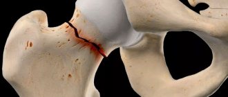
Cruvelier's joint is located in the region of the first cervical vertebra and is formed by the posterior surface of the arch of the atlas and its odontoid process. The first 2 vertebrae of the spine have a special structure. 1 vertebra (C1 or atlas) has a ring shape and its lateral sections are larger than the anterior and posterior ones. These sections form a joint with the occipital bone. The 2nd cervical vertebra or axial (C2) is shaped like a ring. Its lateral surfaces are also thicker, and in front it has a “tooth” - a process that protrudes upward and resembles the phalanx of a finger. The sliding of this tooth along the inner surface of the atlas forms the Cruvelier joint. As a result, the ring has ligaments at the front, and at the back the tooth has its own transverse ligament with the vertebra. The rear ring of the atlas seems to “sag” because it is not connected to anything.
All articular surfaces of the Cruvelier joint are normally covered with a capsule with folds, thanks to which the head can move: turns to the side, rotation of the head and oscillatory movements of the head.
Causes of joint disease
The joint performs a number of the following functions:
- rotational and oscillatory movements;
- flexion and extension.
The lateral fissures (cracks of the Cruvelier joint), measuring from 1.8 to 2.2 cm, allow all of the above movements. The joint experiences repeated stress due to its anatomical location, which takes on the function of supporting the head.
The threat of pathological changes lies in the fact that they may not manifest themselves for a long time, which affects untimely treatment. This increases the risk of arthrosis of the Cruvelier joint.
Common causes of joint disease in adults:
- aging of the body (patient’s age over 50 years);
- inflammation that occurs both in the joint and in the body;
- daily head pressure, which affects the cervical vertebrae;
- previous subluxations and injuries to the neck and spine (road accidents, impacts, sudden head movements, etc.);
- genetic predisposition;
- excess body weight;
- excessive physical activity;
- infectious diseases in the body;
- congenital anomalies of the cervical region, leading to deformation (impaired functioning of the cartilage leads to increased friction between the bones, destroying them);
- dysfunction of the endocrine system;
- heavy sports (boxing, powerlifting, etc.).
Diet
The diet includes the following:
- refusal of smoked meats, refractory fats and switching to vegetable oils;
- refusal of fried foods and canned food, seasonings;
- reducing salt, alcohol and soda;
- exclusion of baked goods and sweets;
- more cereals, fresh vegetables and fruits, and herbs are shown;
- water regime - at least 2.5 liters of clean water per day.
Signs of pathology
Medicine has recorded 4 stages of the disease:
- Degenerative changes are barely present and symptoms are mild. X-rays may not yet reveal abnormalities in the soft tissues, ligaments and lining of the joint.
- The appearance of aching pain that is inconsistent in nature, as well as a feeling of fatigue in the cervical region. There may be stiffness in neck movements. The photo taken during the ultrasound shows obvious joint changes.
- The formation of growths on the joint due to a local inflammatory process. The work of tendons and ligaments is disrupted. Increasing pain occurs, difficult to relieve with analgesics. An X-ray will show all changes in the joint and nearby tissues.
- An active increase in the size of growths, which has a direct negative effect on the functioning of the joint. Upon visual examination, the doctor discovers asymmetrical outlines of the neck. Rotating the head is difficult. Stage 4 processes can have irreversible consequences.
Main signs of pathology:
- pain in the neck;
- stiffness observed when moving the head;
- sleep disorder;
- When the neck moves, there is a characteristic crunching sound.
In rare cases, the following symptoms are present:
- dizziness;
- noise in ears;
- visual impairment;
- headache;
- increased blood pressure;
- the contours of the neck are not symmetrical;
- disturbances in coordination.
Therapeutic measures
Depending on the stage of the disease, therapeutic therapy is divided into the following types:
- Patients who are overweight are prescribed a special diet aimed at bringing their body weight back to normal. This means that BMI (depending on a person’s height) should not exceed acceptable values. To do this, it is recommended to reduce the amount of food consumed and eliminate foods and drinks that have a negative effect on the body.
- Physical therapy (physical therapy) is applicable, consisting of exercises aimed at performing oscillatory and rotational movements of the head. With the help of this treatment, blood circulation in the cervical region is enhanced, and the condition of the cartilage tissue is normalized. First, the exercises must be performed under the guidance of a specialist. Movements should be carried out smoothly, without pain. Crunching of joints is allowed during exercises.
- The doctor prescribes physical therapy (ultrasound, electrophoresis), which helps relieve pain, improve blood circulation and motor function of the joint. The course lasts from 7 to 10 days.
- A specialist may prescribe conservative treatment, including nutritional correction, massage, and rest in a specialized sanatorium. This therapy is effective at the initial stage of the disease.
- Symptomatic therapy. The action of the medications is aimed at eliminating spasms, relieving the inflammatory process and pain.
Drug treatment includes the following measures:
- Signs of development and treatment of arthrosis of the Cruvelier joint
- taking medications in tablet form;
- course of injections;
- use of gels and ointments.
Symptomatic treatment consists of taking medications from various groups:
- Non-steroidal anti-inflammatory drugs. They are used to relieve acute symptoms of the disease. A commonly used drug prescribed by doctors is Ibuprofen.
- Chondroprotectors. They are used to regenerate damaged cartilage tissue and nourish it.
- Glucocorticosteroid hormones.
- Drugs to improve blood circulation.
- Analgesics. Medicines are taken to relieve and relieve pain of varying severity.
- Muscle relaxants.
- Complexes of minerals and vitamins.
- Preparation Vitreous humor (from cattle eyes). In medical practice, they resort to prescribing it as a substance that promotes the resorption of scar tissue.
Sometimes it becomes necessary to perform surgery to eliminate the pathology. Surgery is rarely used and only in cases where other treatment methods have failed.
Drug treatment
First stage:
- The use of NSAIDs: Ibuprofen, Nimesulide, Diclofenac - relieve pain and reduce inflammation.
- Muscle relaxants that relax muscles and relieve spasm.
- Chondroprotectors - to strengthen cartilage tissue, which include chondroitin sulfate and glucosamine - substances that restore cartilage.
- In advanced cases, intra-articular administration of drugs is practiced - these are primarily GCS (corticosteroids) - Hydrocortisone, Diprospan, Dexamethasone. After the inflammation is removed, hyaluronic acid is injected there, which acts as a lubricant and reduces rough friction of intra-articular surfaces, eliminates pain, increases mobility and causes the synthesis of its hyaluronate.
- Since blood flow is impaired, warming ointments can correct the situation: “Bishofite”, “Capsicam”, “Dimexide”. All of them improve blood flow and relieve pain.
The second stage is aimed at improving metabolic processes:
- Taking Riboxin for 2 weeks or ATP intramuscularly.
- To improve the microcirculation process - “Actovegin”, “Trental”, “Curantil” for a month.
- As antioxidants - vitamin-mineral complexes with selenium, vitamins E, C.
What provokes the development of pathology?
Damage to the joints of the cervical spine is very dangerous, since untimely treatment can lead to disability.
Arthrosis is considered an age-related disease, but it also occurs among young people. Damage to the Cruvelier joint is most often influenced by the following factors:
- excessive loads on the cervical spine;
- professional sports;
- neck injuries;
- infectious diseases;
- sedentary lifestyle;
- excess weight;
- pathologies of the thyroid gland;
- heredity.
Return to contents
The main symptoms of arthrosis of the Cruvelier joint
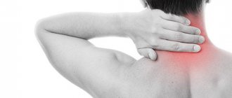
The pain is localized in the back of the head and neck.
Since the Cruvelier joint is responsible for flexion, extension and rotation of the neck, its defeat by arthrosis provokes discomfort when performing these movements. Displacement of the vertebrae can provoke disruption of blood circulation and innervation of the joint. A characteristic sign of damage to this particular part of the cervical spine is parallel damage to the facial joint. In addition, the following symptoms are observed:
- pain at the base of the head;
- swelling and redness in the affected area;
- frequent dizziness;
- partial loss of vision;
- noise and ringing in the ears;
- in advanced form, complete immobilization of the head.
Return to contents
Surgical intervention
In advanced cases, when there are already growths on the vertebrae, there is no effect from conservative treatment, surgical methods are used. During surgery, osteophytes are removed and the affected joint is replaced with an implant. To relieve pain, thermal destruction of the nerve endings in the affected joint - denervation - can be used.
During surgery, the spinal disc is restored. Movements of the neck and head resume and the pain goes away.
Diagnostic measures
If one or more symptoms of the disease are detected, you should consult a doctor. The doctor, by interviewing the patient, finds out the clinical picture of the disease. To confirm the diagnosis of arthrosis in the Cruvelier joint, the following additional studies are prescribed:
- Arthritis of the cervical spine: what it is, causes, treatment, symptoms, signs
- A general blood and urine test will help identify diseases of the systems involved in blood circulation and metabolism.
- X-rays taken in two projections show deformation, displacement of the vertebrae or the formation of osteophytes.
- Vascular angiography determines the degree of circulatory disturbance in the affected area.
- CT or MRI are prescribed to study cartilage tissue or if the formation of malignant tumors is suspected.
Return to contents
How to treat?
NSAID drugs will relieve pain and inflammation.
Therapy for arthrosis of the Cruvelier joint should be comprehensive. For this purpose, conservative treatment methods are used. With the help of medications, pain is relieved and the inflammatory process is neutralized. For this purpose, analgesics and non-steroidal anti-inflammatory drugs are used. But the abuse of these drugs is not recommended, as they can cause disorders in the functioning of other organs and systems. In addition, medications are used to stimulate blood circulation. Muscle relaxants are prescribed only in combination with other methods of therapy.
The selection of medications and determination of dosage should be carried out by a doctor, since self-medication leads to a worsening of the patient’s condition.
The use of drugs is not enough in the treatment of arthrosis. In addition, you should develop a set of exercises together with your doctor to restore full motor activity. Massage and physical therapy also have a beneficial effect on improving blood circulation, restoring cartilage tissue and relaxing neck muscles. In severe cases, doctors resort to surgery. This is a complex operation, so it is better not to bring the disease to this stage, but to seek advice from the hospital in time and begin treatment.
Cruvelier's joint is located in the cervical spine. This is a specific articulation, which is formed by the arch of the first (atlas) and the tooth of the second (epistrophy) cervical vertebrae. It is involved in the rotation of the spine around its axis, and is also necessary for the correct connection of two adjacent vertebrae. Normally it is strong and mobile, but with some diseases, including arthrosis, it cannot perform its functions. This is a common pathology that occurs in adults and children of any age. The disease must be identified and treated in the early stages, as it can lead to dangerous changes in the future.
Cruvelier's joint is located between the first and second cervical vertebrae, at the top of the spine. The second vertebra (epistrophy) has a specific growth - a tooth that slides along the first vertebra (atlas) during movement. It is this area where the epistropheus tooth slides that forms the Cruvelier joint. Despite its small size, it performs important functions:
- various rotational movements in the upper part of the cervical spine (flexion, extension of the neck, turns and tilts of the head);
- support for the first cervical vertebra to reduce the load on it.
All movements in the cervical spine have a small amplitude. The most convenient and natural movement is turning the head around a vertical axis. You can increase the amplitude through regular training and gradual stretching of the ligaments of the neck and head joint.
Normally, the Cruvelier joint has a gap of 1.8 to 2.2 mm. This distance allows full movement in the cervical spine, but with some diseases it may narrow.
Normally, the joint remains mobile, and the area where it is located is painless both at rest and in motion. However, there are a number of diseases that affect the functionality of this joint. They appear in connection with injuries, age-related and degenerative changes in tissues, and also act as a symptom of other pathologies.
Arthrosis is a degenerative disease in which the articular cartilage wears out, becomes brittle and brittle. The causes may be injuries, age-related changes and other factors.
Symptom (subluxation) of the Cruvelier joint is a condition associated with weakness of muscles and ligaments, abnormalities in the development of the second cervical vertebra. This is not an independent disease; it manifests itself as a sign of Down syndrome, rheumatoid polyarthritis and other pathologies. Subluxation can also be caused by blows to the head and face, or headstands.
- What is uncovertebral arthrosis of the cervical spine?
Asymmetry of the lateral joint spaces is a disease associated with subluxation of the atlas and at the same time its displacement to the side. It accounts for up to a third of all neck diseases and requires timely treatment. If the vertebra is severely displaced, any head movements are impossible and are accompanied by acute pain.
The health of the Cruvelier joint is of great importance. Its pathologies are dangerous not only due to painful sensations, but also serious complications. Displacement of the vertebrae or narrowing of the joint space leads to impaired blood circulation in the brain tissue and deterioration of the innervation of the upper extremities.
The Cruvelier joint connects the first cervical vertebra with the second and ensures full motor activity of the neck and head
Arthrosis of the Cruvelier joint is a common disease. It is associated not with inflammatory, but with degenerative processes that develop in articular cartilage. It gradually wears off and cracks appear in it. This leads to a narrowing of the joint space, as well as a decrease in depreciation of the vertebrae during movement. The process is considered irreversible, but it can be slowed down and the destruction of the joint can be stopped.
Depending on the causes, predisposing factors and developmental characteristics, several types of arthrosis of the Cruvelier joint are distinguished:
- primary (also called idiopathic) - associated with natural age-related changes;
- secondary - develops as a complication after injury or inflammatory diseases;
- deforming - manifests itself in combination with a change in the shape of the joint; bone growths are considered its characteristic symptom.
Arthrosis may not show any symptoms for a long time. It develops gradually and is noticeable only in the later stages, when acute headaches and dizziness begin. Over a long period of time, the composition of the synovial (intra-articular) fluid changes, and substances necessary to maintain its elasticity are washed out through the cracks of the cartilage. When the cartilage tissue becomes thin and fragile, head movements are accompanied by friction of the bone elements against each other. As a compensatory mechanism, various growths appear on them.
REFERENCE. Arthrosis of the Cruvelier joint is considered deforming. In most cases, patients experience neoplasms and bone growths in the area of the first cervical vertebrae.
Previously, it was believed that arthrosis was a disease of older people. Over time, tissues do lose strength and elasticity, but the disease can manifest itself at any age. Its causes are both external and internal factors that affect the condition of cartilage tissue and can cause its gradual thinning. These include:
Read the article: Deforming cervical spondylosis
- spinal injuries, especially in the cervical region;
- hereditary predisposition, a history of arthritis in relatives;
- infectious diseases that can spread to joint tissue;
- diseases of the endocrine system, including the thyroid gland - they affect the course of metabolic processes;
- congenital anomalies of the structure of the cervical spine;
- liver diseases;
- pathologies of bone tissue, including osteoporosis - increased porosity and weakness of bones;
- heavy physical activity and sports;
- incorrect posture, uncomfortable head position during sleep and work;
- obesity, muscle weakness;
- age over 50 years.
For arthrosis to develop, it is necessary to be exposed to one or more causes over a long period of time. Poor lifestyle and nutrition, excess weight, frequent hypothermia, lack of physical activity or excess are factors that stimulate the destruction of cartilage in the cervical spine.
Wearing an orthopedic collar will not cure arthrosis, but it will reduce pain and relieve discomfort in everyday life.
The main danger of arthrosis of the Cruvelier joint is that it appears only in the later stages. Initially, compensatory mechanisms allow the spine to function normally, so the disease does not bother the patient. Over time, the first complaints begin, which may worsen in the future.
Arthrosis can be determined by the following symptoms:
- short-term pain in the neck and back of the head, which at first only bothers during physical activity, and then continues constantly;
- migraines for no apparent reason, dizziness;
- fainting;
- toothaches;
- a characteristic crunch in the neck, which is clearly audible when turning the head;
- a feeling of numbness and tingling in the neck, shoulders and arms, decreased sensitivity in the fingers;
- deterioration of hearing and vision.
In the later stages, symptoms of arthrosis of the Cruvelier joint reduce the patient’s quality of daily life. Dangerous symptoms are observed, including unsteadiness of gait and deterioration of balance, frequent attacks of dizziness and fainting. Redness and swelling may appear on the back of the neck, which interferes with normal blood circulation and innervation in the neck and head.
Osteoarthritis of the Cruvelier joint develops in stages. It is important to pay attention to its first signs, as they will help to detect the disease in time and take all necessary measures to treat it. In total, there are 4 main stages, the first of which is considered the easiest and safest, and the fourth is a complicated stage.
At the first stage, degenerative processes have already begun, but there are no symptoms of the disease. It can only be determined on the basis of radiography or MRI.
At the second stage, the joint space narrows, and characteristic bone growths appear at the edges of the vertebrae. During physical activity and when turning the head sharply, painful sensations appear in the neck and head. Movements become constrained, but the discomfort goes away at rest.
The third stage is accompanied by inflammatory processes, and the bone growths are large and affect the ligaments. Patients complain of chronic pain that persists in any condition. Head movements are difficult, and in some cases there is joint immobility.
The fourth stage is the most dangerous. The bone fragments are deformed, so the joint is completely immobilized. At this stage, ankylosis appears - immobility as a result of complete fusion of the articular surfaces of the bones.
At the first stage, treatment of arthrosis will take about 2 weeks, and the patient’s condition can be stabilized. At later stages, the processes are irreversible; in some cases, surgical intervention may be necessary.
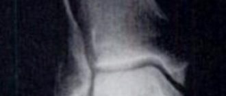
The diagnosis can be made based on the symptoms of the disease and additional tests. X-rays of the cervical spine will be informative - they can determine the width of the joint space of the Cruvelier joint. For a more complete picture and assessment of the condition of surrounding tissues, magnetic resonance or computed tomography is prescribed.
Treatment necessarily includes simple exercises for the cervical spine, which can be performed at home.
Treatment tactics are selected individually, depending on the stage of the disease, the patient’s age, symptoms of the disease and its complications. There are several techniques that are used separately, but are more often used in combination.
- Orthopedics - useful for fixing the neck in a comfortable position and stretching the vertebrae to widen the joint space. The method is to wear a Shants collar.
- Drug treatment is an important component at any stage. It includes pain-relieving ointments and tablets, as well as injections of anti-inflammatory drugs in the presence of complications.
- Physiotherapy is a set of techniques to improve blood circulation and nutrition of cartilage tissue. It includes magnetic and microwave therapy, phonophoresis and other procedures that are carried out in the course. In addition, it is important to perform simple exercises from a complex of physical therapy.
- Proper nutrition is aimed not only at weight loss, but also at preventing the formation of salt deposits in the joints. The diet should be dominated by plant-based foods, and fried foods, carbonated drinks, alcohol and fatty animal products will have to be abandoned.
- Surgical intervention is a radical method of treating deforming arthrosis, which is used only in advanced cases. During the operation, one of the following methods can be chosen: removal of bone growths, replacement of a joint with an implant, or destruction of nerve endings to relieve pain.
Cruvelier's joint, despite its small size, plays an important role in the human body. The functioning of the cervical spine and upper extremities, blood supply and innervation of the brain depends on its health. It is important to identify diseases in the early stages and begin their treatment, as well as pay attention to their prevention: proper nutrition, exercise regimen and periodic examinations of the spinal column.
Arthrosis of the Cruvelier joint is not such a common disease, and therefore few people have heard of this term. If such a diagnosis is made, this means that this pathology has developed in the cervical spine.
Accordingly, treatment should be applied in accordance with the characteristics of the anatomical units.
Structure and functions
Cruvelier's joint is located between the first and second cervical vertebrae, at the top of the spine. The second vertebra (epistrophy) has a specific growth - a tooth that slides along the first vertebra (atlas) during movement. It is this area where the epistropheus tooth slides that forms the Cruvelier joint. Despite its small size, it performs important functions:
- various rotational movements in the upper part of the cervical spine (flexion, extension of the neck, turns and tilts of the head);
- support for the first cervical vertebra to reduce the load on it.
All movements in the cervical spine have a small amplitude. The most convenient and natural movement is turning the head around a vertical axis. You can increase the amplitude through regular training and gradual stretching of the ligaments of the neck and head joint.
Normally, the Cruvelier joint has a gap of 1.8 to 2.2 mm. This distance allows full movement in the cervical spine, but with some diseases it may narrow.
Features of the disease
Cruvelier's joints refer to the posterior surface of the arch of the atlas, as well as the odontoid process. This part is located at the first vertebra of the cervical spine.
With arthrosis of this articular unit, tissue changes and their subsequent deformation are manifested. This leads to compression of not only the blood vessels, but also the nerve roots.
When tissues are traumatized, cartilaginous surfaces gradually begin to deteriorate. Due to the characteristics of this section, symptoms are often absent or mild and are not often associated with the neck. Therefore, treatment is most often postponed until arthrosis has fully developed.
The risk category usually includes people over 50 years of age, as well as those who lead a sedentary lifestyle with a corresponding load on the neck. Since the Cruvelier joint is located at the first vertebra, which is responsible for supporting the head, those who often sit reading books or at the computer usually suffer from the disease. The disease was assigned an ICD code - M19.8.
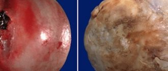
Pictured is the Cruvelier joint
Causes
There are many reasons for this condition:
- Static frequent preservation of a tilted head;
- Neck injuries, including dislocations and subluxations;
- Infectious pathologies;
- Surgical interventions in the area;
- Inflammation of the joint or nearby structures;
- Aging of the body;
- Genetic predisposition;
- Endocrine disorders.
Third-party factors also play a big role, including excess weight, unhealthy diet, improperly equipped places of work and rest, hypothermia, and so on.
Symptoms of Cruvelier arthrosis
Signs may not appear at first. Symptoms are usually temporary or mild. Often patients do not even suspect that all the problems come from the cervical spine. Main symptoms:
- Pain in the back of the head;
- Toothache or headache;
- Migraine;
- Dizziness;
- Fainting conditions;
- Hearing and vision impairments;
- Shoulder pain;
- Feeling tired and weak in the neck;
- Crunch in the neck;
- Sleep disorders.
Symptoms may be temporary, but gradually the frequency of manifestations will increase. Accordingly, internal degenerative processes will progress.
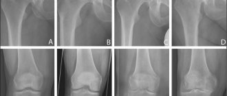
Osteoarthritis of the Cruvelier joint on x-ray
Types of cervical arthrosis
Spondylosis is called arthrosis of the entire spine, and arthrosis of the Cruvelier joint of the cervical spine is considered uncovertebral. There is also cicovertebral arthrosis, which is associated with wear and tear of cartilage. It is characterized by headaches and dizziness.
Any arthrosis can be:
- Primary or idiopathic is age-related.
- Secondary – does not depend on age and develops as a result of injury, existing diseases, dysplasia or inflammation, etc.
- Deforming - with the classic development of degeneration processes, which causes changes in the shape of the joints, disrupts their functions and manifests itself in severe pain.
Arthrosis of the Cruvelier joint is a type of deforming arthrosis.
It is typical for the elderly, since cartilage tissue loses its natural elasticity and synovial fluid decreases in volume. Growths (osteophytes) form on the posterolateral surfaces.
Over time, osteophytes impinge on the nerve roots and neuritis may develop. Without treatment at an early stage, the pathology progresses quickly and becomes irreversible.
Diagnostics
Diagnosis is usually ineffective at the first stage of pathology. In order to identify it, an X-ray examination is performed to determine the size of the gap and how widened or narrowed the space in the joint is.
If possible, MRI is also used. Typically, depending on the stage, we can distinguish:
- There are no obvious signs of the disease, but minor changes in the ligamentous apparatus and soft tissues may appear.
- Osteophytes begin to form, which gradually compress the soft tissues and structural units in the neck. Ultrasound may show obvious joint changes.
- Osteophytes grow significantly and put pressure on blood vessels and nerves, giving corresponding symptoms. Local inflammation develops, which can be seen on ultrasound and MRI. Bone growths appear on x-rays.
- The growths actively increase, disrupting the functionality of the joint. The doctor can visually determine a violation of the symmetry of the neck in the affected area. Rotating the head is significantly difficult.
At the fourth stage, the consequences may already be irreversible. As a rule, in this case, surgical intervention is already required.
Treatment
Therapy involves the use of medications, exercise therapy, and physiotherapy. The first ones combine:
- NSAIDs;
- Analgesics;
- Chondroprotectors;
- Vitamin dietary supplements;
- Hormonal agents;
- Blockades.
The specific range of drugs is selected depending on the patient’s condition. They may also prescribe topical medications that have irritating properties.
Physiotherapy usually includes manual manipulation, acupuncture, electrophoresis, amplipulse, paraffin therapy, ozokerite therapy, medicinal baths, and so on. Good results are also obtained when massage is introduced into the course of treatment. Moreover, ideally it is better to combine such an effect with exercise therapy.
Surgical intervention is used only if conservative treatment does not produce the required results. Accordingly, bone growths may be removed to restore the functionality of the vertebra.
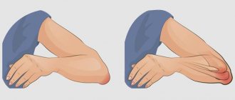
Neck exercises
Forecast
The prognosis for arthrosis of the Cruvelier joint is relative. On the one hand, the disease itself is detected in an advanced state. Accordingly, treatment requires long-term exposure, which not every patient agrees to.
On the other hand, if the doctor’s recommendations are followed, then the chance of restoring functionality or maintaining the condition that has already developed is much higher.
How to properly treat arthrosis with chondroprotectors, watch our video:
A common disease of the cervical spine is arthrosis of the Cruvelier joint. The disease occurs due to dysfunction of the atlantodentate joint, which is responsible for rotation, flexion and vibration in this area. In this case, the front connecting element is destroyed. The disease is degenerative in nature and leads to the destruction of not only the articular structure, but also the cartilage and ligament.
The most common joint pathologies
The most common disease is arthrosis of the Cruvelier joint. Any person during his active life receives many minor injuries, which after 20 years can begin to manifest themselves. This means arthrosis.
In women, arthrosis appears 2.5 times more often. By the age of 50, every 3 people have joint changes, and by the age of 60, everyone, regardless of gender. It is impossible to prevent this, like old age.
Cruvelier's sign may also occur - this is a subluxation of the Cruvellier joint. It was first described by a French doctor. It occurs due to weakness of the cervical ligaments and muscles, improper development of the C2 tooth, and the presence of a gap between the tooth and the body of C2. The symptom can develop with Down syndrome, Morquio's disease, rheumatoid polyarthritis. This is not an independent pathology.
In addition, the disease often occurs in children:
- when landing on the head or face;
- hitting your head;
- headstand or somersaults.
Its danger is that the blood supply and the passage of impulses in this area are disrupted due to compression. The result is muscle hypotonia, paresthesia occurs and the sensitivity of the fingers decreases, paresis of the limbs occurs and unilateral paralysis can develop.
Asymmetry of the lateral spaces of the Cruvelier joint is a rotational subluxation of the atlas, in which not only damage to the vertebra occurs, but also the development of degenerative changes in it. In this case, the vertebra moves to the side. This phenomenon occurs in 31% of all neck injuries. Incorrect position of the vertebra can cause compression of the spinal cord, making it impossible to move the head.
Causes
Arthrosis is provoked by various factors, which include:
- the patient's age is more than 50 years;
- previous injuries to the spine, and in particular to the cervical spine;
- genetic predisposition;
- overweight;
- excessive physical activity;
- infectious and inflammatory diseases in the body;
- disrupted functioning of the endocrine system;
- congenital anomalies of the cervical region.
In total, in medical practice there are 4 stages of development of the disease:
- Degenerative changes are just beginning, and symptoms practically do not appear. Violations of the joint membrane, soft tissues and ligaments are observed.
- Intermittent pain and fatigue appear in the cervical spine. Stiffness of movements is noted.
- A local inflammatory process and characteristic growths occur. The functioning of ligaments and tendons is impaired.
- The growths significantly increase in size, and the ongoing processes become irreversible.
Return to contents
Stages of arthrosis
There are 4 stages of arthrosis in total:
- Degeneration is just beginning. There are no symptoms. Initial changes in the articular membrane and ligaments are noted.
- Fatigue and pain are not constant, they only go away with exercise and go away with rest. Movements become more constrained, the gap of the Cruvelier joint is narrowed, cartilage is already being destroyed and growths begin to appear on the edges of the vertebrae.
- The growths are obvious. An inflammatory reaction develops in the vertebrae with disruption of the ligaments, and deformation occurs. The joint may become immobilized.
- The growths increase even more. The process becomes irreversible - ankylosis.
Main symptoms
Most often, the disease manifests itself as pain in the cervical spine.
The disease has a primary and secondary form, which differ in causes and symptoms. The main manifestations are characterized by:
- pain syndrome localized in the cervical region;
- stiffness of head movements;
- worsening sleep;
- presence of a characteristic crunch;
- dizziness, headaches and loss of coordination.
The development of pathology of the facial joint occurs over a long period of time, which is why it is detected at later stages.
Return to contents
The mechanism of arthrosis development
Arthrosis is a non-inflammatory chronic pathology of joints due to premature wear of intra-articular intervertebral cartilage (disc).
If arthritis occurs acutely and for a short time, arthrosis begins after 20 years and increases throughout life. Doesn't show itself for a long time. It causes degenerative-dystrophic changes in the joints. The cartilage tissue wears away and cracks appear in it due to the narrowing of the intervertebral space.
Through the cracks, the composition of the nourishing intra-articular fluid changes, and proteoglycans, substances necessary to maintain the elasticity of the cartilage, gradually leak out of the joint.
When the full value of cartilage is lost, bones rub against each other, which causes severe pain at the slightest movement. Together, all this leads to pinched spinal nerves, etc. - a vicious circle.
Treatment of the disease
A special collar will help reduce the load on the cervical vertebrae.
It consists in the effective use of both traditional and non-traditional methods. A course of medication and physiotherapy, physical therapy, a specially designed diet and traditional medicine recipes are required. It is important to immediately reduce the load on the cervical spine by wearing a special collar.
Return to contents
Drug treatment
It is compiled exclusively by a doctor, taking into account the patient’s individual indicators, characteristic causes and symptoms. The course of medications includes taking:
- non-steroidal anti-inflammatory drugs;
- chondroprotectors;
- glucocorticosteroid hormones;
- drugs to improve blood circulation;
- analgesics;
- muscle relaxants;
- complexes of vitamins and minerals.
Return to contents
Physiotherapy and exercise therapy
Exercises in general are aimed at rotating movements of the head.
They have a high degree of efficiency. Physiotherapeutic procedures must be carried out on a hospital basis and exclusively by specialists. Thanks to them, pain is reduced, blood circulation and the functioning of the cervical spine are normalized. The course of physiotherapy is 7-10 days. Physical therapy should include simple rotational and oscillatory exercises with the head.
Return to contents
Traditional medicine methods
Should be used only after consultation with your doctor. It is highly not recommended to start treatment on your own, as you can cause irreparable harm to the body and significantly worsen the course of the disease. When selecting ingredients for recipes, you should be sure to check their quality and expiration dates. Before use, test for an allergic reaction.
Bibliography:
- https://ortocure.ru/pozvonochnik/spondiloartroz/sustav-kryuvele.html
- https://osteokeen.ru/ar/formy/artroz-sustava-kryuvele.html
- https://surgicalclinic.ru/info/sustav-kryuvele
- https://gidmed.com/ortopediya-i-travmatologiya/sustavu-kosti/artroz/kryuvele.html
- https://etosustav.ru/ar/tipy/artroz-sustava-kryuvele.html
Expert equipment
The study is carried out on the latest generation X-ray system DIGITAL DIAGNOST (PHILIPS, the Netherlands). This is a completely digital x-ray station. Its advantages:
- Maximum data transfer and processing speed
- Obtaining the highest resolution (quality) image possible
- Lowest radiation exposure comparable to a short flight
The results of radiation diagnostics of the Yauza Clinical Hospital are accepted anywhere in the world, which is important for patients planning to undergo certain stages of treatment abroad.