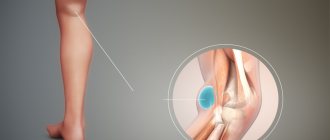
Inflammations localized in the joint of the lower extremities can provoke the formation of a neoplasm in the popliteal tissues, which is commonly called a Baker's cyst. Clinical manifestations of the neoplasm can be traced only after the cyst reaches a certain size, at which nerves and blood vessels are compressed.
Identification and approval of the diagnosis is carried out based on the results of laboratory and instrumental studies. Often the pathology is recorded in the fair sex, but it can also be observed in men.
What is a Baker's cyst?
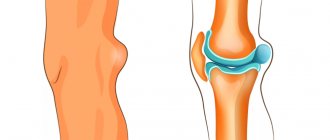
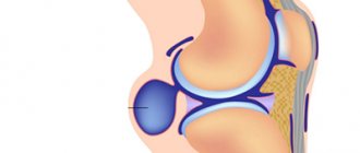
Baker's cyst is a pathological neoplasm that involves the accumulation of varying volumes of fluid in the synovial sac, which is localized on the back of the knee joint.
The morphological signs of the existing pathology allow us to assert the benign nature of the formation, without classifying it as a cancerous tumor.
Popliteal synovial cysts most often occur when the patient has an intra-articular disease, such as osteoarthritis. The formation can have a different size (from 2 mm or more) and occurs at any age.
Diagnosis of Becker's cyst
To determine the disease, the doctor conducts a detailed examination of the patient’s knee. It is also necessary to exclude infectious inflammation. Then an ultrasound examination is performed, which is the “gold standard” for this pathology. Sometimes MRI or pneumoscintigraphy is prescribed. In case of combined pathology of the knee joint, an operation is often prescribed - arthroscopy, which is both a diagnostic and therapeutic procedure, including for this pathology, in cases where the cyst communicates with the joint cavity.
Baker's cyst symptoms
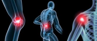
According to average statistical data, Baker's cyst is accompanied by the manifestation of a symptomatic picture of meniscal or chondral disorder, which usually includes:
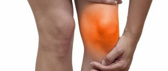
- pain localized in the knee or popliteal fossa, occurring both when performing various types of loads and in a calm state;
- extraneous clicks, crunches inside the joint;
- feeling of stiffness when moving.
- pain;
- swelling of the arteries of the popliteal region;
- loss of joint functionality due to mechanical blocking of its mobility.
The actual symptoms of a Baker's cyst, as such, appear extremely rarely and are often associated with progressive growth and an increase in the size of the tumor (from 3 cm in diameter). These include:
When a Baker cyst reaches a noticeable size, it can be noted that the formation often has a hard structure when the knee is fully extended and softer when the leg is bent. This phenomenon is called “Foucher's sign” and is observed in a situation where the cyst is compressed between muscle tissue.
It is also worth paying attention to the fact that patients with this diagnosis may experience signs and general symptoms of thrombophlebitis, which include pain and mild swelling of the lower extremities.
Causes and features of pathology
Becker's cyst in a child
It is customary to classify a neoplasm under the knee, which is called a Becker's cyst in a child, as benign, but many are still arguing about this. The reason for the controversy is the fact that such formation is caused by atypical cell proliferation.
However, the disease can be asymptomatic and in no way threaten the health and well-being of the child. But it must be recognized and controlled in time, treated in a certain way, according to the doctor’s recommendations, and if it grows, it should be removed. The cyst itself is a small sac filled with fluid that protrudes from the back of the knee.
While the cyst is small, it does not cause concern or restrictions. But when it grows, it interferes with movement and causes unpleasant and even painful symptoms. Even a small cyst under the knee can be determined by palpating the area under the knee if it is present. Most often it occurs in adults, and even more often in women. But children also experience similar pathological protrusions in the form of a stretched joint capsule with articular fluid under the knee. When the cyst is already quite large, it is difficult to bend the leg, but there may be no pain.
The cause of this pathology in childhood can be any inflammatory process in this area, pathology of the musculoskeletal system:
- Arthritis.
- Rheumatism.
- Injuries to the components of the knee joint.
The fact remains that the exact causes of tendon hernia in either children or adults cannot be established. In childhood, most likely this will be a traumatic injury to the knee: tendons, cartilage, meniscus, and so on. In babies under one year of age, inflammatory processes can lead to this disease.
Pathogenesis of Baker's cyst
The pathogenesis of Baker's cyst under the knee is based on the non-standard anatomical location of the articular tissues, as well as the popliteal fossa.
Statistics show that in more than 40% of cases, when examining healthy people of different ages and genders, there is an intertendinous mucous bursa between the tendons of their muscular frame of the knee joint, which is not a pathology, but is considered an acceptable deviation from the norm.
The progression of joint inflammation and the formation of fluid in the joint tissues lead to an increase in the size of the cyst. The presence of a valve mechanism allows liquid to leak into its cavity, but does not allow reverse flow to occur.
Causes
The main cause of popliteal cysts in children is knee injuries. In the absence of restorative treatment and rehabilitation measures, inflammation can become chronic, which is the basis for the development of synovial hernias.
The next most important cause is systemic connective tissue diseases, in particular juvenile rheumatoid arthritis. With this disease, the knee joint is damaged by its own immune system, which for some reason mistook it for a foreign agent. As a result of the high activity of the process, inflammatory exudate may begin to accumulate in the joint cavity.
Long-term infectious arthritis, especially with widespread tuberculosis, can also lead to the formation of synovial hernias.
In some cases, Baker's cyst may be idiopathic. In this case, the cause remains uncertain and surgical treatment is preferred.
Baker's cyst classification
Baker's cyst has several variations in manifestation and development, which makes it possible to classify cases according to their location in the popliteal fossa and distinguish two types:
- typical - located between the muscles of the knee joint;
- atypical – posterolateral cyst.
In addition, it is worth noting that Professor Raushing’s research made it possible to prove the existence of two forms of cysts:
- symptomatic – is a symptom of a pathogenic joint disease;
- idiopathic - a cyst in which no pathological articular processes are detected.
Diagnosis of Baker's cyst
Clarification of the clinical picture is realized using methods such as:
- laboratory blood test (clinical and biochemical analysis;
- instrumental research methods.
The process of diagnosing Baker's cyst involves a differential approach that excludes the possibility of such pathologies as:
- popliteal artery aneurysm;
- soft tissue tumor;
- meniscal cyst;
- hematoma of various origins;
- thromboembolism;
- seroma.
In order to confirm existing symptoms, instrumental research methods such as:
- radiograph;
- arthrography;
- ultrasonography;
- Magnetic resonance imaging.
Let's look at each of the methods used in more detail.
X-ray examination
X-ray studies are indicated for use in the initial stages of assessing the patient’s condition, since they can be used to identify other pathological processes accompanied by the presence of popliteal neoplasms. Among these diseases:
- arthritis of various groups;
- free bodies of cartilage and bone type.
Arthrography
Arthrography involves intra-articular injection using a high-contrast agent with mandatory mobilization of the limb. The use of this diagnostic method can improve the effectiveness of x-ray examination.
Ultrasound examination (ultrasound)
Ultrasound examination falls into the category of preliminary diagnostic techniques for Baker's popliteal cyst.
Among the key advantages of the presented method are:
- availability;
- non-invasive;
- safety.
Magnetic resonance imaging (MRI)
MRI is a key diagnostic method in identifying Baker's cysts and differentiating them from other pathological phenomena.
Using MRI, it is possible to fully cover and study the entire existing spectrum of pathological processes.
Baker's cyst treatment
Treatment of Baker's cyst involves the use of both conservative and radical methods based on orthopedic and traumatological treatment.
The key purpose of conservative treatment methods is the implementation of therapeutic measures aimed at eliminating the pathological process. Among the main approaches in the treatment of conservative therapy are such approaches as:
- maximum relief of the load on the affected joint;
- prescription of physiotherapeutic treatment;
- taking a puncture followed by evacuation of the contents of the cyst and administration of specialized drugs.
The determination of the method of therapeutic treatment is determined by such criteria as:
- the degree of inflammation present;
- volume of accumulated liquid;
- the exact location of the pathological cavity.
In order to treat Baker's cyst as effectively as possible, it is recommended to use an integrated approach and combine drug therapy with physical therapy (physical therapy) and other methods.
Drug treatment for Baker's cyst
The use of drugs in the treatment of Baker's cyst is used to enhance the results of conservative methods and can reduce or completely eliminate inflammation inside the joint.
The following groups of drugs may be prescribed as drug treatment:
- NSAIDs (nonsteroidal anti-inflammatory drugs);
- painkillers;
- antispasmodics;
- vitamin and mineral complexes;
- hormonal;
- chondroprotectors.
Particular attention is paid to the group of chondroprotectors that help accelerate regeneration processes. Artracam is considered to be one of the most effective drugs in this group.
IMPORTANT! Drug treatment should be carried out exclusively under the supervision of the attending physician, taking into account the individual characteristics of the clinical picture of a particular patient. Self-medication is unacceptable.
Physical therapy (physical therapy) as a method of treating Baker's cyst
In order to restore the functionality of the knee joint, therapeutic physical education is used as a component of complex treatment.
Performing a number of exercises allows you to speed up the rehabilitation process, the duration of which is determined by the characteristics of the body. Before starting classes, consultation with a treating specialist is required.
Complexes of exercise therapy are based on certain principles and pay special attention to muscle groups such as:
- calf;
- quadriceps;
- gluteal
Among the general recommendations for performing exercises are:
- Be sure to warm up your muscle fibers before exercising and do quality stretching.
- Avoid using gymnastic exercises with increased intensity.
- Limit your loads, making sure to eliminate the possibility of muscle fatigue.
- Carefully prepare the location of the classes by preparing the necessary clothing, surface and equipment.
Surgical intervention
Radical therapy is used in situations where conservative treatment methods are not effective enough or are not applicable at all in the current situation.
Surgical intervention involves an open operation, the key task of which is to isolate the tumor from the surrounding tissue.
Despite the obviously successful solution to the problem, the result of surgical intervention is not in all cases capable of producing a guaranteed positive result. The recovery period may be complicated.
Treatment
Sometimes no treatment or simple supportive measures lead to spontaneous resolution or reduction of symptoms. If this does not happen, it may be worth using invasive and surgical instruments.
The asymptomatic condition often improves and over time, Baker's cyst disappears on its own. If the cyst is symptomatic, rest may ease the pain. You can also take non-steroidal anti-inflammatory drugs (NSAIDs) and ice to reduce pain.
If pain persists, steroids with an anesthetic solution may be recommended, which may relieve the pain but will not prevent the cyst from recurring. This is only a temporary solution. In the presence of a popliteal cyst of inflammatory origin, this is sufficient to treat the underlying disease. When the underlying disease is not treated, Baker's cyst may recur. Arthroscopic examination and treatment of all pathological conditions should be carried out before considering dissection of the popliteal cyst.
The cyst may be removed surgically if it becomes very large or causes symptoms such as discomfort, stiffness, or painful swelling. There are three surgical approaches to treat the cyst: the standard posterior approach, the posteromedial approach, and the medial intra-articular approach. The first two methods are cyst removal methods. The latter method involves creating an opening in the cyst, which is later closed.
Physical therapy
Using ice for 15 minutes every 4 to 7 hours will reduce inflammation. Treatment is based on the principles of RICE (rest, ice, compression and elevation), followed by some muscle training exercises.
A rehabilitation program can improve knee control through a series of therapeutic exercises. This will increase the mobility of the joint and also increase flexibility. A physical therapist can develop a program to improve mobility and stretch the hamstrings, which may also include exercises to strengthen the quadriceps. This will result in less pain in about 6-8 weeks.
There is an experiment comparing ultrasound-guided corticosteroid injections with horizontal therapy (hardware physiotherapy). The 60 people were divided into three groups, injections only (Group A), horizontal therapy only (Group B) and (Group C) in which patients received both injections and horizontal therapy. Horizontal therapy was performed using a custom commercial device, and the investigators followed the manufacturer's instructions. In patients in groups A and C, pain decreased after a month. Group C had the lowest VAS scores and also the best WOMAC scores. In groups B and C, stiffness and disability scores were also improved to the greatest extent.
Possible complications if Baker's cyst is left untreated
Uncontrolled development of a popliteal Baker's cyst can cause a large number of complications, the most likely of which are:
- development of an infectious disease;
- rupture of the cyst membrane;
- forceful effect on the neurovascular bundle, leading to thrombophlebitis.
In rare cases, untreated Baker's cyst under the knee can cause compression of the popliteal vein or even an artery. If a complication of this type is detected, urgent hospitalization and surgical intervention are indicated.
Disease prevention
The best treatment for any pathological condition is to prevent its occurrence.
In order to prevent the formation of a Baker's cyst, the following preventive measures should be followed:
- prevention of injuries to the joints of the lower extremities;
- tracking optimal body weight;
- complete cessation of bad habits;
- balanced diet;
- timely treatment of pathological processes found in articular tissues;
- the use of specialized structures to reduce the load on the knee joints.
Having information about what a Baker's cyst is, pathologists can promptly recognize it and begin its treatment, preventing the likelihood of its rupture.
Preventive measures
Proper nutrition as preventative measures
Important preventive measures will be those aimed at maintaining the overall health of the child:
- Proper nutrition containing all the minerals, vitamins, and microelements necessary for a growing body.
- For those who play sports, it is necessary to properly warm up the muscles before training and adhere to the load and rest regime. Every time before training you need to do a warm-up and then rest.
- Timely and competent treatment for injuries and any diseases of the bone and joint systems.
Since the causes of the pathology are not completely clear, and the disease occurs at a very early age, up to a year, prevention will be by following the general rules of a healthy lifestyle and hardening, which children’s doctor Komarovsky talks about a lot. The doctor believes that hardening is the main preventive measure against all diseases!