MRI diagnosis of synovial folds of the knee joint
Filisteev Pavel Anatolyevich, head of the MRI department of the MED-7 diagnostic center, doctor of the X-ray diagnostics and tomography department of the Federal State Budgetary Institution "Central Clinical Hospital with Clinic" of the Administration of the President of the Russian Federation
Synovial folds of the knee joint are part of the normal anatomy of the knee joint and can sometimes cause clinical manifestations. Magnetic resonance imaging (MRI) and MR arthrography allow non-invasive assessment of the condition of synovial folds and differential diagnosis of the cause of pain in the knee joint. Synovial folds appear on MR images as linear, hypointense bands against surrounding fluid and fat. T2- and proton-weighted pulse sequences with suppression of the MR signal from adipose tissue have been recognized as optimal for visualizing synovial folds.
Synovial fold syndrome (SPS) is accompanied by pain in the knee joint and impaired function, and should be considered by clinicians and radiologists as one of the possible diagnoses. A diffusely thickened fold, usually accompanied by local synovitis and erosions of articular cartilage, is the most common manifestation of CVS. Treatment of synovial fold syndrome consists of conservative management of the patient at the first stage; if ineffective, arthroscopic resection of the pathological fold is performed.
Introduction
The fold of the knee joint is a duplication of the synovial membrane lining the joint cavity. The structure of synovial folds is thin, membranous, with zones of vascularization. From an embryogenetic point of view, the folds are partially vestigial synovial membranes and are found in the knee joints of healthy people. The exact function of synovial folds is unknown. A number of authors believe that synovial folds, like the eyelids, help improve the lubrication of the joint with synovial fluid.
In cases of direct trauma to the knee joint, with repeated stress or inflammation, clinical symptoms may occur due to the mechanical effect of the folds on other intra-articular structures. In particular, upon movement, the thickened and fibrotic synovial plica causes irritation of the synovial membrane at the edges of the bone condyles, which ultimately leads to reactive synovitis and thinning of the articular cartilage (synovial plica syndrome - SPL).
Changes in the mediopatellar fold occur most frequently, but an accurate assessment of the prevalence of CVS is difficult due to the lack of clear diagnostic criteria.
On MRI, intact synovial folds appear as thin, hypointense linear structures arising from the synovial lining. Their visualization is significantly improved in the presence of fluid in the joint (articular effusion, blood, or contrast agent during MR arthrography).
Mediopatellar fold syndrome
The mediopatellar fold is a thickening of the synovium that lines the inner surface of the knee joint and secretes lubricating fluid.
It can cause discomfort if it enlarges and grows. This process is called mediopatellar fold syndrome and practically does not occur as an independent disease. As a rule, it occurs against the background of injuries (damage to the menisci, ligaments, cartilage) and certain diseases, such as synovitis (inflammation of the synovium, accompanied by fluid accumulation), arthritis. In some cases, this pathology may be a congenital feature of the joint structure.
Mediopatellar fold syndrome is manifested by the following symptoms: pain in the front of the knee, especially when standing up from a sitting position, painful clicks during flexion-extension movements. If at least one of them causes you discomfort, we recommend that you undergo examination at the Odrex Medical House, whose specialists have extensive experience in treating diseases of the musculoskeletal system.
The mediopatellar fold is a normal anatomical formation, but it is detected only in 30-50% of people. If this fold is present in the knee joint, it is not a problem in itself. The need for treatment arises only when it thickens, becomes inflamed and grows.
Often, hypertrophy of the mediopatellar fold is an incidental finding during the diagnosis of diseases of the knee joint. It can only be detected using MRI. Due to the complexity of the knee joint, it is difficult to examine this pathology with ultrasound, as well as with computed tomography.
Head of the Department of Orthopedics and Traumatology Konstantin Palagnyuk
Treatment of mediopatellar fold syndrome can be conservative or surgical.
Conservative therapy is prescribed to eliminate inflammation. It involves wearing an unloading knee brace, taking non-steroidal anti-inflammatory drugs, performing physical procedures (magnetic therapy, electrophoresis) and blocking (a medical procedure that is carried out to relieve pain and restore the functions of the limb) of the knee joint.
If conservative therapy is unsuccessful or the fold is greatly enlarged, it is advisable to resort to arthroscopic intervention , during which it is excised. This is a low-traumatic operation: only two small punctures are required, into one of which a mini-camera is inserted. Thanks to this, all manipulations can be performed with maximum efficiency and minimal risk of complications, and the patient is discharged from the hospital after 1-2 days. The recovery period is also much faster compared to open surgery - from 3 weeks to 1.5 months.
Who is most susceptible to mediopatellar fold syndrome?
At risk are primarily athletes (running, cycling, football) and dancers. Constant physical activity and frequent microtrauma can cause damage to various parts of the knee, including mediopatellar fold syndrome. As already mentioned, pinching of this fold also occurs due to the congenital features of the anatomical structure of the joint.
Can relapse occur after surgery?
The recurrence rate after arthroscopic excision of the mediopatellar fold is very low. In our clinic, thanks to the extensive experience of traumatologists, modern equipment and a competent approach to postoperative rehabilitation, such cases have not yet arisen.
What does postoperative rehabilitation include?
The rehabilitation period is very important; the final result of treatment largely depends on it. After arthroscopy, massages, physical therapy, and exercise on a special exercise bike are prescribed. The patient should approach physical activity with caution and follow all doctor’s recommendations.
Embryogenesis
Currently, it is widely believed that the knee joint initially consists of three compartments - medial, lateral and suprapatellar, separated by synovial septa. Incomplete resorption of these septa leads to the formation of well-developed synovial folds occupying the space between the distal parts of the epiphysis of the femur and the proximal parts of the tibia. Intrauterine movement in the fetal knee joint can simultaneously contribute to the resorption of folds and the formation of a joint cavity. This theory explains the presence of suprapatellar and infrapatellar folds, but is unfounded for the lateral and medial folds, since the patellofemoral joint is not divided into cavities in the plane of these folds at any stage of embryogenesis (that is, in the frontal one). Embryological work by S. Ogata and HK Uhthoff showed that the mediopatellar fold is not a remnant of the synovial septum. It is probably of mesenchymal origin and is associated with intrauterine lateralization of the patella, as evidenced by the predominance of mesenchyme in the mediopatellar zone.
There are no precise data in the literature on the prevalence of these persistent embryological structures in the population. In an anatomical study of the knee joints, T. Jouanin et al. in 11% of cases three main folds were present, in 10% no folds were identified. The suprapatellar and infrapatellar folds are the most common but are usually clinically intact. The lateral fold of the knee joint is extremely rare. Due to its clinical manifestations, the mediopatellar fold is the most studied to date.
Possibilities of MRI in the diagnosis of synovial folds
On MR images, synovial folds appear as hypointense cords with peripheral fluid and fat. The most informative pulse sequences in the diagnosis of synovial folds due to the high signal from the fluid are recognized as gradient T2-weighted pulse sequences, as well as T2- or proton-weighted sequences using fat suppression. MR arthrography is applicable in case of insufficient amount of intra-articular fluid and the presence of pronounced clinical symptoms of the manifesting fold. The contrast agent provides high intertissue contrast of the articular surfaces, stretches the joint capsule, which together helps to visualize all folds of the knee joint, verify or exclude their pathological changes.
The size and morphological restructuring of synovial folds are not a reliable sign of their clinical significance. In the presence of persistent clinical symptoms and the absence of other possible causes of a similar symptom complex (for example, damage to the medial meniscus or internal collateral ligament, patellofemoral arthrosis, arthrosis of the medial parts of the joint), characteristic signs of pathological fold syndrome are local synovitis, erosion of the articular cartilage of the patella and the patellar surface of the femur bones.
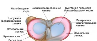
Damage to the lateral ligaments of the knee joint
Ligament injuries occur when there is a sharp excessive deviation of the tibia outward (tibial collateral ligament) or inward (fibular ligament), as well as during rotation, which can occur when jumping with a parachute, from a car, or when dismounting from gymnastic apparatus.
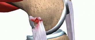
The tibial collateral ligament, starting from the medial epicondyle of the femur, descends in a rather wide band to the medial condyle of the tibia, where it is attached. The ligament is closely connected over a long distance to the capsule of the knee joint and, accordingly, part of its fibers - to the medial meniscus. The frequent combination of injuries to the medial meniscus and tibial collateral ligament is explained by the peculiarities of their anatomical structure. The fibular collateral ligament originates from the lateral epicondyle of the femur and attaches to the head of the fibula. It is not connected to the articular capsule and is separated from it by loose fiber. Usually the ligaments are partially damaged. Their complete ruptures are observed relatively rarely. The tibial collateral ligament is damaged much more often (usually partially), the fibular collateral ligament is damaged less often (usually completely). The most common site of injury to the collateral ligaments is their superior insertion points.
Complete ruptures of the medial collateral ligament are treated conservatively by applying a plaster cast. Complete rupture of the lateral collateral ligament requires surgical treatment, which should be performed in the first days after the traumatic injury. They are tightened and stitched or plastic surgery is performed using the biceps femoris tendon. If an avulsion fracture of the apex of the head of the fibula occurs, the fragment is fixed to the fibula with a screw. Sometimes the ligament not only ruptures, but also separates into individual fibers. Then the ligament is reconstructed using grafts.
Suprapatellar fold
In the literature, there are several synonyms for the suprapatellar fold: suprapatellar septum, superior fold, patellar fold.
The suprapatellar fold is localized between the suprapatellar bursa and the cavity of the knee joint, has an obliquely descending course, starting from the synovium on the anterior surface of the femoral metaphysis and ending in the posterior part of the quadriceps tendon in the area of attachment to the patella. Until the end of the 4th month of intrauterine development of the fetus, the suprapatellar fold completely separates the cavity of the knee joint from the suprapatellar bursa, and by the end of the 5th month its shape does not differ from that of an adult.
It is believed that the key factor ensuring the degree of fold reduction is mechanical. Depending on the range of motion in the knee joint, either a small perforation in the fold or its partial or complete absence may form. T. Zidorn classified the degree of “development”, or more precisely, reduction, of the suprapatellar fold into 4 groups depending on the morphological features:
MR tomogram of the knee joint in T2 V.I., sagittal plane
- type I (septum completum) - closed suprapatellar bursa - the suprapatellar fold completely separates the patellar recess from the cavity of the knee joint;
- type II (septum perforatum) - the suprapatellar fold has one or more holes connecting the suprapatellar zone with the joint cavity;
- type III (septum residuale) – a residual connective tissue cord is visualized, often on the inside;
- type IV (septum extinctum) – characterized by complete involution of the suprapatellar fold.
In type II folds, the zones of its perforation, which provide circulation of articular fluid to the area of the inversion, are called the hilum.
The suprapatellar fold persists very often - up to 89% of cases according to autopsy data. On MRI of the knee, the fold is best identified in the sagittal plane as a linear hypointense band posterior and superior to the patella.
During arthroscopy, a completely unreduced suprapatellar fold can be suspected only by a decrease in the volume of the suprapatellar recess. Sometimes the diagnosis is made by chance during joint puncture, when the injection needle ends up in the suprapatellar bursa and not in the joint cavity.
Infrapatellar fold
The infrapatellar fold, or ligamentum mucosum, is the most common fold of the knee joint. Its formation occurs from 8 to 12 weeks of gestation and depends on the degree of involution of the primitive embryonic membrane separating the medial and lateral sections of the joint. The shape of the fold formed the basis for its arthroscopic classification by S. Kim and W. Choe:
- 0—no fold;
- 1 - complete persistence of the fold, the fold is separated from the anterior cruciate ligament;
- 2 - complete persistence of the fold, the fold is separated from the anterior cruciate ligament and has several bundles;
- 3 - vertical fold - complete persistence of the fold, which is associated with the anterior cruciate ligament and divides the joint cavity into medial and lateral sections;
- 4 - fenestrated vertical fold - a vertical fold with a hole/defect.
The infrapatellar fold begins in the anterior parts of the intercondylar recess of the femur, expands forward and downward, and attaches to the lower pole of the patella. The thickness of the fold varies widely from submillimeter to the size of the anterior cruciate ligament.
The infrapatellar fold is easily identified on sagittal MR images as a linear, hypointense structure running through the Hoffa fat pad in a plane parallel to the anterior cruciate ligament. In some cases, especially in patients with complete rupture of the anterior cruciate ligament, a well-developed infrapatellar fold can simulate its preserved bundles. It is also necessary to carry out a differential diagnosis of the infrapatellar fold with local nodular synovitis, postoperative changes and a loose body in the area of the Hoffa fat pad. The awareness of radiologists about the existence of this anatomical structure allows in most cases to make the correct diagnosis.
Anterior cruciate ligament (ACL) injury
ACL ruptures are more common in athletes and are often accompanied by damage to other structures of the knee joint.
Along with this ligament, the menisci, collateral ligaments, etc. are damaged. The anterior cruciate ligament (ACL) acts as a stabilizer, keeping the lower leg from moving forward and rotating. In case of damage to the ACL, an unnatural forward displacement of the tibia occurs and a disruption of the physiological axis of rotation in the joint, which leads to damage to the cartilage and menisci. In addition to its mechanical function, the ACL serves as a sensory organ, providing a sense of movement and a sense of the position of the knee in space). ACL injuries are more common in athletes of play (CONTACT) sports, alpine skiers, jumpers, and wrestlers. The main complaint made by patients with an ACL rupture is instability of the knee joint, that is, there is a feeling of “SHIFTING” of the bones relative to each other in the knee joint - subluxation or “givability.” This sensation most often occurs during awkward movement or during acceleration AND ROTATION.
An accurate diagnosis of an ACL rupture can be determined by MRI, CT -1000 and a mandatory examination by a doctor.
Treatment is complex, including surgery and equally important rehabilitation, which begins immediately, while surgery can be postponed until the condition of the damaged knee joint normalizes.
ACL reconstruction with autograft
Diagnostic arthroscopy is performed, during which damage to other intra-articular structures is determined. Arthroscopy is performed through 2 small puncture incisions in the area of the knee joint measuring 1-1.5 cm. The transplant from the patellar ligament is collected through the same incision enlarged to 4-5 cm. If a hamstring muscle graft is used, an additional small incision (about 3 cm) is made on the posterolateral surface of the thigh. Our clinic uses all types of transplants according to indications.
In our stabilizing operations, we use a variety of fixation structures from global manufacturers Mitek, Smith & Nephew, Karl Shtorz, and other autografts in a position that closely resembles the ACL. All fixation structures consist of tricalcium phosphates and lactic acid and have the property of dissolving, which does not require repeated operations.
Now our clinic uses a two-bundle ACL plastic technique, which, according to numerous studies, largely replicates the anatomy of the ACL.
Mediopatellar fold
The mediopatellar fold is also called the medial fold, synovial patellar fold, pterygoid fold, patellar meniscus, and synovial protuberance. The mediopatellar fold begins in the area of the medial wall of the knee joint, goes obliquely downwards and is woven into the synovium around the Hoffa fat pad. It can be connected to the suprapatellar fold, but more often it has a separate attachment. If overdeveloped, the mediopatellar fold may extend to the medial surface of the femoral trochlea and the medial facet of the patella.
MR tomograms, axial plane, proton-weighted mode with signal suppression from adipose tissue
J. Sakakibara, based on the shape and size, identified 4 types of mediopatellar folds:
- type A – the fold is represented by a linear rope-like protrusion of the synovial wall;
- type B – the fold is represented by an elongated linear cord with uneven contours, but does not reach the medial condyle of the femur;
- type C – the fold is elongated, thickened, with uneven fringed contours, extending to the area of the medial condyle of the femur.
- type D – the fold extends to the area of the medial condyle of the femur, is thickened, uneven, and has a central defect (fenestrated fold).
This classification has received universal recognition and approval, as it is easy to use and clinically relevant. Types A and B of the mediopatellar fold are asymptomatic. Types C and D may impinge between the medial femoral condyle and the patella with subsequent thickening and induration, damaging the cartilage at the patellofemoral joint. The main mechanism of action of the fold is compression of the articular cartilage of the medial facet of the patella during flexion and the patellar surface of the medial femoral condyle during extension. A number of authors are of the opinion that the fenestrated mediopatellar fold (type D) more often causes damage (impingement) of the articular cartilage of the medial parts of the patellofemoral joint.
For visualization of the mediopatellar fold using MRI, T2- or proton-weighted images in the sagittal and axial planes with or without fat suppression are considered the most informative. The mediopatellar fold has a low MR signal and is easily recognized in a typical location against the background of a small amount of intra-articular fluid. On the other hand, one should always pay attention not only to the size of the fold and its location relative to the medial condyle, but also to the condition of the articular cartilage in the medial facet of the patella, femoral condyle, and the patient's complaints. With massive intra-articular effusion, lateral displacement of the fold may occur, complicating the differential diagnosis of types B and C. The large size of the mediopatellar fold may make it difficult to examine the medial parts of the joint during arthroscopy.
Lateral patellar fold
Schematic representation of the anatomical structures of the lateral recess of the knee joint (normal)
The lateral patellar fold is the most rarely encountered fold of the knee joint. Most often, it has a longitudinal shape, is very thin and is located 1-2 cm lateral to the patella. The lateral patellar fold originates on the lateral wall and is attached to the infrapatellar fat body. S. Ogata and HK Uhthoff hypothesized that rare persistence of the lateral patellar fold is associated with lateral displacement of the patella, leaving no room for it in the lateral recess. The lateral fold may make it difficult to perform knee arthroscopy through the anterolateral approach.
The lateral patellar fold should not be confused with the more commonly encountered pterygoid, superolateral, and transverse arcuate folds.
The arrows indicate: in Figure A - lateral patellar fold (white thin arrow), lateral pterygoid fold (black thin arrow), transverse arcuate fold (black thick arrow); in Figure B - superolateral fold (white thick arrow).
The lateral pterygoid fold is located close to the patella along the outer wall, the superolateral fold is a type of suprapatellar fold dislocated outward. A transverse arcuate synovial fold is often found at the junction of the vertical and anteroposterior portions of the lateral recess.
Posterior cruciate ligament injuries
Posterior cruciate ligament (PCL) injuries are one of the most serious injuries to the capsular ligament system of the knee joint. They are much less common than anterior cruciate ligament (ACL) tears. PCL ruptures can be isolated or combined with injuries to other ligaments and structures of the knee joint (menisci, ACL, collateral ligaments, joint capsule, popliteus tendon, arcuate ligament).
Due to the difficulty of distinguishing between ACL and PCL injuries in making a correct diagnosis, PCL injuries are often underdiagnosed, leading to the development of posterior instability and secondary changes in the knee joint.
To diagnose damage to the PCL, in addition to the clinical examination of the patient, special additional research methods are used: ultrasound, MRI, plain radiography, functional radiographs with stress.
In case of acute injury to the knee joint, arthroscopy becomes particularly relevant, especially in cases where there is a distorted clinical picture and it is impossible to properly examine the knee joint due to severe pain. Arthroscopy allows you to identify the entire volume of intra-articular damage and apply surgical methods to repair damaged structures.
Contraindications for arthroscopy of the knee joint are acute dislocation of the lower leg, open injuries of the knee joint, since due to rupture of the joint capsule, fluid is transuded into the soft tissues of the lower leg, which can lead to the development of a complication such as compartment syndrome. In these cases, arthroscopy can be performed after 2 weeks using low pressure and careful monitoring of the limb. Also contraindications to arthroscopy of the knee joint are fibrous ankylosis, various septic conditions of a general and local nature.
Treatment
In the acute period of injury (up to 2 weeks), when the PCL is separated from the internal femoral condyle, it is possible to refix the ligament stump to the site of anatomical attachment using arthroscopic techniques.
In the case of the development of chronic posterior instability of the knee joint in a compensated form, conservative treatment is carried out, which includes: therapeutic exercises aimed at strengthening the muscles that prevent pathological posterior displacement of the tibia, massage, and electrical stimulation of the quadriceps femoris muscle.
Subcompensated or decompensated posterior instability of the knee joint can only be eliminated surgically. For this purpose, intra-articular autoplastic stabilizing operations are performed.
In the department of sports and ballet trauma of the CITO, in case of damage to the PCL, arthroscopic intra-articular stabilizing operations are performed using a single or double-bundle autograft from the patellar ligament, tendon stretch of the 4th femoris muscle, tendons of the gracilis and semitendinosus muscles, for fixation of which in the joint bioabsorbable inferential screws are used .
Pathogenesis and clinical manifestations
Synovial fold syndrome (shelf-, plica-syndrome or their most common variety - mediopatellar fold syndrome) describes a complex of painful dysfunctions of the knee joint, in which the only finding is a thickened and fibrous-changed fold.
MRI scan of the knee joint
Patient A., 18 years old. MR imaging of the knee, proton-weighted fat suppression (axial plane) and arthroscopic slides. Complaints of pain in the internal parts of the knee joint. There is elongation, thickening, and fibrillation of the medial patellar fold, which reaches the medial femoral condyle (type C according to Sakakibara). The arrow indicates the area of “cracking” and chondromalacia of the articular cartilage of the medial facet of the patella.
The onset of pathology is usually an injury to the knee joint (direct blow, repeated load, twisting, leading to stretching of the fold), less often inflammatory and degenerative changes. Post-traumatic, inflammatory swelling of the fold leads to loss of elasticity, thickening and clinical manifestations if it reaches the patellofemoral joint during movements. The result of fibrosis and hypertrophy is reactive mechanical synovitis, as well as erosion of the articular cartilage of the femur and patella. In the early stages of the disease, the cause of pain is often the fold itself, while in a later period, chondromalacia of the articular cartilage and synovitis come to the fore. It should be remembered that the size, shape, and rigidity of the folds of the knee joint directly depend on the hereditary and constitutional characteristics of a particular person, however, most experts agree that a thick fold manifests itself clinically much more often than a thin one.
Mediopatellar fold syndrome is more often diagnosed in adolescents than in adults due to the rarer occurrence of concomitant lesions of the menisci and ligaments. The most typical occurrence of symptoms is after blunt trauma to the knee. The main complaint of patients is pain in the medial parts of the joint, which can occur both with load and at rest, and intensifies with repeated flexion/extension. The pain is localized medially from the patella, above the line of the joint space of the knee joint. Nonspecific signs include crepitus, popping, clicking, pseudojamming, and effusion. It is most often necessary to differentiate CVS from a medial meniscus tear and patellar instability. If a dense, painful cord is palpated medial to the patella, the diagnosis of mediopatellar fold syndrome becomes more likely.
There are two main mechanisms for the pathological effects of the suprapatellar fold. The first is compression of the knee joint cavity by the suprapatellar bursa with impingement of the articular cartilage of the superomedial parts of the femur during flexion. The second is inflammation of the suprapatellar bursa.
MRI scan of the knee joint (sagittal plane)
MR tomogram of the knee joint (sagittal plane) in T2-weighted mode with fat suppression before contrast (A) and T1-weighted after intravenous administration of a contrast agent (B). A completely unreduced suprapatellar fold is identified with the formation of a closed suprapatellar bursa. There is thickening of the synovial membrane of the bursa (signs of bursitis), prolapse of the lower pole of the bursa into the upper parts of the patellofemoral joint with compression phenomena.
As a rule, both pathological conditions occur with a completely unreduced fold (type I) with the formation of a closed suprapatellar (subquadricipital) cavity.
The lateral patellar fold is extremely rare and is usually asymptomatic. Clinical and MRI signs of lateral CVS are identical to those of mediopatellar fold syndrome. Manifestations of lateral CV joint may include pain in the lateral area of the joint and clicking. A palpable tourniquet-like painful cord of appropriate localization is a pathognomonic symptom.
Anterior knee pain syndrome
Anterior knee pain syndrome includes, among others: mediapatellar and suprapatellar fold syndrome and anterior pain syndrome associated with traction apophysitis
Mediapatellar and suprapatellar fold syndrome
The synovial membrane lining the joint has many folds (synovial duplications), which fill all the free articular space formed due to the incongruence of the articular ends.
There are three permanent synovial folds: plicae suprapatellaris, infrapatellaris (ligamentum mucosum), mediapatellaris. The first and last of them are in fairly close contact with each other, depending on the position of the joint.
When flexed, hypertrophied mediapatellar and (or) suprapatellar folds can come into close contact with the articular surface of the medial femoral condyle, first causing irritation from friction, and then chondromalacia in it and along the medial surface of the patella.
According to domestic researchers, the most common causes of clinical symptoms are:
- Pathological mediapatellar fold – 72.4%
- Pathological suprapatellar fold – 3.3%
Reasons for development
According to Miller, in a number of cases, the cause of the development of fold syndrome was a previously crudely performed medial arthrotomy, during which the folds were damaged, with the subsequent development of fibrosis, thickening, scarring and the development of a symptom complex of a peculiar impingement syndrome, called shelf syndrome.
In most cases, the development of the syndrome is associated with direct trauma, accompanied first by hemorrhages and then by aseptic inflammation of the synovium and its folds.
Clinical manifestations
This syndrome is clinically expressed in pain along the anterior surface of the joint, sharply increasing with flexion; clicks during movements caused by sliding of folds from the femoral condyle. More common in children and adolescents.
Diagnostics
The diagnosis should be made as early as possible, before the development of chondromalacia .
Clinically, media and suprapatellar fold can be suspected as follows: in the position of extension in the joint with the first finger, the researcher detects a painful compaction in the lower segment along the inner surface of the patella, subsequent displacement of the patella outward leads to the disappearance or sharp reduction of pain.
For diagnosis, MRI and arthroscopy , the latter method is 100% informative.
Before the widespread use of the above methods, the diagnosis in the vast majority of cases was made during exploratory arthrotomy.
Treatment
The main method is arthroscopic resection of folds . Important: the operation should consist of excision, and not intersection of the fibrous fold, in order to prevent its subsequent scarring and relapse of symptoms.
Anterior pain syndrome associated with traction apophysitis
A number of orthopedists consider it necessary to carry out a differential diagnosis between patellar tendinopathy and Sinding-Larsen-Johansson disease, attributing the latter to traction apophysitis of the lower pole of the patella.
The disease occurs in adolescents aged 11-15 years, more often in boys during a period of intensive growth.
In rare cases, the disease ends with the formation of osteophytes in the area of former periosteal layers.
Clinical manifestations
It manifests itself as pain in the area of the lower pole of the patella and the presence of a painful area and compaction along its anterior surface in the lower third.
Diagnostics
X-ray reveals periosteal layers of varying density, sometimes with areas of their fragmentation.
Ultrasound can be used for diagnosis.
Classification
Neuschwander proposed a classification of Sinding-Larsen-Johansson disease based on radiological data. They identified 5 stages of the disease:
- Stage 1 (initial) – no changes.
- Stage 2 – irregular calcification is detected in the area of the lower pole of the patella.
- Stage 3 – fusion of calcification areas.
- Stage 4A – inclusion of calcifications in the cortical layer (there are no changes on the lateral radiograph).
- Stage 4B – calcification is clearly separated from the patella.
Treatment
In most cases, clinical symptoms disappear without special treatment after excluding sports overload.
Treatment
The first stage of treatment for CVS usually involves conservative techniques such as restriction of physical activity, massage, physical therapy, stretching of the hamstring muscles, and strengthening of the quadriceps femoris muscle. Drug therapy consists of cutaneous, oral or intra-articular administration of non-steroidal anti-inflammatory drugs and corticosteroids.
If conservative therapy is ineffective, arthroscopic resection of the synovial fold is performed. Radical surgery involves complete removal of the fold from the base. With a non-radical operation, for example, when dividing a fold with scissors, its independent repair may occur with the recurrence of symptoms.
An interesting study by DS Tearse et al., who, based on data from 3000 arthroscopy of the knee joint, found lateral folds in 21 patients. Of these, 14 were thickened, fibrous and correlated with the severity of complaints. In 13 patients, resection of the fold was accompanied by complete recovery.