Arthropathy is usually understood as a secondary damage to articular tissues that develops against the background of other diseases and pathological conditions.
The progression of such an unpleasant disease can be caused by allergic reactions, the acquisition of infections, as well as disruptions of the endocrine system, the active phase of chronic diseases and metabolic disorders.
Among the key symptoms of arthropathy, it is customary to highlight pain, asymmetry of the affected area, as well as tracing a clear dependence of the disease on the characteristics of the course of the primary disease.
Confirmation of the diagnosis is confirmed only in the absence of symptoms of gout or rheumatoid arthritis.
The treatment regimen for arthropathy is determined on an individual basis, taking into account the characteristics of the main diagnosed disorder.
Prognosis for recovery is ambiguous and depends on the time of seeking medical help and the effectiveness of the chosen treatment strategy.
General information
Arthropathy is a secondary lesion that occurs against the background of concomitant non-rheumatic diseases. Activation of pathological processes can occur in diseases of various etiologies.
The key manifestation of arthropathy is painful conditions without affecting the shape and functionality of the joint. In some cases, reactive arthritis occurs.
Localization is in the joints mainly of the lower extremities, therefore in medical practice arthropathy of the knee joint is most often diagnosed. The manifestation is inflammation, swelling, as well as rapid fatigue and difficulty in making movements.
It is important to note that the key difference between the condition under consideration is the dependence of the existing syndrome on the course of the “main” pathological process.
Severe pathologies develop extremely rarely. With a competent approach to eliminating the underlying disease, arthropathic symptoms are significantly reduced or completely disappear.
Effective diagnostic methods
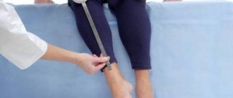
As you know, the hip joint is the most difficult to access for examination, because it is anatomically complex. Therefore, inspection and palpation, which are often used to detect signs of arthritis in other joints, will not be enough in this case. To diagnose a lesion in the hip joint, the doctor may prescribe:
- OAC and OAM, biochemical and immunological research: allow to identify rheumatoid, gouty and allergic forms of the disease;
- general clinical analysis of synovial fluid: displays the processes occurring in the cartilage tissue and synovial membrane;
- arthroscopy is a diagnostic and therapeutic method during which the doctor can carefully examine the articular cavity, femoral head, acetabulum, and cartilage tissue;
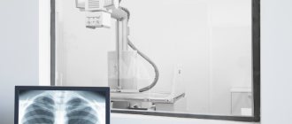
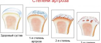
- X-ray examination - will help establish the stage of the disease and assess the degree of destruction of bone elements;
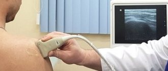
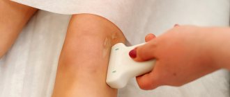
- ultrasound diagnostics – helps to identify pathological processes even in the early stages;
- computed tomography is a non-invasive method that allows you to obtain layer-by-layer images of various areas of the skeletal system;
- magnetic resonance imaging with signal suppression from adipose tissue - will help to see inflammatory changes that precede the formation of structural damage.
Some methods make it possible to differentiate inflammatory changes in the hip joint area even in the absence of clinical manifestations.
Varieties
Today, it is customary to distinguish several types of secondary diseases. Arthropathy of the knee, hip, elbow, etc. can be:
- reactive - occurs against the background of a specific tissue reaction to the development of systemic pathologies of the body. Occurs in leukemia and endocrine system disorders;
- dystrophic - progression is due to insufficient nutrition of cartilage tissue. Occurs in people over 55 years of age when natural disorders affecting the functioning of the entire body are detected;
- pyrophosphoric (chodrocalcinosis) - has a connection with metabolic disorders, namely, the exchange of salts and calcium in the tissues of the human body. Often, the provocateurs of this type of illness are injuries to large joints;
- allergic – provoked by allergic reactions;
- psoriatic – occurs with psoriasis;
- idiopathic - defined in a situation where it is impossible to establish reliable causes for the development of a pathological condition of a secondary type;
- hereditary form - transmitted at the genetic level, established on the basis of the collected anamnesis.
Arthropathy in Reiter's syndrome
Reiter's syndrome is a triad that includes damage to the organs of vision, joints and the genitourinary system. Most often, the cause of development is chlamydia; less commonly, the syndrome is caused by salmonella, shigella, yersinia, or occurs after enterocolitis.
Persons with a hereditary predisposition suffer. Typically, symptoms appear in the following sequence: first - acute genitourinary infection (cystitis, urethritis) or enterocolitis, soon after this - eye damage (conjunctivitis, uveitis, iridocyclitis, retinitis, keratitis, iritis) and only after 1-1.5 months - arthropathy . In this case, eye symptoms may appear within 1-2 days, be mild and go unnoticed.
Arthropathy is the leading symptom of Reiter's syndrome and is often the first reason for seeking medical help. Typically, asymmetric arthritis affects the joints of the lower extremities: ankles, knees and small joints of the foot. In this case, the joints, as a rule, are involved in the inflammatory process sequentially, from bottom to top, with an interval of several days.
A patient with arthropathy complains of pain that worsens at night and in the morning. The joints swell, local hyperemia is noted, and in some patients effusion is detected. Sometimes pain occurs in the spine, sacroiliitis develops, heel bursitis is possible with the rapid formation of a heel spur and inflammation of the Achilles tendon.
Symptomatic picture
Among the general symptoms of arthropathy (regardless of its type), it is customary to highlight pain of a paroxysmal nature, as well as inflammation and swelling of the affected joint, which are not accompanied by visual changes.
In addition, there are:
- soreness;
- disorders of the genitourinary system;
- noticeable stiffness of movement;
- increase in body temperature.
There are also specific symptoms that are observed exclusively in women. For example, nagging pain in the lower abdomen, intermenstrual bleeding, purulent discharge, etc.
It is also important to pay attention to the fact that symptoms of arthropathy may also include signs of secondary damage, which usually include:
- pain of varying intensity (dull/aching);
- limited mobility;
- painful palpation.
Diagnosis of hip arthritis
When examining a patient with inflammatory lesions of the hip joints, one cannot limit oneself to making a syndromic diagnosis; It is imperative to determine the root cause of arthritis. For this purpose, the nature and intensity of pain, the duration of the disease, and concomitant pathology are clarified from the anamnesis. The patient is examined in a lying position, standing and while walking. In this case, special attention is paid to the shape of the joints and the position of the limbs, the presence of muscle atrophy and contractures, gait, the ability to perform and the amplitude of passive and active movements.
Along with the clinical examination, radiation diagnostic methods play a decisive role in the diagnosis of arthritis: radiography of the hip joint, ultrasound, MRI, contrast arthrography, etc. To assess the nature of the inflammation, a diagnostic puncture of the hip joint is performed under ultrasound guidance. In some cases, in order to confirm the diagnosis, it becomes necessary to perform diagnostic arthroscopy and biopsy of the synovium of the hip joint.
Etiology and list of the most likely causes of joint arthropathy
Arthropathy of the knee joint or other joints of the musculoskeletal system (MSA) of the human body can be diagnosed in people of different ages, in particular in children.
Considering the stages of the course of secondary pathology, we can distinguish:
- acute period, the duration of which can reach two months;
- a protracted form is observed throughout the year;
- The chronic form involves a long course with remission and the occurrence of periodic relapses.
It is important to note that regardless of the type or form of the disease being considered, there is a list of the most likely causes of its development. Each cause has certain consequences, which largely determine the form of the disease being diagnosed.
Among the most common causes of secondary pathology of this type are:
- allergic reactions;
- associated painful conditions;
- infectious and parasitic lesions;
- inflammation of blood vessels;
- overweight;
- disruption of internal organs.
Main types and symptoms
The ability to quickly recognize the symptoms of hip arthritis is the key to promptly prescribing the correct treatment and reducing the risk of disability. The main clinical manifestations of arthritis of the hip joint, regardless of its form, can be considered:
- pain in the groin, outer thigh or buttock;
- swelling and pain when touched;
- difficulty walking, lameness;
- crunch;
- redness;
- pain that gets worse when walking or lifting heavy objects;
- feeling of stiffness in the morning.
Acute arthritis develops rapidly: sharp and often throbbing pain intensifies when trying to move the leg. It can radiate to the groin or buttock. Sometimes a person cannot even walk. All this is accompanied by weakness, fever, lack of appetite and nausea.
The chronic form is characterized by slow development: at first, the joints only ache from time to time after significant stress. Subsequently, the pain increases and bothers the patient even at rest, preventing him from living and moving fully.
There are more than a hundred different forms of this disease. For example, with osteoarthritis, symptoms usually develop gradually: pain with movement can be felt anywhere in the thigh, but usually in the front, and does not radiate to the tissues that are located above. Some patients complain of pain in the area of the iliac wing.
Rheumatoid arthritis can manifest itself in different ways. Patients most often complain that they experience joint stiffness and swelling in the morning. The danger is that this disease can also damage a person’s internal organs: lungs, kidneys and heart. The infectious form is characterized by a severe course: the pain is usually severe and acute. The affected area swells and becomes hot. The patient's body temperature rises to 38-40 °C.
Risk factors for joint arthropathy
There are certain factors that indicate an increased risk of secondary pathological disease. These include:
- autoimmune diseases;
- hemophilia;
- excessive stress on the joints;
- genetic predisposition;
- alcohol abuse;
- tendency to allergic reactions;
- chlamydia infection;
- impaired metabolism;
- accumulation of uric acid in joint tissues.
Causes of the inflammatory process
Arthritis may result from:
- infectious diseases: brucellosis, tuberculosis, gonorrhea, syphilis, dysentery, salmonellosis, yersiniosis, etc.;
- diseases caused by rubella, herpes, Epstein-Barr, hepatitis C viruses, etc.;
- parasitic infestation;
- breakthrough of an abscess located next to the hip joint;
- non-communicable diseases: lupus erythematosus, psoriasis;
- injuries of the ligamentous apparatus or surrounding tissues;
- endocrine disorders: diabetes mellitus, thyroid pathologies;
- age-related changes in the body.
A special role in the occurrence of this painful disease is played by genetic predisposition, as well as chronic focal infection - inflammation of the tonsils, paranasal sinuses and other factors that weaken the body's protective functions. The reasons may be: allergic reactions, malignant tumors, or even excessive growth of microflora in the intestines after operations.
Excess weight, bad habits, a sedentary lifestyle, hypothermia, and stress also increase the risk of developing the disease. Studies have been conducted that have shown that with a decrease in weight of just one kilogram, the load during movement on each hip joint is reduced by 4 kg.
When is it time to seek medical help?
Despite the extensive symptomatic picture and seemingly obvious signs of a secondary disease, identifying it in the initial stages is not an easy task.
Only over time does a feeling of a sharp deterioration in health begin, which makes it possible to track vivid symptoms.
You should visit a traumatologist or orthopedist in a situation where:
- swelling of the periarticular tissues is observed;
- body temperature rises “unreasonably”;
- there is a feeling of extreme fatigue, weakness is present;
- lymph nodes increase in size;
- movements are severely limited or completely impossible.
Self-medication in such a situation is not the best solution. Wasting time reduces your chances of recovery.
Preparing to visit the doctor
To identify inflammatory arthropathy, it is necessary to undergo tests, undergo diagnostics and visit an orthopedic traumatologist. This is a specialized specialist who will help determine the root cause, select appropriate therapy and overcome the disease.
No special preparation is required before visiting a doctor. You just need to call and make an appointment at JSC “Medicine” (clinic of Academician Roitberg). Our clinic is located at 2nd Tverskoy-Yamskaya lane, 10 (TsAO, not far from the Mayakovskaya, Belorusskaya, Tverskaya, Chekhovskaya, Novoslobodskaya metro stations).
You must have extracts and certificates of previous and chronic diseases with you.
Main events
In order to make a correct diagnosis, everyone who seeks medical help must be prepared for such manipulations as:
Additionally, if signs of inflammation in the joints are detected, the patient is referred for examination to an ophthalmologist.
As a result of passing all diagnostic measures, the attending physician determines further treatment, taking into account the degree of damage, the presence of concomitant diseases, age and other characteristics.
Why does reactive arthritis occur?
The main reason is pathogens that have entered the body. But if you examine the joint fluid in the acute period, you will not be able to detect bacteria or antibodies in it. This fact is explained by the following phenomenon: blood leukocytes, aimed at fighting bacteria, begin to combine with them, forming immune complexes that mistake joint cells for foreign bodies that need to be destroyed. Thus, a malfunction in the immune system leads to inflammation of the joints.
The causative agents of the disease are transmitted by airborne droplets and airborne dust, during birth from the mother. Domestic animals are also carriers.
Features of arthropathy treatment
Treatment of arthropathy is determined individually based on the characteristics of the existing disease and the results of the diagnosis.
In order to achieve the most effective result, a complex of physiotherapeutic and medicinal methods is used. At the rehabilitation stage, therapeutic physical education (PT) is used.
In case of severe inflammation, as well as a recurrent course of the pathology, transfer of the patient to inpatient treatment is allowed.
Strategy
The entire treatment strategy consists of following the simplest rules:
- Rejection of bad habits
- Eating balanced food.
- Maintaining optimal body weight.
- Following the instructions of the attending physician (compliance with the intended plan and features of drug therapy).
The effectiveness of a particular treatment regimen for arthropathy is determined by the onset of recovery or positive dynamics of the clinical picture of a particular case. The use of therapeutic methods should help reduce pain and restore functionality.
Physiotherapy
Physiotherapy is a special branch of medicine that involves the use of a methodological complex that can have a beneficial effect on human health. The methods used differ in the influence of natural or artificial factors (light, water, etc.) on the body.
In the treatment of arthropathy, radon baths, climatotherapy, exposure to electric current, etc. are actively practiced.
The intensity and duration of therapy is determined solely by the attending physician and depends on a large number of factors (features of drug therapy, general health, age and much more).
Drug therapy
Drug treatment of arthropathy involves taking such pharmacological groups of drugs as:
- antibiotics - used to eliminate agents that provoke the progression of the disease;
- non-steroidal anti-inflammatory drugs (NSAIDs) – relieve inflammation, eliminating visual manifestations;
- chondroprotectors – activate tissue regeneration, thanks to the enrichment of articular tissues with building material.
The most important role is played by chondroprotectors, which largely help the body overcome diseases of the musculoskeletal system. Artracam is considered to be one of the best drugs of this pharmacological group.
In order to improve the effectiveness of the prescribed therapy, a combination of medication and physiotherapeutic treatment is used, which makes it possible to significantly speed up recovery and achieve such positive results as:
- relieving inflammation;
- reduction in intensity or complete relief of pain;
- improving blood circulation in the affected joint.
Surgical intervention
Like any other pathology of the musculoskeletal system, arthropathy can lead to the need for complex surgical intervention.
The main goal of the radical method is to eliminate the consequences of the progression of pathologies and restore the functionality of the site of its localization.
Exercise therapy as a method of rehabilitation
Exercise therapy is an excellent way to speed up the restoration of the previous level of health and consolidate the results of treatment of pathological conditions of the musculoskeletal system.
A set of exercises is selected individually for each patient, which allows for targeted action and activation of the full functioning of the muscle frame, which in turn has a positive effect on the stability and functionality of the bone joint.
Complications and prognosis
Delayed consultation with a doctor, self-medication, as well as violation of the established regimen for taking medications or visiting physical therapy can lead to a number of complications, for example:
- limitation or complete loss of joint mobility;
- infection;
- the need for surgical intervention;
- disability.
With a timely, high-quality approach, we can talk about positive forecasts. Despite the impossibility of completely getting rid of the disease, there is an elimination of unpleasant sensations and the ability to control the existing disease.
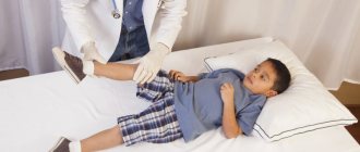
Joint pain in children
There is an opinion that normally children’s joints do not hurt, and arthralgia is a manifestation of joint diseases, the cause of which is always visible on magnetic resonance imaging (MRI). In this case, how can we explain from an instrumental point of view the presence of arthralgia against the background of hypermobility and carriage of a chronic nasopharyngeal infection or the post-exertional nature of pain? But what if the experts’ versions of the cause of joint pain in a child do not coincide? How not to miss the onset of juvenile arthritis in a child with joint pain syndrome? And can everything really be explained using MRI?
It is known that joint pain in children is a common problem for a growing body. The relevance of the problem of arthralgia in childhood is due to the high frequency of occurrence, polymorphic genesis, diffuse nature of complaints and anxiety of parents [1, 2]. Often, parents independently describe or present their own joint complaints as complaints of their child, which often complicates the timely diagnosis of arthropathy. It is believed that a child with complaints of joint pain should be comprehensively examined, since the nature of arthralgia can be identical both in the presence and absence of an anatomical “substrate” [3]. Looking ahead, I would like to note that there is no causeless pain in the joints in children.
The child's body is exposed to numerous external and internal aggressive factors. Nature, verticalizing the child, took care of a mechanism that gradually proportionally distributes the load, does not inhibit its growth activity and protects the baby from banal traumatic injuries. We are talking about age-related anatomical features of the osteoarticular system, which in young children are characterized by hypermobility, immaturity of the sensory innervation of the capsular-ligamentous apparatus and imbalance of the muscles of the lower extremities [4]. With age, strengthening of the capsular-ligamentous apparatus, perfection of sensory and proprioceptive sensitivity contribute to the balanced function of the muscles of the lower extremities and uniform distribution of the load [5].
There are “harmless” and “pathological” arthralgias of childhood. The nature of “harmless” pain (not causing harm) may be associated with the physiological characteristics of childhood. Arthralgia acquires a pathological connotation in the case of hyperreactivity or insufficiency of the immune system (immunodeficiency state), the presence of an arthrotropic infection, imperfect interaction between the central and peripheral nervous systems, irrational or unadapted loads, as well as genetically determined diseases of the osteoarticular system. It is known that joint pain can be acute and chronic, short-term and long-term and, depending on the time of day, its occurrence is classified into morning, morning and afternoon, day and evening, evening and night, scattered throughout the day or “constant”. Depending on the influence of the causative factor (stress, infection, etc.), arthralgia can be linked or unmotivated. Detailing pain sensations allows us to determine their degree of intensity, localization, fixity, as well as the presence of signs characteristic of inflammation. Instrumental diagnostics makes it possible to confirm the presence or absence of an anatomical “substrate” for arthralgia. The final verification of the cause of arthralgia should be based on a combination of clinical, anamnestic, instrumental and laboratory data.
When starting to diagnose arthralgia in childhood, it is necessary to keep in mind that the innervation of the joint is carried out through sympathetic and sensory nerve fibers. Sensory receptors (nociceptors and mechanoreceptors) permeate all joint structures, with the exception of cartilage tissue. Normally, in children and adults (in the absence of signs of inflammation or arthrosis), everyday movements are not accompanied by pain, despite irritation of joint receptors. This occurs due to the natural decoding of the afferent fiber signal into the central nervous system (CNS). However, if the frequency of impulse generation in the afferent nerve fiber increases (potentially dangerous movements, trauma, inflammation), the central nervous system interprets the increased nociceptive activity as pain. It is believed that in children two main types of pain can be distinguished: nociceptive, due to irritation of receptors, and neuropathic, as damage to the nerve fiber [6].
Diagnosis of arthralgia in childhood should be based on the study and assessment of the onset, dynamics of the articular syndrome, instrumental data, and the conclusions of other specialists [7]. In addition to the anatomical and physiological characteristics of the osteoarticular system of childhood, the diagnosis of arthralgia should take into account gender and age characteristics, as well as the psycho-emotional status of the child. The questions that the doctor should always answer are usually the same - these are pathological or causeless pain, inflammatory or non-inflammatory in nature, requiring treatment or requiring only dynamic observation. The characteristics of pain in children with “harmless” and pathological pain are clearly presented in Table.
Non-arthritic pain in children
Meniscal pain - as a rule, pain is always of one joint with a previous fact of injury. Children over 10–12 years of age are most often affected. Painful sensations are strictly load-bearing, detailed in nature and are localized in the projection of the joint space of the damaged meniscus. Blocks in the joint are possible. In most cases, there is a positive clinical test for damage to the lateral or medial meniscus [8]. Signs of the inflammatory process, as a rule, appear at the time of acute injury and are short-term in nature. The reliability of MRI (more than 1.5 T) in diagnosing damage or malformation of the meniscus (atypical discoid form) is more than 90–95%. Damage to the posterior horn of the medial meniscus is considered an MRI finding and cannot be a source of pain [9].
Osteochondral pain is usually pain in one joint, usually the lower limb, and occurs in children over 8–10 years of age. Previous irrational physical activity can serve as a trigger mechanism for the formation of osteochondropathy, but often the cause remains unclear. Pain sensations are stressful, detailed and limited to the joint area. This is typical both in the case of damage to the epiphysis and in the case of damage to the apophysis (tuberosity of the calcaneus, tuberosity of the tibia). The true focus of aseptic necrosis is always located within the loaded joint-forming zone of the bone (Fig. 1). Cases of detection of such a focus of non-loaded zones of the epiphyses indicate its dystrophic nature or are even a feature of ossification (Fig. 2). Often there is a reaction of the synovial membrane to the phenomena of osteonecrosis (first stage) or fragmentation of the epiphysis (second stage) in the form of a mild exudative component (Fig. 3). Synovitis itself can contribute to increased pain or transformation of pain with the appearance of morning stiffness. In the case of “unlacing” of the osteochondral fragment from the focus of necrosis, sensations of a free intra-articular body may appear. In children with osteochondropathy of the femoral head, symptoms of synovitis can often be protracted and persistent, requiring a long continuous course of anti-inflammatory therapy. The reliability of MRI (more than 1.5 T) and radiography in the diagnosis of osteochondritis (apophysitis), osteochondropathy is 100% [10].
Pain in the projection of the patellas, except in cases of osteochondropathy (apophysitis, osteochondropathy of the tibial tuberosity) and pathological dislocations, can occur with chondromalacia, high standing patellas and mediopatellar fold syndrome. Among orthopedic pathologies, the source of stress-related foot pain, with the exception of cases of osteochondropathy, can be a number of bone coalitions (talocalcaneal, talonavicular) and congenital malformations of the feet. In addition, systemic skeletal diseases, clinically manifested by joint stiffness syndrome, can also have manifestations such as arthralgia.
Enthesitic and tendon-muscular pain can occur in children as an acute (transient, episodic, post-traumatic) or chronic pathology. Primary chronic enthesopathy is a manifestation of SEA syndrome (seronegative enthesoarthropathy) or juvenile arthritis. Secondary chronic enthesopathy, as a rule, is concomitant or secondary-reactive in nature against the background of the underlying orthopedic pathology [11].
The following predisposing factors are identified:
- nonphysiological hypermobility or stiffness of joints;
- excess body weight;
- puberty (period of rapid extension);
- irrational or unadapted physical activity, chronic trauma;
- frequent acute respiratory diseases, persistence of chronic foci of nasopharyngeal infection;
- presence of the HLAB27 gene.
Clinical variations of the joint syndrome, as well as the severity of pain, may vary. The knee, ankle, and hip joints are most often affected, and less commonly, the shoulder and elbow joints. Painful joint syndrome is limited in nature and is localized in the projection of the tendon-muscular bundle or the area of attachment of the tendon to the bone tissue (enthesis), with clinical signs of local inflammation. However, due to the anatomical and physiological characteristics of childhood, pain can often have a diffuse, non-detailed nature. More typical is the presence of starting pain, which intensifies against the background of physical activity, and less commonly, with everyday movements. The instrumental picture is characterized by inflammatory changes in the area of attachment of tendons to bone tissue, tendonitis/tenosynovitis, less often with the development of osteophytes, erosions, usually with mild symptoms of synovitis. However, this type of change is visualized exclusively in adults. In children, the diagnosis of enthesopathy is based on clinical symptoms [12]. Perhaps, only against the background of the phenomena of Achilles bursitis in the structure of juvenile arthritis can erosive-dystrophic changes in the heel bone occur. The prevalence of the enthesitic nature of the articular lesion determines a relatively “favorable” variant of the course of the disease, provided there is low laboratory activity, the absence of an erosive articular component, signs of sacroiliitis and carriage of the HLAB27 gene.
Pain due to hypermobility in children of preschool and primary school age is often diffuse, symmetrical in nature and localized exclusively along the anterior surface of the legs, less often involving the knee and ankle joints or feet. The pain is usually of varying intensity, post-exertional, often in the evening and at night. Synovitis and signs of laboratory activity are always absent. Pain is relieved by intense stroking and the use of local or oral non-steroidal anti-inflammatory drugs (NSAIDs). The pain component is associated with the phenomena of myalgia due to overstrain of the leg muscles (anterior portion of the muscles) against the background of physiological weakness of the capsular-ligamentous apparatus. This type of pain is described in many literature sources as growing pains [13].
N.B. True arthralgia in children of this age is always pathological in nature.
N.B. Night pain requires special attention if it occurs exclusively in one segment of the limb (tumor formation of the bone, Fig. 4) or occurs in the form of ossalgia with fever and profuse sweating (oncohematological syndrome).
Pain due to hypermobility in older children is more often diffuse, non-detailed, exclusively in the joints of the lower extremities. Pain affects one or more joints. Pain of varying intensity associated with physical activity can be presented as stress or post-exert (daytime and evening). According to MRI, signs of mild exudative synovitis can be detected, often of a protracted nature, without signs of proliferation of the synovium. Pain is always relieved by reducing the intensity of physical activity, and children rarely need long-term use of NSAIDs. Positive tests for joint hypermobility are clinically mandatory. The absence of signs of inflammatory laboratory activity confirms the benign form of arthropathy. The mechanism of formation of the pain component and transient symptoms of synovitis is associated with microtraumatization of intra-articular structures, stretching of the capsular-ligamentous apparatus and overstrain of the patellofemoral joint. In addition, decreased proprioceptive sensitivity of the knee joints and imbalanced calf muscles also further contribute to exercise tolerance. Overstrain of the patellofemoral joint is characterized by the presence of localized pain in the retropatellar region, often of a unilateral nature [14].
Some children with joint hypermobility, in addition to symptoms of vegetative-vascular dystonia, have the so-called increased anxiety syndrome. This syndrome, according to some researchers, may be associated with interstitial duplication on chromosome 15 (Gratacos et al., 2001, duplication of 15q24-q26, called DUP 25). Accentuation of pain, the associative way of thinking of a “sick child” and stereotypical behavior are more common in girls of puberty. With this syndrome, a “colorful” picture of pain is often described, hysteria and crying are noted during the examination. Pain sensations can vary depending on the weather, time of day and the mood of the young lady herself. Arthralgia is often accompanied by episodes of false blockade of the joint, cases of pseudoluxations of the patella or instability of the entire limb; Sometimes there is an inability to fully support the foot. Clinically, imaginary pain is detected upon palpation of the joint area, a “grimace of pain” is present during examination, false contracture, stiffness of the joint or, less often, stiffness without anatomical prerequisites is possible. During the examination, as a rule, a discrepancy between the clinical and instrumental picture is revealed, there are always no signs of laboratory inflammatory activity, and the presence of neuropathy is possible. The presence of local neurological symptoms, impaired sensitivity and motor function of the limb, signs of hyperalgesia requires the exclusion of complex regional pain syndrome, as well as tumor processes in the lumbosacral spine [15].
Diagnosis of non-arthritic arthropathy consists of deciphering the etiological factor of arthralgia. Despite the apparent similarity of pain, the described variants of arthropathy have some characteristic distinctive features that allow them to be differentiated. However, true difficulties always arise with a mixed version of articular pathology, namely in the case of enthesopathy in a child with a syndrome of increased anxiety or hypermobility. As is known, the main treatment of aggressive enthesopathy is the prescription of NSAIDs, in contrast to the benign form of hypermobility or arthralgia due to the syndrome of increased anxiety. In the latter types of arthropathy, the emphasis is on restoring impaired function of a limb or joint and gradually increasing physical exercise, overcoming habitual stereotypes of “catastrophic thinking” regarding possible damage and pain, creating a positive attitude and increasing self-esteem, and to a lesser extent, emphasis is placed on drug treatment of NSAIDs .
Arthritic pain in children
• Infectious arthritis:
- bacterial (septic); - viral; - specific (tuberculosis); — Lyme arthropathy.
• Post-infectious arthritis:
- infectious-allergic, infectious-toxic; - reactive arthritis, Reiter's syndrome.
• Chronic arthritis:
- primary; - secondary.
Arthralgia due to a viral infection, as a rule, develops in children in the prodrome or during the height of the disease and coincides with the period of skin exanthema. Joint pain lasting from several hours to 1–2 weeks can less often be accompanied by symptoms of synovitis, which disappears without a trace. Viral arthritis or arthralgia can occur against the background of current hepatitis B, rubella, chickenpox, parvovirus B19 infection, entero- and adenovirus and some other viral diseases. The tropism of the group of herpes viruses to joint tissues is minimal. Children usually require a short course of NSAIDs [16].
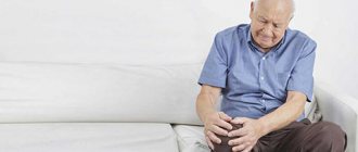
Arthralgia of the hip joint in the post-infectious period is interpreted as infectious-toxic (infectious-allergic, post-infectious) coxitis or transient coxopathy. Sudden onset, pain and inability to support the lower limb, lameness, as a rule, colorfully characterize this pathology. Reversibility of symptoms occurs 2–3 days from the onset of the disease. Less common is recurrent (more than 3-4 episodes) or protracted arthropathy (more than 2 weeks) with the formation of osteoporosis and reversible neurodystrophic changes in the femoral head. True short-term coxitis or arthralgia of the hip joint disappears without a trace [17]. Irradiation of pain to the anterior surface of the thigh and to the knee joint in the acute period of coxitis is associated with transient neuropathy of the obturator nerve. Children aged 2 to 8 years are most often affected; the onset of the disease is often preceded by a nasopharyngeal infection, sometimes the causative factor remains unclear. In addition to the clinical picture, imaging methods (radiography, ultrasound, MRI) are of diagnostic importance.
N.B. Osteomyelitis can occur under the mask of acute coxitis, and osteochondropathy of the femoral head can occur under the mask of a protracted course of acute coxitis (Fig. 5).
Arthropathy of post-streptococcal etiology in children can occur in the form of arthralgia or acute arthritis. Joint syndrome occurs after a nasopharyngeal infection caused by group A β-hemolytic streptococcus (clinically proven episode). The duration of arthralgia is 2–4 weeks, the course of arthropathy is non-aggressive and does not cause permanent joint deformities in children. The ASL-O level (the total amount of IgA, M, G for streptococcal toxin) does not always play a decisive role in the diagnosis of arthropathy and the choice of treatment tactics, in contrast to the isolation of group A β-hemolytic streptococcus [18].
N.B. The course of acute arthritis must be differentiated from rheumatic arthritis.
Arthralgia of post-staphylococcal etiology in children, as a rule, is of a mild nature and is often characterized by the phenomena of sluggish synovitis or enthesopathy. Clinically, this may manifest itself as a slight decrease in the child’s motor activity, evening or night pain. The presence of a focus of chronic nasopharyngeal infection in the oral or nasopharyngeal area is of decisive importance in diagnosis. Sanitation of the source of infection, as a rule, relieves the child of joint pain.
In most children, articular syndrome with reactive arthritis (ReA) occurs within 1–4 weeks after a urogenital (Chlamydia trachomatis) or intestinal infection (Enterobacteriaceae family). It is believed that this is an acute non-purulent inflammation of the joints, in which the infectious agent or its antigens are not detected in the joint cavity, and the articular syndrome is associated with a number of immune disorders. However, the identification of pathogen DNA by the polymerase chain reaction method made it possible to somewhat transform this theory. Children who carry the HLAB27 gene and some other cross genes (B7, B13, B40) are at high risk for ReA. CD8+ HLAB27+ T cells are now thought to have a more robust immune response with overexcretion of TNF-α. In particular, chlamydia contributes to the chronicity of infection due to inhibition of the expression of HLA antigens on the surface of infected cells, reducing the potential for apoptosis of T cells with stimulation of local synthesis of TNF-α. And the lipopolysaccharide layer of gram-negative microorganisms is a powerful activator of tissue macrophages, synovial fibroblasts and osteoclasts, and CD8+ HLAB27+ T cells are less effective in eliminating the pathogen. The course of arthropathy is possible against the background of episodes of fever without systemic manifestations. As a rule, monoarthritis of a large joint or asymmetric oligoarthritis of the joints of the lower extremities (knee, ankle) is characteristic, sometimes in combination with the phenomena of dactylitis. Less commonly, articular syndrome can be represented by polyarthralgia. The duration of ReA can range from 1 to 3 months; the course of arthropathy is mainly acute, which dictates the need for long-term antibacterial and anti-inflammatory therapy [19].
Juvenile arthritis (JA) is a chronic inflammatory disease with more complex pathogenetic mechanisms, which usually leads to joint deformation and rarely goes away without a trace. Currently, several heterogeneous forms of arthropathy in the structure of JA are distinguished [20]. Chronic progressive inflammation of the inner lining of the joint capsule (synovial tissue), which has a high degree of aggressiveness and a tendency to spread to all structures of the joint, including the capsular-ligamentous apparatus [21]. Painful joint syndrome is multicomponent and has its own peculiarity, namely, it occurs exclusively during passive or active movements in the joints; at rest, children do not complain of pain in the joints. A characteristic feature of JA is morning stiffness, defined as short-term lameness accompanied by sensations of numbness and tenderness in one or more joints. Painful sensations that appear in the morning subside only in the evening as the intensity of the load decreases. The child spares the limb, protects the joint or joints subject to chronic inflammation from excessive physical exertion and injury. The severity of the pain syndrome depends on the aggressiveness of the disease, the type of joint involved, the amount of intra-articular fluid, as well as the reaction of the periarticular soft tissues and tendon-ligamentous apparatus. There is no point of maximum pain, and pain occurs both during palpation in the area of the projection of the joint space and in the area of the hypertrophied, inflamed synovium. Often, young children are not able to localize pain in the joint; swelling of the joint area may be poorly visualized against the background of a physiologically excess subcutaneous fat layer, and the first signs of an inflammatory process in the joint may be limited movement or lameness. It is known that any joint can be a target of arthritis. Rapid reversibility of inflammatory changes and resolution of contracture indicate the acute nature of arthritis [22].
This review was devoted to one of the current problems in pediatrics, namely joint pain in children. The most common forms of childhood arthropathy were presented, the leading symptom of which may be joint pain. Of course, any researcher or practitioner who sometimes faces similar problems will be able to name at least a dozen more nosological forms in which joint pain is not uncommon. However, this article is devoted to the most common forms of joint pathology in children in pediatric practice.
And in conclusion, I would like to say that joint pain is just a symptom, not a disease. A child with complaints of joint pain should be comprehensively examined. Joint pain does not lead to joint deformation and the formation of arthrosis. And “harmless” pain in the joints of childhood should remain in childhood.
Literature
- Alekseeva E.I., Bzarova T.M. Joint damage in childhood // Attending Physician. 2010; 6:46–51.
- Tsurikova N. A. Differential diagnosis and treatment of oligoarthritis in children. Thesis... Ph.D.: 01/14/08. M., 2021. 147 p.
- Asif Naveed and Peter Heinz. Joint pain in children // Paediatrics and child health. 2014; 2 (24): 45–50.
- Bird HA Joint hypermobility in children // Rheumatology. 2005, vol. 44, Issue 6, 1 June, p. 703–704. DOI: 10.1093/rheumatology/keh639.
- Nazarova T. I., Petruk N. I. Anatomical and physiological features, examination methods, semiotics of damage to the skeletal and muscular systems in children. Characteristics, research methods, semiotics of the lesion. Educational and methodological manual for studying the course “Childhood diseases”. M.: RUDN, 2012. 52 p.
- Sakovets T. G. Features of neuropathic pain in joint damage // Practical Medicine. 2014; 4 (80): 103–106.
- Kozhevnikov A. N., Pozdeeva N. A., Konev M. A., Maricheva O. N., Afonichev K. A., Novik G. A. X-ray diagnosis of chronic oligoarthritis in children // Bulletin of Siberian Medicine. 2017; 16(3):224–234.
- Bryanskaya A. I., Baindurashvili A. G., Arkhipova A. A. et al. Arthroscopic treatment of diseases of the knee joint in children // Orthopedics, traumatology and reconstructive surgery of children. 2014, vol. II, issue. 3:18–23.
- Gumerov R. A., Abzalilov A. A., Valiullin D. R., et al. Diagnosis and treatment of post-traumatic synovitis of the knee joint in children // Pediatric surgery. 2012; 5:25–28.
- Abalmasova E. A. Osteochondropathies // Children's arthrology. M.: Medicine, 1981. pp. 284–303.
- Slizovsky N.V., Masalova V.V., Zinchenko M.A. Arthritis in children. St. Petersburg, 2004. 76 p.
- Weiss PF Diagnosis and treatment of enthesitis-related arthritis // Adolesc Health Med Ther. 2012; 3:67–74. DOI: 10.2147/AHMT.S25872.
- Uziel Y., Hashkes PJ Growing pains in children // Pediatr Rheumatol Online J. 2007; 5: 5. DOI: 10.1186/1546–0096–5-5.
- Fatoye F., Palmer S., Macmillan F. et al. Proprioception and muscle torque deficits in children with hypermobility syndrome // Rheumatology. 2009; 48: 152–157.
- Weissmann R., Uziel Y. Pediatric complex regional pain syndrome: a review // Pediatr Rheumatol Online J. 2016; 14:29.
- Miller LM, Cassidy TJ Postinfectious. Arthritis and Related Conditions. In: Behrman RE, Kliegman RM, Jenson HB (eds.). Nelson Textbook of Pediatrics, 18 th end. Philadelphia, WB. Saunders 2007; cap: 147, 519–522.
- Young Dae Kim, Alan V. Job, Woojin Cho. Differential diagnosis of juvenile idiopathic arthritis // J Rheum Dis. 2017; 24 (3): 131–137. https://doi.org/10.4078/jrd.2017.24.3.131.
- Uziel Y., Perl L., Barash J., Hashkes PJ Post-streptococcal reactive arthritis in children: a distinct entity from acute rheumatic fever // Pediatr Rheumatol Online J. 2011, Oct 20; 9 (1): 32.
- Plesca D., Luminos M., Spatariu L. et al. Postinfectious Arthritis in Pediatric Practice // Maedica (Buchar). 2013, Jun; 8 (2): 164–169.
- Novik G. A., Abakumova L. N., Letenkova N. M., Slizovsky N. V., Slizovskaya N. N. Juvenile arthritis - experience in diagnosis and treatment // Attending Physician. 2008; 4:23–27.
- Kirkhus E., Flatø B., Riise O. et al. Differences in MRI findings between subgroups of recent-onset childhood arthritis // Pediatr Radiol. 2011, Apr; 41(4):432–440. DOI: 10.1007/s00247–010–1897-y.
- Kozhevnikov A.N., Pozdeeva N.A., Konev M.A. et al. Juvenile arthritis: features of the clinical and instrumental picture and differential diagnosis // Attending Physician. 2016; 4:58–62.
A. N. Kozhevnikov*, 1, Candidate of Medical Sciences N. A. Pozdeeva*, Candidate of Medical Sciences M. A. Konev* M. S. Nikitin* A. I. Bryanskaya*, Candidate of Medical Sciences E. V. Prokopovich* , Candidate of Medical Sciences K. A. Afonichev**, Doctor of Medical Sciences G. A. Novik**, Doctor of Medical Sciences, Professor
* Federal State Budgetary Institution Scientific Research Institute for Children's Orthopedics named after. G. I. Turner, Ministry of Health of the Russian Federation, St. Petersburg ** Federal State Budgetary Educational Institution of Higher Education, St. Petersburg State Pediatric Medical University, Ministry of Health of the Russian Federation, St. Petersburg
1 Contact information
Joint pain in children / A. N. Kozhevnikov, N. A. Pozdeeva, M. A. Konev, M. S. Nikitin, A. I. Bryanskaya, E. V. Prokopovich, K. A. Afonichev, G. A. Novik
For citation: Attending physician No. 4/2018; Issue page numbers: 50-55
Tags: children, joints, pain, diagnosis
Prevention of joint arthropathy
According to available data, arthropathy is a secondary pathology and develops against the background of other diseases. The main goal of prevention is to prevent the development of any pathological processes or at least their timely elimination.
To significantly reduce the risk of developing joint arthropathy, it is recommended:
- systematically monitor your general health;
- maintain at least a minimal level of physical activity;
- adhere to the basics of proper, nutritious nutrition;
- monitor body weight and prevent obesity;
- give up bad habits;
- reduce the likelihood of infection;
- attend preventive examinations with your doctor;
- promptly seek medical help and treat pathologies, infections and other diseases in accordance with the scheme approved by the doctor;
- carry out prophylactic administration of drugs from the pharmacological group of chondroprotectors (Artracam, etc.) in order to maintain the quality of joint tissues.
Health is something that cannot be bought, but can be maintained. Maintain an optimal level of quality of life by following simple recommendations for the prevention of various diseases.
Arthropathy in other infectious and parasitic diseases
Arthropathy often appears against the background of various pathologies that are associated with infections and parasites. When a person has suffered from Lyme pathology, brucellosis and trichinella, he develops flying arthralgia. Rubella occurs in combination with symmetric polyarthritis. And arthropathy in combination with mumps is similar to rheumatoid arthritis. The following signs are observed:
- unstable inflammation in the joints;
- the character is migratory;
- inflammation of the pericardium occurs.
From chicken pox, mononucleosis with infections, arthropathy appears, which is an unstable arthritis. It quickly disappears if the symptoms of the main pathology disappear.
Artopathy accompanied by meningococcal infection is observed a week after the development of the pathology. Most often this is accompanied by monoarthritis of the knee joint, but sometimes polyarthritis of large joints appears. Viral hepatitis also provokes the appearance of arthropathy, while the pathology manifests itself as arthralgia or volatile arthritis, joint damage occurs symmetrically. Artopathies make themselves felt at the beginning of the development of the disease, when there is not even jaundice.
HIV infections are accompanied by various signs of joint disorders:
- It could be arthritis, arthralgia.
- AIDS arthritis of the lower leg and knee joints may also occur. In this case, the function of the limbs is noticeably impaired, and pain is felt.
If the underlying disease is treated, then the signs of the affected joints disappear.