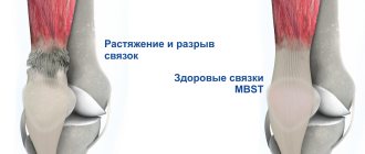
Interfibular syndesmosis is the connection of the tibia and fibula bones in the distal part of the ankle and ankle joint. It can be a consequence of excessive external rotation or dorsiflexion of the foot with simultaneous traumatic exposure. Excessive rotation of the foot almost always leads to a primary rupture of the connections between the bones. If this injury is ignored, then in the future there may be complete separation of the distal condyles of the tibia and fibula. This leads to rapid deformation of the ankle joint and the development of osteoarthritis with loss of the ability to move freely in space.
People who lead an active lifestyle and engage in sports such as volleyball, football, high and long jumps, running, tennis, etc. are susceptible to this type of traumatic injury. In young people, a rupture of the syndesmosis may be associated with a fracture of the ankle bones.
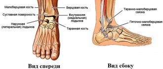
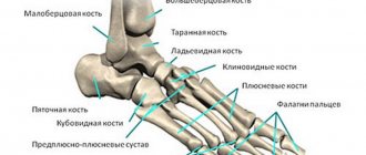
The distal tibiofibular syndesmosis includes the following anatomical structures:
- anterior ligament between the tibia and fibula;
- posterior inferior ligament between the condyles of the tibia and fibula;
- transverse ligament between the tibia and fibula;
- interosseous membrane;
- inferior transverse and interosseous ligaments.
In its normal state, the syndesmosis resists excessive axial shock-absorbing load and does not allow displacement of the talus. When walking, due to the elasticity of all tissues of the syndesmosis, the tibiofibular gap can increase by up to 10 mm in width. The deltoid ligament stabilizes and reduces the amplitude of expansion.
With minor damage to the tibiofibular syndesmosis, a poor clinical picture is observed. The person does not lose the ability to move independently. All symptoms (pain, weakness of the lower leg muscles, limited mobility of the foot) quickly disappear even in the absence of adequate treatment. But over time, this leads to the formation of scar deformation. If numerous minor injuries of the same type occur to the tibiofibular syndesmosis, then gradually this entails its complete rupture.
Even after a minor injury, comprehensive rehabilitation is recommended. To do this, you should contact a manual therapy clinic. In Moscow, you can make an appointment for a free appointment with a doctor at our center. Experienced doctors work here who have every opportunity to assist patients with any problems of the musculoskeletal system.
In order to make an initial free appointment with a doctor, fill out the feedback form located below on this page. A specialist from our clinic will contact you and agree on a time convenient for your visit.
The mechanism of occurrence of tibiofibular syndesmosis or its tear - causes of injury
In the disease under consideration, the center of localization of the pathological process is the ligaments of the distal tibiofibular syndesmosis , through which the tibia is connected to the fibula.
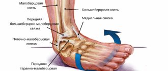
Stabilization of the ankle joint is provided by 3 ligaments: the anterior inferior tibiofibular, posterior inferior tibiofibular, and interosseous tibiofibular. In the event of a strong external impact on the specified element of the musculoskeletal system, the ligaments are not able to cope with their main function - they are damaged, even to the point of complete rupture.
When the syndesmotic ligament ruptures, the tibia and tibia move significantly away from each other - diastasis .
Often, the type of injury in question is combined with a fracture of these bones. Less commonly, such injuries are limited to minor sprains.
In such situations, people do not always seek qualified medical help - which, in the future, can lead to the development of exacerbations.
If you believe the statistical data, every 4th case of ligamentous damage to the ankle joint is associated with injury to the tibiofibular syndesmosis.
As a rule, the disease in question is diagnosed in people who play sports professionally. Football players, acrobats, hockey players, skiers, dancers, and track and field athletes are at particular risk.
“Unsportsmanlike” injuries to the tibiofibular syndesmosis often occur as a result of an ankle fracture.
In addition, wearing shoes with high, unstable heels can cause this injury.
Closed reduction with further plaster immobilization
This is a common treatment method used primarily for ankle fractures. Its essence lies in the manual reposition of bone fragments and subsequent fixation of the ankle with a plaster cast. This method is accessible and low-traumatic, but has its drawbacks.
Long-term plaster immobilization disrupts trophism and impairs blood circulation in the tissues of the limb, causing irreversible changes in the nerves and muscles of the lower leg, foot, and ankle joint. It does not provide sufficiently stable fixation and does not guarantee complete immobility of the fragments. All this can lead to improper bone fusion and the formation of a false joint.
In case of concomitant dislocations and subluxations of the ankle, it is recommended not to use closed reduction. The reason is the high risk of unsatisfactory treatment results.
The most common of them:
- postoperative contractures;
- false joints;
- severe deforming arthrosis.
Therefore, many specialists prefer a different approach to the treatment of displaced fractures. For patients of working age, they recommend open reduction with fixation of bone fragments with metal structures. And for patients in the older age group, reposition under image intensifier control and fixation with Kirschner wires.
Symptoms of injuries to the tibiofibular syndesmosis of the ankle joint
The pathological condition in question cannot be diagnosed through a physical examination alone: it has similar symptoms as with dislocations or sprains.
This analogy is the reason for the late diagnosis of a rupture of the tibiofibular syndesmosis: people hope for self-healing, so they do not go to a medical institution. However, over time, the pain syndrome intensifies and becomes chronic, and the muscle mass of the lower leg decreases.
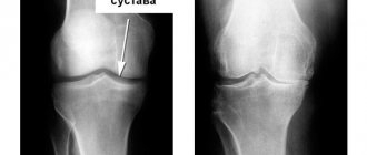
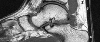
In order to make an accurate diagnosis, an x-ray examination of the damaged area is performed.
The symptomatic picture of this disease is characterized by the following manifestations:
- Aching pain in the damaged area. With axial pressure, as well as in the case of palpation of the lower leg, the pain increases.
- Swelling. It is often unexpressed. In rare cases, swelling may increase and bruising may appear in the area of the injury.
- The appearance of small subcutaneous hemorrhages in the area above the ankle joint.
- Non-standard ankle position. If the distal membrane is severely damaged, the person is unable to place the foot on the floor: it turns outward.
Characteristics/Clinical picture
It has been noted that damage to the ankle syndesmosis is accompanied by less swelling than a sprain of the lateral malleolus, as well as severe limitation of plantar flexion and inability to put weight on the foot. Ecchymosis may appear several days after injury due to damage to the interosseous membrane. Difficulty or inability to walk on toes is often noted. History: chronic pain, long recovery, recurrent sprains, formation of heterotopic ossification within the interosseous membrane. The most common mechanisms are when the leg is in external rotation and excessive dorsiflexion.
Types and degrees of damage to the tibiofibular syndesmosis
Adequate and accurate classification of this pathology makes it possible to prescribe the most appropriate treatment and minimize the risk of developing exacerbations in the future.
Based on the severity of the clinical picture, as well as the duration of the course, the type of injury under consideration is divided into three groups:
Acute injuries
The symptomatic picture in the first 3 weeks after injury is quite pronounced, and a preliminary diagnosis is possible through functional tests. Based on radiographs in various variations, rupture of the tibiofibular syndesmosis is classified as follows:
- Without separation of the fibula and tibia.
- With hidden diastasis.
- With the formation of a pronounced tibiofibular space.
Subacute injuries of the tibiofibular syndesmosis
Characteristic of injuries that are more than 3 weeks old.
Chronic ruptures of the syndesmosis
The condition in question becomes chronic if more than 3 months have passed since the injury.
Diagnosis through functional tests in this situation is ineffective, and the overall picture may be supplemented by ankle deformity, osteoarthritis of the ankle joint, as well as some other complications.
Depending on the extent of the damage, there are 3 degrees of severity of injury to the tibiofibular syndesmosis:
- I – mild degree . It is characterized by microscopic destruction of some fibers of the syndesmotic ligament. Patients complain of minor pain and mild swelling in the affected area. The injury does not affect the functionality of the ankle joint in any way. There are no subcutaneous hemorrhages.
- II – average degree . Instrumental diagnostic measures reveal ligament tears. The ankle joint functions with certain deviations: the patient is not able to fully move the foot. When examining the patient, swelling and subcutaneous hemorrhages are revealed.
- III – severe degree . In this case, the ligament is torn across, which negatively affects the functionality of the ankle joint: they are almost completely lost. The symptomatic picture is represented by severe pain, severe swelling, and significant subcutaneous hemorrhages.
What is this?
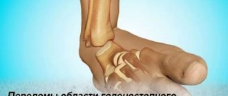
A rupture of the distal tibiofibular syndesmosis is a traumatic injury that most often occurs in factories, in car accidents, and during falls from low heights. This injury is combined with damage to the membrane (its integrity). Fractures or displacements often occur. Damage to the ligament is the basis for the formation of serious violations of the integrity of the ankle joint.
The injury is accompanied by quite significant pain, and it is very difficult to move. Urgent and highly qualified medical care should be provided, because without it, complete loss of limb functionality is very likely.
Conservative treatment of rupture of the tibiofibular syndesmosis - is it possible to do without surgery?
Therapeutic tactics for the type of injury under consideration will be determined by the degree of damage to the syndesmotic ligament:
For mild severity
Treatment lasts 3-5 days and includes the following activities:
- Apply cold compresses every half hour. You need to hold this compress for 15 minutes.
- Peace. Physical activity should be completely avoided during the treatment period.
- Prevention of swelling. Achieved by elevating the injured limb.
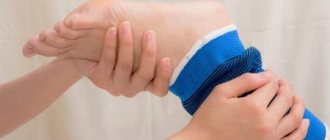
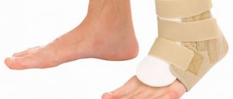
- Compression. This is achieved using a fixing bandage or elastic bandage.
For moderate injuries
The procedures described above are carried out over 7-10 days.
At the end of this period, the patient is prescribed to wear a soft orthosis.
With a transverse rupture of the syndesmotic ligament
- The patient is given a polymer or plaster cast, and in order to unload the joint when walking, it is recommended to use crutches.
- After 10 days, the plaster is replaced with a semi-rigid brace, which must be worn for about a month.
- After this, the functionality of the joint is restored through physiotherapeutic procedures, exercise therapy, and massage. All exercises are performed in a soft fixing bandage, and their main goal is to restore joint mobility, strengthen the muscles in the area of damage, and restore muscle sensitivity.
Treating this pathological condition with a conservative method takes a lot of time, and the end result is not always positive.
Pain is relieved by short-term use of non-steroidal anti-inflammatory drugs : Aspirin, Nurofen, Diclofenac, etc.
In the absence of the required effect, corticosteroids - in the form of intra-articular injections, or opioid analgesics .
Why is this dangerous?
If the syndesmosis ruptures, treatment must be carried out quickly and without fail. In the absence of timely assistance, a more serious violation of the integrity of the membrane is formed, which in the future will become the basis for a decrease in motor activity.
In certain cases, damage can cause loss of motor function of the limb. When such an injury occurs, bone structures are destroyed. This, in turn, can cause internal injuries to soft tissues, become the basis for the formation of inflammation processes, etc.
Indications for surgery and features of surgical treatment of injury
Surgical intervention for the injury in question is used in the following cases:
- Lack of positive effect from conservative therapy, which is manifested by the presence of pain, as well as instability of the ankle joint.
- Violation of the anatomical integrity of several ligaments at once.
- Advanced conditions: from 3 weeks after injury. This often happens in the absence of timely medical care: when the patient self-medicates for a long period.
Today, 2 methods of surgical treatment of rupture of the tibiofibular syndesmosis are actively practiced:
- Tendoplasty . Makes it possible to completely renovate a destroyed area. The new ligament is formed from an artificial graft, preserved tendon, or from the fascia of a healthy thigh taken from the patient himself. To attach the new ligament, holes are drilled in the shin bones. This technique leads to complete recovery in 92% of cases.
- Use of a special tightening mechanism. Such a mechanism can be a special screw or tie bolt made from a special metal alloy.
Surgery in the treatment of tibiofibular syndesmosis - different options for metal clamps
Thanks to this procedure, it is possible to fix the bones of the lower leg at the correct distance: they will not move or grow together in the future, which completely eliminates the risk of developing ankle joint contracture. These devices are removed after 2-3 months. Patients should wear a fixation bandage for some time after the screws are removed.
Among other things, damage to the syndesmotic ligament is fraught with disruption of the integrity of the vascular network , which will provoke the formation of blood clots in the future. In order to prevent this phenomenon, medications are prescribed that help thin the blood, as well as medications to strengthen the walls of blood vessels and ensure their elasticity.
Surgical intervention for ruptured disc joint
According to a meta-analysis, surgical therapy reduces the incidence of relapses, chronic symptoms and improves quality of life. However, the studies assessed were also able to show the disadvantages of surgical treatment: recovery takes longer than usual.
With a total rupture of the syndesmosis, surgical intervention is an inevitable procedure. The doctor will first stitch up the torn syndesmosis and then stabilize it with a set screw. The screw sets the correct distance between the different muscles. After about six weeks, the doctor will remove the screws so the patient can continue exercising.
Recovery after injury and surgical treatment - recommendations for patients
After surgical treatment of the injury in question, patients are sent home the next day.
The first two weeks after surgery, the damaged area is fixed with plaster. During the specified period of time, the load on the operation area should be minimized. To do this, you need to ensure rest, and when moving, use crutches.
To reduce swelling, the foot should be kept elevated, but only in a horizontal position.
After the swelling has partially subsided, the cast is replaced with an orthopedic boot , and the patient is allowed to take short walks and perform simple exercises for flexion/extension of the foot.
Until the wound is completely healed, the foot must be kept cool and dry, otherwise infection may occur.
It is recommended to start driving a car no earlier than 6 weeks after the operation.
It is mandatory to wear an orthosis after removing the screws, as well as during physiotherapeutic procedures - their use will ensure stabilization of the ankle joint.
Regardless of what treatment the patient received - conservative or surgical - he should undergo a course of rehabilitation . The duration of this course will depend on the degree of damage to the ligament: from 1 month to 1 year.
Symptoms
A rupture of the distal tibiofibular syndesmosis is manifested by the following symptoms:
- A nagging or very sharp pain occurs almost instantly. It extends to the entire affected area.
- When moving the limb or when palpating, the pain syndrome becomes even more pronounced.
- A very significant swelling appears, which has a blue or burgundy tint.
- The foot is in an incorrect position; visually it turns outward.
- A person cannot stand on his leg as a result of a sufficiently severe damage to the membrane, since the lower leg is very deformed.
- Multiple foci of internal hemorrhage occur.
If left untreated, all symptoms become even more pronounced. The best way to check for injury is to take an x-ray.
Prevention
As in most cases, in order to protect yourself from any injury, including rupture of the tibiofibular syndesmosis, you need to choose the most comfortable shoes for constant movement. If a woman loves heels, then they should be stable and wide.
It is important to monitor your actions, namely the process of support when a person moves on ice, or runs and jumps. Also, proper and balanced nutrition and playing feasible sports play no small role in strengthening the ligamentous apparatus. If a person is professionally involved in sports, then he needs to use special fixing bandages during training.
It is very important to start treatment in a timely manner if a syndesmosis injury has been sustained, since in the initial stage it is possible to manage with conservative therapy, but advanced cases are subject to exclusively surgical treatment.
With the right medical approach and the patient’s absolute adherence to all the specialist’s recommendations, the functions of the ligamentous apparatus in the injured area will be restored in full, and in the shortest possible time.
This injury can significantly affect a person’s quality of life and cause partial disability. In case of injury, it is very important not to self-medicate, but to contact a medical institution to receive qualified assistance.
Prevention
- You should choose shoes for movement wisely; they should be as comfortable and convenient as possible. When it comes to shoes with heels, you should give preference to options that have increased stability parameters.
- It is very important to exclude the formation of injuries. Try to use special attachments on your shoes that prevent slipping during icy conditions.
- You should also work on strengthening the ligamentous apparatus. Firstly, organize a proper and balanced diet, and secondly, give preference to systematic training to strengthen muscle mass. If you play sports professionally, use special fixing bandages.
- If an injury has been sustained, then you need to immediately consult a doctor and begin timely treatment. In such a situation, success from conservative therapy will be guaranteed. In other cases, you will have to resort to a surgical method to eliminate unpleasant symptoms.
Therapy
Currently, depending on the degree of complexity of the tibiofibular syndesmosis rupture diagnosed, treatment can be carried out either by a conservative method or by surgical intervention. Let's take a closer look at both options.
Conservative
The presented treatment method is preferable when the patient has an uncomplicated or partial rupture of the tibiofibular syndesmosis. You can resort to this method of therapy only in situations where the injury was received no later than 20 days from the date of going to the hospital. First of all, the victim undergoes manipulations to eliminate painful sensations. Most often, the principle of novocaine blockade is used for this.
Subsequently, the doctor performs actions aimed at immobilizing the damaged area of the lower limb as completely as possible, compresses the tibiofibular fissure widened as a result of the injury and gives time for the damaged ligamentous fibers to recover on their own.
Often, a plaster cast is used to immobilize. The patient must wear a specific “boot” without removing it for a month; sometimes it may take six weeks for the ligaments to heal. After the allotted time, the plaster is removed, and instead the doctor recommends that the patient wear a removable splint, with which he will have to walk for about 14 days.
Also, in order to speed up the recovery process, during this period the therapeutic complex is supplemented with various physical procedures, including exercise therapy, massage, etc.
The patient must understand that the period of conservative treatment is always long, but no one guarantees the result of complete recovery. For six months after removal of the cast, the patient may suffer from painful and uncomfortable symptoms.
Operational
In situations where a complicated rupture of the tibiofibular syndesmosis has occurred, surgery may be the only correct solution and the right direction in therapy. This technique is also used if the clinical case has a certain statute of limitations (20 or more days from the date of receipt), that is, when the patient does not immediately go to the clinic, but tries to recover on his own, thereby aggravating his situation.
Surgical intervention is also necessary if conservative therapy has not given the desired effect, and it was not possible to close the gap between the bone structures of the leg by applying a plaster cast. In medical practice, there are two main methods of surgical treatment of injury, which we will consider in more detail.
In the first case, the patient can undergo an operation that doctors call tendoplasty. The intervention consists of transplanting a certain part of the fascia lata of the thigh, a preserved tendon or a Dacron tape to the place where there is a rupture of the syndesmosis. In this method, the damaged area is completely renewed.
Definition
The main components of the element of the musculoskeletal system under consideration are the interosseous membrane, transverse, posterior and anterior ligaments. Rupture of the tibiofibular syndesmosis, in most cases, occurs as a result of some kind of injury to the ligamentous apparatus.
The main provoking factor in violating the integrity of the fibers is always a strong physical impact or tension in the area of the articular joint. As a result of this, rupture of the distal tibiofibular syndesmosis or direct one may occur. Often this type of injury is typical for people who are professionally involved in sports.