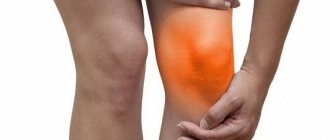
Why is synovitis dangerous?
The synovial membrane performs an important function in the joint - it ensures the separation of hard tissues. Due to this, the joint moves with minimal bending of the components. The membrane is capable of changing shape, thereby adapting to supporting surfaces and providing shock absorption. The quality and quantity of synovial fluid in the joint depends on its condition, the same fluid whose deficiency develops coxarthrosis or gonarthrosis.
The connective tissue that forms this membrane can become inflamed. Swelling forms in the joint, during movement the person feels pain, and bleeding is possible. If the disease is not treated, the synovial membrane thickens and new blood vessels appear in it. In the future, this will lead to frequent joint bleeding.
Synovitis without treatment can lead to joint dysfunction
Physiotherapeutic procedures
NSAIDs and glucocorticosteroids quickly eliminate pain and inflammation, but also have many side effects. Rheumatologists reduce their dosages, and to maintain clinical effectiveness, prescribe physiotherapeutic measures to patients. After 5-10 sessions, blood circulation is restored in the affected tissues, and exudate stops accumulating in the joint capsule. By stopping the inflammatory process, the intensity of pain and swelling decreases, and joint mobility increases. In the treatment of left-sided and right-sided synovitis of the hip joint, the following physiotherapy procedures are used:
- electrophoresis with analgesics, chondroprotectors, NSAIDs. A sterile cotton-gauze swab soaked in drug solutions is applied to the joint area. A metal plate is placed on top of it, through which weak discharges of electric current are passed. Under their influence, drug molecules penetrate the inner lining of the synovial bursa;
- sinusoidally modulated currents. They irritate basic receptors, providing a distracting effect, reducing the severity of pain. Blood begins to flow to the damaged tissues, saturating them with molecular oxygen and nutrients. The use of sinusoidally modulated currents promotes the removal of products of the inflammatory process from the joint capsule;
- UHF therapy. The area of pain and inflammation is affected by ultra-high frequency electromagnetic fields. Heat penetrates the hip joint, accelerating regenerative processes. Swelling decreases, range of motion in the joint increases. UHF therapy, unlike other physiotherapy procedures, can be carried out even during an acute inflammatory process.
The use of magnetic therapy in the treatment of large joints, especially the hip, has been proven to be highly effective. The joint is exposed to a magnetic field - high or low frequency, alternating or constant. As a result, lymphatic vessels contract, promoting tissue detoxification. The removal of excess fluid accelerates, the condition of blood vessels, nerve and muscle fibers improves.
What is tenosynovitis
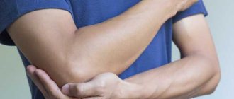
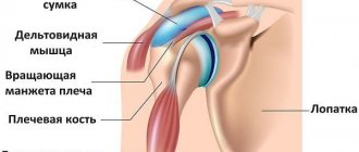
Tenosynovitis is an inflammation of the tendon sheaths. The latter are tubular-shaped connective tissue filled with liquid lubricant. The sheaths surround the tendons, which are made of flexible fibrous collagen tissue. It is what attaches muscles to bones. If the outer covering of the tendons becomes inflamed, it is called tenosynovitis. Both inflammatory diseases are accompanied by pain when moving, so only a doctor can make an accurate diagnosis.
Tenosynovitis most often affects the hands, wrists and feet
Joint damage in childhood
Hemorrhagic vasculitis (Henoch-Schönlein disease)
Joint syndrome: arthralgia or arthritis, polymorphic, predominantly hemorrhagic rash on the lower extremities, large joints, buttocks. Joint syndrome is unstable.
Features: combined with abdominal and renal syndrome.
Chronic ulcerative colitis and Crohn's disease
Articular syndrome: peripheral asymmetric arthritis, with predominant damage to the joints of the lower extremities.
Features: spondylitis, sacroiliitis, associated with the activity of the underlying disease. High detection rate of HLA B27.
Tuberculosis
Articular syndrome: severe arthralgia, spinal damage, unilateral drive, coxitis. Diffuse osteoporosis, marginal bone defects develop, and rarely, a limited bone cavity with the presence of sequestration; destruction of the articular ends of bones, their displacement and subluxation. There is also reactive polyarthritis that develops against the background of visceral tuberculosis. Characteristic damage to small joints.
Features: combined with positive tuberculin tests.
Lyme disease (systemic tick-borne borreliosis)
Articular syndrome: mono-, oligo-, symmetrical polyarthritis. Erosion of cartilage and bones may develop.
Features: combined with tick-borne erythema, damage to the nervous system, heart. Detection of antibodies in blood serum to Borrelia burgdorferi of the IgM and IgG class by indirect immunofluorescence.
Viral arthritis
Arthritis may be associated with viral infections.
Joint syndrome is short-term, completely reversible.
Features: found in acute viral hepatitis, rubella, mumps, chickenpox, arbovirus infection, infectious mononucleosis, parvo- and adenovirus infection, etc. Determination of antibodies to viruses is rarely indicated, since rapid spontaneous recovery usually occurs.
Hypertrophic osteoarthropathy (Marie-Bamberger syndrome)
Articular syndrome: defiguration of fingers in the form of “drumsticks”, hypertrophic periostitis of long tubular bones, arthralgia or arthritis with effusion into the joint cavity. Symmetrical damage to the distal joints of the upper and lower extremities (wrist, tarsus, knee joints).
Features: found in tuberculosis, fibrosing alveolitis, lung cancer, sarcoidosis.
Hemophilia
Joint syndrome: accompanied by bleeding in the joints followed by an inflammatory reaction and effusion. The knee joints are affected, and less commonly, the elbow and ankle, wrist, shoulder and hip joints. Relatively rare - joints of the hands, feet and intervertebral joints.
Features: begins in early childhood.
Leukemia
Joint syndrome: ossalgia, flying arthralgia, asymmetrical arthritis with sharp constant pain in the joints, exudative component and painful contractures.
Features: must be excluded in case of systemic variants of juvenile rheumatoid arthritis (JRA).
Neoplastic processes. Neuroblastoma, sarcoma, osteoid osteoma, metastases in leukemia
Articular syndrome: may be accompanied by myalgia, ossalgia, arthralgia, monoarthritis. Characterized by severe pain in the periarticular areas, a severe general condition that does not correlate with the activity of arthritis.
Features: combined with typical hematological and radiological changes.
Osteoid osteoma
Osteoid osteoma is a benign bone tumor.
Usually the pain is moderate and constant. Typically, the pain occurs in the middle of the night and is severe enough to wake the child.
In these cases, it is necessary to conduct a thorough examination of the child to exclude malignant neoplasms of bone tissue, which have similar symptoms and cause chronic bone pain that can wake the child at night. With osteoid osteoma, taking paracetamol or ibuprofen is usually sufficient to relieve pain. In all cases where a malignant bone tumor is suspected in a child, as well as in cases where the pain is not relieved by paracetamol or ibuprofen, it is necessary to consult the child with an experienced orthopedist familiar with this pathology.
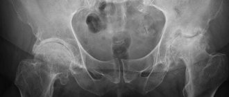
Osteoid osteoma is more common in boys than in girls. In most cases, clinical manifestations appear in adolescence, however, in some cases, the onset of pain and associated anxiety occurs at an earlier age. The usual location of osteoid osteoma is in the hip joint (see below), but sometimes the tumor is located in the knee area. When localized in the hip joint, the diagnosis of osteoid osteoma is not difficult and is easily detected on a regular x-ray. In most cases, once osteoid osteoma is definitively diagnosed, no treatment is required. However, in some cases the pain syndrome can be quite pronounced. In the most severe cases, surgical removal of the area of bone damaged by osteoid osteoma is possible.
Hypothyroidism
Articular syndrome: arthralgia with slight swelling of soft tissues and non-inflammatory effusion into the joint cavity. The knee, ankle and hand joints are affected, and carpal tunnel symptoms may develop.
Features: impaired skeletal formation, slower growth of long tubular bones and ossification, osteoporosis. Muscle weakness and myalgia are evident.
Congenital dislocation of the femur
Congenital dislocation of the femur is detected in newborns during a thorough examination of the hip joints in the maternity hospital and during the first subsequent examination. Immediately after diagnosing the dislocation, an abduction splint should be applied. Congenital dislocation of the femur occurs in approximately 0.7% of newborns. Family cases are common. The hip joints should be examined at the first regular medical examination after birth. If there is a dislocation, the femur returns to its normal position with an audible click. A provocative test is performed by dislocating the femoral head while pressing on the medial sides of the thighs. Different lengths of the lower limbs may indicate a dislocation. It is best detected by bending the legs at the hip and knee joints at an angle of 90°. The presence of asymmetrical inguinal folds suggests congenital dislocation of the femur, however, such a sign is detected 10 times more often than the dislocation itself. The calcaneal-gluteal test for dislocation of the femur is often positive: with the child lying on his stomach, the legs are bent at the knee joint and the hips are extended so that the heels touch the buttocks. The heel on the side of the dislocation crosses the midline and touches the buttock of the opposite side. Congenital dislocation can be bilateral. In such cases, asymmetry tests do not detect dislocation.
Structural abnormalities of the limbs
Clubfoot (talipes equinovarus) can always be diagnosed immediately after birth. With congenital calcaneovalgus clubfoot (talipes calcaneovalgus congenitus), the newborn’s foot is turned into a valgus position and is in dorsiflexion. For this condition, treatment is rarely prescribed; parents are advised to develop the foot. The adductor clubfoot of the metatarsus (talipes metatarsus adductus) is characterized by internal rotation of the forefoot, visible as an inverted curvature of its medial part. Sometimes a plaster cast, adhesive tape, or surgery is indicated.
Flat feet are considered benign if the forefoot can be moved up and down freely and the valgus angle disappears when the child stands on tiptoes.
Walking with internal or external rotation of the foot is a normal condition that does not require treatment.
O-shaped curvature of the legs (genu varum). Consultation with a specialist is indicated for unilateral damage or worsening of the deformity after the second year of life.
X-shaped curvature of the legs (genu valgum). Consultation with a specialist is indicated for unilateral lesions (Blunt's disease), as well as if there is a minimum distance of 10 cm between the ankles at the start of school.
Toes that overlap each other rarely require surgery.
In case of syndactyly of the fingers, the condition is assessed by a pediatric surgeon immediately after birth, if the first and second fingers are involved in the process. In other cases, surgical correction must be performed at the age of 4–5 years. In all cases of syndactyly, consultation with a pediatric surgeon is necessary. For toe syndactyly, treatment is usually not indicated.
Aseptic synovitis of the hip joint
Aseptic synovitis of the hip joint is diagnosed based on typical symptoms, and unnecessary tests should be avoided. The most common cause of acute lameness in children under 10 years of age. In addition to lameness, the child complains of pain in the hip or knee joints and fixes the hip in a position of flexion and external rotation. A typical clinical sign is limited range of motion and pain during internal rotation of the hip. A slight increase in ESR is possible (not higher than 35 mm/h). Effusion into the cavity of the hip joint is easily detected by ultrasound. If possible, ultrasound should always be performed to confirm the diagnosis. The disease is rarely bilateral. Simultaneous damage to other joints excludes aseptic synovitis of the hip joint.
The child is prescribed to rest with his legs bent at the hip joints. Aspiration of the contents of the joint cavity in a hospital setting is necessary only if septic arthritis is suspected or with severe pain. The prognosis is favorable.
Epiphysiolysis of the femoral head
Early diagnosis and surgical treatment of the disease are necessary. If left untreated, medial and posterior displacement of the femoral head progresses and treatment outcome worsens. With moderate and severe diseases, the likelihood of early onset of secondary osteoarthritis increases. The epiphysis of the femoral head slips to the side relative to the femoral neck. The condition is considered stable if the epiphysis is fixed in a new position. An unstable condition means that the pineal gland is not fixed and pain often occurs. The disease occurs between the ages of 10 and 16 years (a little earlier in girls), often in combination with obesity. The disease occurs 2.5 times more often in boys than in girls. Bilateral lesions are noted in 20–30% of cases.
Symptoms: lameness and pain in the knee, hip and groin area occur when supporting the affected limb. Typically, the patient holds the lower limb in a slightly externally rotated position.
Diagnosis: X-ray reveals posterior displacement of the epiphysis. If a disease is suspected (symptoms of hip damage appear at a typical age), it is necessary to evaluate the condition of both limbs in the anteroposterior projection and Lauenstein projection. Indentation caused by recent displacement can be seen on ultrasound. If an effusion is detected on ultrasound, then epiphysiolysis is unstable.
Treatment: Treatment is always surgical. The epiphysis is usually stabilized using a single screw. The smaller the volume of surgical intervention, the better the outcome. It is necessary to consider the possibility of prophylactic fixation of the opposite unaffected hip if the patient is young or if there is an underlying endocrine disease or metabolic disorder (in 7% of cases).
Perthes disease (Legg–Calvé–Perthes disease, pseudocoxalgia)
This is aseptic necrosis of the femoral head, leading after 4–8 months to a clinically manifested subchondral fracture due to physical activity. Fracture and delayed ossification of the cartilage of the femoral head lead to softening of the epiphysis. If left untreated, the femoral head may flatten. It is necessary to diagnose the disease early and treat the tendency to subluxation. The prognosis is favorable if the round shape of the femoral head is preserved. The disease occurs mainly in boys aged 2 to 12 years. The incidence ratio among boys and girls is 5:1. In 10% of cases, the disease is bilateral, but both femoral heads are usually not affected at the same time. The main manifestation is lameness. The disease resembles aseptic synovitis of the hip joint, but begins gradually and becomes protracted or recurrent. The pain is localized in the area between the groin fold and the knee joint. ESR, C-reactive protein level and the number of leukocytes in the blood were not changed. Bone age lags behind passport age by 2 years. Diagnosis is based on radiography: the first sign is a subchondral disorder of the bone structure, subsequently accompanied by the formation of cysts and flattening of the epiphysis. The prognosis for young children (up to 6 years) is favorable.
Osguth–Schlatter disease
Osgut-Schlatter disease is repeated damage to the apophysis of the tibial tuberosity during physical activity. The onset of manifestations coincides with the period of rapid growth in adolescence. Spontaneous healing should be promoted by avoiding excessive stress. Pain occurs in the upper third of the lower leg or knee when running or jumping. While running, the pain is more severe when the heel touches the supporting surface; when the heel is lifted from the surface, the pain is less pronounced (biomechanics of the knee extensor apparatus!). The tibial tuberosity is prominent and sensitive to palpation. Radiography reveals fragmentation of the tuberosity. In typical cases, there is no need for x-rays.
Spondylolisthesis
The most common cause of long-term or recurrent back pain in adolescents. Lower back pain that occurs after exercise and often radiates to the hips may be a sign of spondylolisthesis. Clinical signs:
- excessively pronounced lumbar lordosis;
- the displacement of the vertebra is palpated (palpable “threshold”);
- prominent spinous process.
The diagnosis is based on identifying the displacement of the vertebra on a lateral x-ray (an anteroposterior x-ray is not necessary). The course of spondylolisthesis is monitored by performing radiographs 2-3 times a year (consultation with a pediatric surgeon is also necessary) in children with characteristic manifestations. Excessive stretching (lifting weights, gymnastic exercises) should be avoided.
Scheuermann's disease
Scheuermann's disease is an osteochondritis affecting the anterior part of the vertebral cartilage, occurring in late puberty (ages 13–15 in girls and 15–17 years in boys). The disease occurs 4 times more often in girls than in boys. The most common manifestations are pain in the thoracic spine and back stiffness, and stiffness of the hamstrings.
Diagnosis is based on radiographic findings.
- Wedge-shaped deformity of the vertebral bodies with a flattened anterior part.
- Deformed marginal vertebral plate; Schmorl's hernia (a depression towards the vertebral body) develops in the later stages.
Scoliosis
Scoliosis is idiopathic, progressive in 85% of cases and functional in 15%. Scoliosis is a disease, not bad posture; exercise therapy is not effective for this disease. The disease develops in a phase of rapid growth (at the age of 10–12 years in girls, in boys several years later). For screening, examination of the spine is used in the position of bending the torso forward with straightened legs. Scoliosis is confirmed if one shoulder blade is higher than the other. In mild or controversial cases, children should be monitored at 6-month intervals. The severity of scoliosis is assessed using radiographs, determining the maximum angle of deformity. X-ray examination is indicated if obvious scoliosis is clinically detected.
Calve's disease (vertebra plana)
A rare disease in children aged 2–10 years, manifested by complete flattening of the vertebral body. The clinical picture includes local pain, increased ESR, and sometimes leukocytosis. When carrying out differential diagnosis, it is necessary to exclude tuberculosis. A patient with suspected Calve's disease should be sent to the hospital for further research.
Discosis
Usually aseptic, but can also be caused by bacteria. Difficulty walking and sitting are typical clinical manifestations in preschool children. The diagnosis is based on local tenderness and pain when moving the spine. The diagnosis can be confirmed by radioisotope bone scanning using technetium. The child must be hospitalized for further examination. In many cases, the condition is benign and resolves spontaneously.
Growing pains (clinically insignificant leg pain)
- Growing pains never appear during the daytime.
- Regardless of the severity of the pain attack at night, if it is growing pains, then in the morning the child always feels good/great.
- Any child who experiences pain in the morning or during the day requires a full medical examination.
Clinically insignificant leg pain (so-called “growing pains”) is diagnosed based on clinical manifestations after ruling out arthritis. In children under 4 years of age, another disease should be considered. It is necessary to evaluate the psychosocial background, especially in the presence of other manifestations (headache, abdominal pain). Recurrent pain in the limbs without organic causes occurs in children during growth. 50% of children experience leg pain at some point during childhood. Leg pain usually occurs around 4 years of age. The condition is most common between the ages of 6 and 11 years. Often the condition is hereditary. Most often the muscles of the legs and thighs and the area of the knee joints hurt. The upper extremities are rarely affected. The child may also have a headache or stomach pain.
The child unexpectedly wakes up at night during deep sleep and complains of pain in his leg. Parents immediately understand that the child has a health problem because he wakes up and cries in bed. Most often, such episodes occur a few hours after the child has fallen asleep, but sometimes the child may wake up late at night. Typical symptoms include pain in the knee, either in the front or back, or pain in the muscles directly above the knee. Usually the pain disappears after ten or fifteen minutes with a light massage and does not remind you of itself in the morning. Most cases of growing pains affect large joints, such as the knee, rather than the fingers or toes. Sometimes a child may wake up in pain for several nights in a row, but more often these episodes occur sporadically over a period of several weeks or several months. Growing pains often appear after days when the child has been especially physically active. Growing pains may disappear for several months or years and then reappear during periods of particularly rapid growth.
The main point when assessing growing pains is the fact that if these are growing pains, then in the morning after waking up the child feels absolutely healthy.
The best explanation for the mechanism of growing pains is the tension of the muscles and tendons as the leg bones rapidly increase in length.
During the day, the child is active, and the information that his brain receives from tense muscles and tendons is lost in the flow of other information from many other events. At night, when the child falls asleep and there are no distractions, the pain impulses that come from the tense muscles and tendons reach the higher nerve centers and the child wakes up in pain. As a result, it can be summarized that growing pains are observed during the period of the child’s most intensive growth and after days of high physical activity.
Growing pains can be a worrying experience for both parents and baby, especially when painful episodes occur over several nights in a row. In most cases, a light massage and conversation is enough to get the child to sleep. Children with more severe pain usually stop complaining after taking the usual dose of paracetamol or ibuprofen, exactly the same as for a headache. If the child wakes up for several nights in a row, then pain medication can be given before the child goes to bed. This will reduce pain sensitivity so your child can sleep peacefully through the night. After two or three nights without pain, you should stop giving the child the medicine.
It is necessary to assume another cause of pain if the child has the following manifestations:
- prolonged pain in one limb;
- combined symptoms (for example, lameness or change in general condition);
- Symptoms occur in the morning or during the day.
If atypical symptoms appear, it is necessary to conduct at least a general blood test and determine the ESR.
Damage to joints and ligaments in children
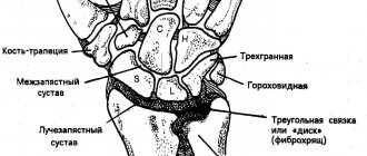
Incomplete dislocation of the radial head
Typical injury in children aged 1 to 5 years.
Injury may occur if the child is pulled or lifted by the arm. A recent injury can be easily reduced by first rotating the arm into a supinated position and then pressing on the head of the radius, pushing it through the annular ligament. At this moment, you can hear a click and the severity of symptoms immediately decreases. In old lesions, reduction is often unsuccessful. In such cases, treatment includes the application of a sling bandage. Symptoms disappear spontaneously.
Foot subluxation
A torn anterior tibiotalar ligament is the most common tendon injury. If the traction test is positive (when you pull the foot forward, there is a feeling of separation of the foot from the lower leg), the application of a support bandage, sometimes a plaster, as well as surgical treatment (rarely) may be indicated. Specialist consultation is required.
Patella dislocation
The most common dislocation in teenagers is patellar dislocation.
If the patella is displaced to the lateral side, it is necessary to carry out reposition as soon as possible.
Literature
- Brewer EJ Jr., Bass J., Baum J. Current proposed revision of JRA criteria // Arthritis Rheum. 1977, 20 (Suppl): 195.
- Cassidy J., Petty R. eds. Textbook of pediatric rheumatology, 5 nd ed. // New York: Churchill Livingstone. 2005.
- Ilona S. Szer, Yukiko Kimura, Peter N. Malleson, Taunton R. Southwood. Arthritis in children and Adolescents. Juvenile Idiopathic Arthritis // Oxford University Press. 2006. 456 p.
- Alekseeva E. I., Litvitsky P. F. Juvenile rheumatoid arthritis. Etiology, pathogenesis, clinical picture, diagnostic and treatment algorithms. M.: Publishing house "VEDI". 2007. 360 p.
- Neeck G., Michels H. Endocrine aspects of pediatric rheumatic diseases // Bailliere's Clinical Rheumatology. 1996. No. 2. P. 349–363.
- Alekseeva E. I., Zholobova E. S. Reactive arthritis in children // Issues of modern pediatrics. 2003, vol. 2, no. 1, p. 51–56.
- Nasonov E. L., Nasonova V. A. Rheumatology. National leadership. Geotar-Media, 2008. 720 p.
- Alekseeva E. I., Bzarova T. M. Rheumatology. Clinical recommendations. Ed. E. N. Nasonova. M.: Publishing group "GEOTAR-Media". 2005, 2006, p. 120–140.
- Alekseeva E. I., Zholobova E. S., Chistyakova E. G., Valieva S. I. Reactive arthritis. Educational and methodological manual. M.: Publishing house MMA im. I. M. Sechenova, 2004. 134 p.
E. I. Alekseeva , Doctor of Medical Sciences, Professor T. M. Bzarova
Scientific Center for Children's Health, Russian Academy of Medical Sciences, Moscow
Contact information for authors for correspondence
Causes of synovitis and tenosynovitis
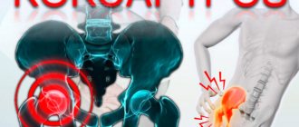
Synovitis is one of the symptoms of rheumatoid arthritis of an autoimmune (unexplained) nature. Chronic inflammation of synovial membranes provokes inflammatory processes in other organs - a full-scale process is launched in the body, causing a person no less pain than arthrosis of the knee joint in advanced forms.
Synovitis can also occur with the following diseases:
- juvenile arthritis;
- psoriatic arthritis;
- lupus;
- rheumatic fever;
- gout;
- tuberculosis;
- injuries.
The causes of tenosynovitis still raise questions. It is believed that inflammation occurs after excessive strain on the tendons, but this is just a guess. Obviously, due to increased physical activity, tendons, surrounding muscles and bones are injured, so the risk of inflammation of the membranes increases.
Basic treatment methods
At the initial stage of treatment, accumulated synovial fluid is removed from the joint cavity using a puncture. Sometimes the procedure is carried out at the diagnostic stage, when the exudate is taken for biochemical research. In severe cases of the disease, the doctor may resort to arthroscopy to determine its cause. He makes an incision in the skin over the joint and inserts a device equipped with a miniature camera and lighting into the joint cavity. Immediately after the examination, excess fluid is pumped out. The joint capsule is treated with antiseptic solutions, and if necessary, intra-articular injections of glucocorticosteroids are performed.
Nonsteroidal anti-inflammatory drugs
Treatment of synovitis of the hip joint, like all pathologies of the musculoskeletal system, is carried out with the use of non-steroidal anti-inflammatory drugs (NSAIDs). For acute pain, the doctor prescribes them in injection form, for nagging, aching pain - in capsules or tablets. The most commonly prescribed NSAIDs contain the following active ingredients:
- ketoprofen;
- nimesulide;
- ibuprofen;
- ketorolac;
- diclofenac;
- meloxicam;
- indomethacin.
To enhance the therapeutic effect of injection solutions and tablets, the simultaneous use of ointments and gels with NSAIDs is recommended. These are Voltaren, Ketorol, Finalgel, Artrosilene, Ortofen. NSAIDs have a pronounced anti-inflammatory, decongestant, and analgesic effect. But the high clinical effectiveness of the drugs is offset by their ability to damage the gastrointestinal mucosa. To minimize complications, rheumatologists, along with NSAIDs, prescribe proton pump inhibitors - Rabeprazole, Omez, Nolpaza.
The use of non-steroidal anti-inflammatory drugs in the form of rectal suppositories - Voltaren, Indomethacin - helps to avoid undesirable consequences.
Antibiotic therapy
Often the cause of inflammation of the synovial membrane is infection. When conducting laboratory tests, the resistance of microorganisms to antibiotics of a certain group is established. In accordance with the test results, patients are recommended to take the drugs for 7-20 days. What antibiotics can be included in the therapeutic regimen:
- semisynthetic protected penicillins - Amoxiclav, Augmentin, Panklav, Flemoklav;
- macrolides - Azithromycin, Erythromycin, Clarithromycin;
- cephalosporins - Cefazolin, Ceftriaxone, Cefotaxime.
To treat specific synovitis of the hip joint, in addition to antibiotics, it is necessary to take antimicrobial agents - Trichopolum, Metronidazole, Co-trimoxazole, Biseptol. The doctor can administer injectable forms of antibacterial drugs immediately after removing the intra-articular fluid.
After antibiotic therapy, it is necessary to take medications to restore the intestinal biocenosis. The simultaneous administration of drugs from the groups of probiotics and prebiotics is practiced: Enterol, Bifidumbacterin, Acipol, Linex together with lactulose, pantothenic acid, and inulin preparations.
Other medicines
If NSAIDs are ineffective in the treatment of synovitis, glucocorticosteroids are used - Prednisolone, Hydrocortisone, Dexamethasone. They are used for oral administration or intra-articular administration. They have anti-inflammatory, analgesic, antipyretic activity. Glucocorticosteroids are not intended for long-term treatment, as they have a negative effect on bone tissue, kidneys, liver and stomach. Other pharmacological drugs are also used in the treatment of synovitis:
- chondroprotectors - Structum, Teraflex, Dona. They are used to gradually normalize the production of synovial fluid. Long-term course use (from several months to 2 years) helps prevent the spread of destructive and degenerative changes in the joints. Taking chondroprotectors at the initial stage of diseases leads to the restoration of damaged tissues;
- multivitamins with microelements. In rheumatology, the use of drugs containing B vitamins is practiced - Combilipen, Milgamma, Pentovit. They improve innervation, normalize blood circulation and microcirculation. Patients are additionally prescribed vitamin complexes with microelements - Centrum, Selmevit, Vitrum. Their course intake has a general strengthening effect, replenishes calcium, zinc, copper, and molybdenum.
To treat diseases that provoke synovitis, specific drugs are used. Therefore, for each patient, a therapeutic regimen is drawn up individually. Some pathologies cannot yet be treated, for example, rheumatoid arthritis or gout. To prevent recurrence of synovitis, maintenance therapy is carried out.
After stopping the inflammatory process, warming ointments and balms can be prescribed - Finalgon, Viprosal, Capsicum. After applying them under the influence of heat, blood circulation in damaged tissues improves: nutrients and biological substances begin to flow into them, accelerating regeneration.
Diagnosis and treatment
During the initial examination, the doctor checks the condition of the joint visually: notes the presence of pain, changes in skin color, difficulties with movement, and an increase in local temperature. MRI or ultrasound can help determine the amount of accumulated fluid. If there is tissue damage in the joint, an x-ray will show it.
To determine the root cause of the inflammatory process, the following is prescribed:
- a blood test to determine the bacterial infection that triggered the inflammation;
- analysis of synovial fluid.
Self-medication of synovitis, especially warm compresses, can lead to dangerous complications
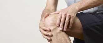
Treatment of synovitis of the knee joint
Let's consider two main types of treatment for the disease. Conservative therapy. Treatment of aseptic type synovitis in most cases is outpatient, includes puncture with fluid evacuation and immobilization of the limb with a plaster splint, knee brace or tight bandage. Immobilization can last a maximum of 7 days, since prolonged absence of walking can cause the development of joint stiffness. Patients are prescribed exercise therapy, microwave therapy, electromagnetic treatment, electrophoresis with dimethyl sulfoxide or hydrocortisone and ultrasound with anti-inflammatory drugs. After eliminating the acute phase, paraffin, ozokerite, mud treatment, and balneotherapy are recommended.
For reactive synovitis, treatment is prescribed with glucocorticoids, calf cartilage extracts, heparin, ibuprofen and indomethacin. During an exacerbation, from the third or fourth day, patients are sent to physical therapy: phonophoresis with corticosteroid medications, UHF, magnetotherapy, electrophoresis with aprotinin, ketoprofen and heparin. Please note that heparin should not be taken immediately after injury or surgery, or if you have clotting problems. If you need help, please contact us. Our clinic employs only qualified specialists; they conduct individual exercise classes, massage, etc.
For chronic synovitis with obvious infiltration of the synovial membrane, inhibitors of proteolytic enzymes are prescribed, aprotinin and corticosteroids are injected into the joint.
The standard treatment regimen includes:
- non-steroidal anti-inflammatory drugs to relieve swelling and reduce pain;
- short-term corticosteroids;
- cold compresses to reduce pain and swelling;
- rest to reduce the load on the joint;
- antibiotic therapy - in the presence of a bacterial infection.
If the joint is severely damaged, sometimes synovitis can only be cured by surgery
Is it worth pumping out fluid from the knee joint? Opinion of a practicing orthopedic traumatologist:
Synovitis is often diagnosed in people with degenerative joint diseases. Damaged cartilage surfaces become inflamed and fluid accumulates in the joint. In such conditions, it is impossible to begin treatment for osteoarthritis. It is necessary to stop the inflammation, and only after that undergo a course of intra-articular injections of synovial fluid prosthesis or resort to other, less effective therapy.
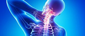
Symptoms
The general clinical picture of the pathology consists of severe pain, weakness/poor health and local hyperthermia. The patient's body temperature may also rise to low-grade levels (37-37.5 °C). Often inflammation is accompanied by swelling and redness of the skin over the joint. The pain syndrome intensifies when leaning on the leg, moving it to the side and during palpation.
With an infectious etiology, the disease begins acutely, with an increase in body temperature and general malaise. Then bursting pain appears in the joints (at the site of suspected synovitis). Within a few hours, severe swelling, hyperthermia and local hyperemia develop. The patient feels a headache, an increase in general body temperature, and sometimes there are other signs of intoxication.
With non-infectious etiology, the pathology does not develop so quickly, but this will depend on the cause. The primary symptom complex is represented by pain when moving. After a couple of days, swelling, hyperthermia, hyperemia, joint deformation and increasing pain are added.
Surgery
If conservative methods of treating synovitis are ineffective, the patient is prescribed a surgical operation - synovectomy. It can be arthroscopic or performed in the traditional open way. The disadvantages of the latter technique include a high likelihood of developing complications: the formation of postoperative hematomas, scars and cicatrices, painful sensations that do not subside for a long time and limited range of movements. As a result, the duration of hospitalization and rehabilitation period increases. Therefore, open synovectomy is usually performed only for complicated synovitis of the hip joint.