From this article you will learn everything about joint pain (including in the knee joint): what kind of pathology it is, why it occurs, how it manifests itself. Its treatment and prevention.
Author of the article: Victoria Stoyanova, category 2 doctor, head of the laboratory at the diagnostic and treatment center (2015–2016).
Article publication date: 07/16/2019
Article updated date: 01/11/2020
A joint mouse is a pathological formation that moves freely within the joint. It could be a fragment of cartilage or bone tissue, a meniscus, or a clot of connective tissue.
X-ray of a knee with a joint mouse. Click on photo to enlarge
The official name of the pathology is arthremfit. In response to a provoking factor (trauma, arthrosis deformans, arthritis, Koenig's disease, hemarthrosis, osteochondritis), a part is separated from the articular tissue, which moves freely in the articular cavity and causes pain.
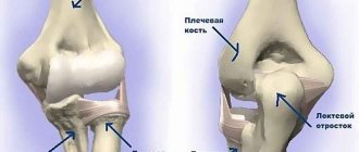
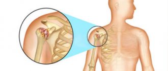
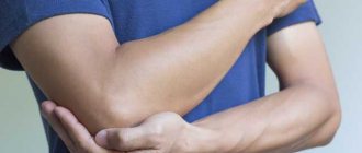
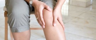
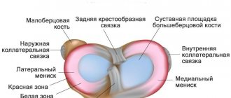
This disease usually affects the knee joint. In very rare cases - elbow, even less often - shoulder, hip.
This article will consider the articular mouse of the knee joint, since in other joints this pathology occurs very rarely - for example, there is almost no chance of pathology developing in the wrist joint.
The disease often occurs in the knee joint due to its complex structure and heavy loads when walking, jumping, and running.
Most susceptible to the development of pathology:
- men and women over 40 years of age;
- athletes;
- persons of heavy physical labor;
- overweight people.
Arthremfit is a serious pathology, the only effective method of treating the disease is surgery, which requires rehabilitation over several months.
With early removal of a joint mouse of traumatic origin, the treatment result is favorable: the functional ability of the joint is completely restored.
If the pathology is latent and there is no pain (when the fragment of cartilage tissue is very small or hidden in the volvulus of the synovial membrane (a pocket of the membrane surrounding the joint), a person can delay treatment. This leads to the progression of the pathology, the following are possible:
- complete loss of limb function;
- blockade;
- ossification of connective tissue.
Click on photo to enlarge
An arthrologist is responsible for the diagnosis and treatment of pathology.
Causes of arthremfit (joint mouse)
Most often, pathology occurs as a result of injury. Due to the impact, a small piece of bone tissue, cartilage or a small part of the meniscus may break off. Also, trauma can lead to the formation of fibrin clots (fibrin is a protein that is the basis of a blood clot), which subsequently becomes denser.
Another possible cause is Koenig's disease (osteochondritis dissecans - separation of part of the cartilage from the adjacent bone and its displacement into the joint cavity). It provokes necrosis and rejection of particles of bone, cartilage or joint capsule. This disease is typical for people constantly engaged in heavy physical labor (loaders, builders, miners).
Joint mouse can also occur against the background of arthrosis. With this disease, osteophytes (bone protrusions to which muscle tendons are attached) grow on the joint, particles of which can break off.
Click on photo to enlarge
The size of the articular mouse ranges from 1–2 mm to 1–1.5 cm. It can be either single or multiple. Sometimes up to 5 freely moving fragments are identified in the cavity of the knee joint.
What could the debris be? He can be:
- Round.
- In the form of rice.
- In the form of a polyhedron.
- Formless.
Examples of articular mouse
What can cause artemphyte - causes of the disease
- Fractures, dislocations, consequences of sudden and maximum amplitude movements leading to injuries to the limbs.
- Koening's disease, as a result of heavy physical activity and work.
- Destruction of cartilage tissue. This occurs with joint diseases caused by inflammatory or degenerative processes.
- Consequences of arthritis, deforming arthrosis, osteochondritis, hemarthrosis.
It is not difficult to identify problems in the knee joint, even without the help of a specialist, because they are obvious.
Characteristic symptoms
The pathology is asymptomatic for a long time. An intra-articular formation makes itself felt only when it is pinched between the surfaces of the joints. Then a so-called joint block occurs.
It manifests itself:
- acute pain;
- limited or complete inability to move.
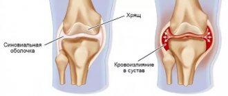
Constant irritation of the joint cavity by the “mouse” threatens the development of inflammatory processes in the joint capsule.
Additional symptoms:
- pain;
- edema;
- accumulation of effusion (accumulation of fluid released from blood vessels) consisting of excess synovial fluid.
What usually bothers you?
It is obvious that a freely moving particle sooner or later gets between the articular surfaces. Until this moment, the joint may not bother you. Patients note that most often pinching of the articular mouse occurs when climbing stairs. The condition when a free element “wedges” into the articular lumen is called joint blockade. The pain is very sharp, the joint is “blocked”, movement becomes impossible or severely limited. It is difficult to predict when the first joint blockade may occur. Quite a long period of time may pass. Depending on the etiology (cause), resorption of small joint formations is possible. If pinching of a joint mouse is periodically repeated, inflammation of the joint capsule occurs and it releases synovial fluid (effusion). The formation of effusion in this case is characterized by a persistent course and often recurs after puncture. If treatment is not resorted to, the chronic inflammatory process leads to the development of arthrosis.
Identifying the presence of a joint mouse is not always easy. A simple x-ray may be sufficient. But more often we have to resort to ultrasound and MRI. Sometimes, especially if the formation is large, the patient himself can feel the articular mouse.
First aid for joint blockade
- Immobilize the knee using a tight elastic bandage. Stretch the bandage and wrap once 10 cm above the knee. Repeat the rotation so that the next layer overlaps the previous one by 2/3. Finish bandaging 10 cm below the knee. Make sure that the bandage lies flat, without folds, secures tightly, but does not pinch the knee (the pain should not intensify; if this happens, loosen the tension).
- Transport the patient to a doctor or call an ambulance.
Diagnostics
If a person complains of sudden pain in the knee or elbow, he needs to be given first aid. To do this, the area of possible damage is tightly wrapped with an elastic bandage. The patient can also wear a special knee brace. Then you can call an ambulance, which will take the patient to the clinic. Here, experienced doctors will be able to establish the correct diagnosis: bruise, injury, sprain or joint strain of the knee joint.
An X-ray will confirm the doctors' fears. With the help of medical equipment, a specialist will be able to examine what size the mouse is, where it is located, and so on. In addition, the patient will undergo a CT scan. With its help, the doctor learns about the condition of cartilage, menisci, and bone tissue. The patient also needs to undergo blood tests: general and biochemical. Sometimes he is recommended to undergo various immunological studies.
Treatment and rehabilitation
The prognosis for recovery is conditionally favorable. With the help of surgery, you can completely get rid of the disease.
Rehabilitation will last from 1 to 3 months. After this, the patient can return to normal daily life.
The only way to completely get rid of the pathology is surgery.
If the fragment moving freely in the joint cavity is large, then open access surgery is necessary.
If the “mouse” is small, then removal is possible using the arthroscopic method - by introducing a camera through punctures into the joint cavity to visualize the surgical field and surgical instruments.
During the operation, doctors can perform joint plastic surgery (plasty is reconstruction, restoration of articular surfaces using one’s own tissues or artificial materials) to prevent recurrence of spalls.
If the operation was performed with open access, a plaster cast is applied to the knee. If the treatment was carried out arthroscopically, it is recommended to wear a rigid orthosis (a knee pad with a soft inner layer and a hard outer layer (metal, plastic)).
Rigid orthosis
Next, postoperative treatment will be necessary:
- physiotherapeutic procedures;
- physiotherapy;
- medicines.
The duration of the rehabilitation period after surgical treatment is from 1 month (with the arthroscopic method) to 3 months (with surgery with open access to the joint).
Rehabilitation: physiotherapeutic procedures
They allow you to recover faster after surgery by improving blood circulation and relieving pain. The following procedures have been successfully used:
- Electromyostimulation – muscle stimulation with electric current; improves blood supply to tissues.
- Electrophoresis is the administration of drugs through the skin using an electric current.
- Phonophoresis is the administration of drugs through the skin using ultrasound.
- UHF therapy – exposure to high frequency magnetic waves; relieves inflammation, increases blood flow in tissues.
- Paraffin applications are a thermal effect on the joint using heated paraffin.
- Magnetotherapy – exposure to magnetic waves; stimulates local immunity, improves blood flow in the tissues around the joint.
Carrying out magnetic therapy
Rehabilitation: therapeutic exercises
Exercise therapy allows you to restore the normal range of motion of the joint and prevent postoperative complications.
During the rehabilitation period, it is recommended to perform the following exercises:
- Starting position – lying on your back with your knees bent. Feet are on the floor. Press your heels firmly into the floor and feel the muscles in the back of your thighs tighten. Hold this position for 5 seconds, relax. Repeat the exercise 10 times.
- The starting position is similar. Bend your healthy leg. Straighten the patient and place her on the floor. Raise your straight leg to a height of 30 cm, pulling your toe towards you. Hold it for 5 seconds and lower it. Repeat the movement 10 times.
- Starting position: standing against a wall, you can lean on it with your hand. Elevate the affected leg 45 degrees. Wait 5 seconds, lower slowly. Do the exercise 10 times.
- Half squats, resting your hands on the back of a chair.
Illustration of exercises above:
Exercises for rehabilitation after surgery to remove a joint mouse. Click on photo to enlarge
The doctor selects the remaining, more difficult exercises individually.
Rehabilitation: medications
To relieve pain and inflammation, you need to take NSAIDs - Ibuprofen, Meloxicam. External use of anti-inflammatory drugs is also possible.
Click on photo to enlarge
If a joint mouse has provoked an inflammatory process with the accumulation of effusion, you must first perform a puncture to remove it and take a course of anti-inflammatory drugs, and only then remove the fragment.
Rehabilitation: folk remedies
Traditional methods are suitable as a complement to other methods of rehabilitation after surgical treatment. Homemade remedies help relieve pain and restore the joint.
Traditional medicine recipes:
- Compresses with red and blue clay. Mix them in a ratio of 1 to 1. Add water, bringing the mixture to the consistency of sour cream. Apply the product to gauze or a piece of cloth. Apply to the sore joint for 2 hours. Rinse off with warm water.
- Jerusalem artichoke baths. For 1.5 kg of crushed plant, 8 liters of boiling water. Wait until the product cools down. Place your leg in it until the liquid covers the knee joint. Keep for 30 minutes.
Treating a sore joint with blue clay
Compresses and baths are contraindicated in the acute stage of the process (immediately after injury, with severe pain during exacerbation). You can use folk remedies after acute inflammation subsides and surgical removal of the fragment. Before starting therapy, it is a good idea to consult a doctor.
Diagnostic and treatment approaches
A visual examination and anamnesis of complaints allows the doctor to highly likely assume the presence of an intra-articular mouse in the patient. To clarify, X-rays, computed tomography or magnetic resonance imaging are prescribed. The data obtained serve as the basis for choosing treatment tactics. However, there is no diversity of approaches here. The main and effective method is surgical removal of the articular mouse. If the operation is performed in a timely manner, the patient recovers completely and mobility returns to the joint.
Treatment after surgery
After the articular mouse has been removed, the patient needs restorative therapy:
- To improve blood circulation in the damaged area, Andekalin, Angiotrophin, and Kallikrein Depot are prescribed.
- If there is an inflammatory process, Gordox or Contrikal is prescribed.
- It is recommended to take medications that nourish the tissues in the joint area: Solcoseryl, Actovegin, B vitamins.
- The patient also needs medications to help restore the remaining part of the articular cartilage. This is, for example, “Glycosamine” or “Chondroitin sulfate”.
- Be sure to prescribe a new drug, but one that has already proven itself to be positive, “Piaskledin”. It is expensive, but very effectively protects cartilage from further destruction.
- It is necessary to undergo a course of hyaluronic acid injections. It acts as a natural joint lubricant and therefore significantly reduces painful friction.
You cannot do without warming medications that increase blood flow to the operated area - compresses with Dimexide or Bischofite.
Physiotherapeutic treatments
After surgery, the patient is required to undergo a series of other procedures to ensure that the knee joint no longer manifests itself. Treatment using physiotherapeutic methods involves the following manipulations:
- Joint massage.
- Ultrasound treatment.
- Mud therapy.
- Adoption of vans: radon and turpentine.
- Impact of dynamic current.
- Electrophoresis of sulfur, lithium and zinc to restore metabolic processes in the joint.
- Galvanization with enzymes that remove “bad” tissue.
- Phonophoresis with analgesics to relieve inflammation.
Similar procedures can be performed in a clinic or hospital, where there is appropriate equipment and highly qualified personnel.
Minimally invasive surgery
The articular mouse is also removed using minimally invasive methods. Surgical treatment can be performed using arthroscopy. This method is less traumatic compared to conventional surgery. The doctor uses a special device - an arthroscope - to make two holes in the knee. One introduces optical technology, using it to examine the internal state of the pathology. In this case, the picture is broadcast on the screen. The doctor inserts a manipulation tool into the second hole. Thus, the damage to the patient is minimal. Within a week you can begin rehabilitation.