In the picture: subluxation of the atlas (first cervical vertebra), view from the back. Atlas (atlas, atlas) is the first vertebra of the cervical spine, the function of which is to maintain the skull in a stable condition and regulate the position of the entire spine.
Displacement of the first vertebra is the name of a very common injury, subluxation of the atlas. The first cervical vertebra of the atlas (C1) is attached to the occipital region of the skull and connects to the second vertebra, the axis (C2), forming the atlantoaxial joint. It ensures head mobility, normal support of the skull, and high-quality blood supply to the brain and spinal cord.
In the picture: the first cervical vertebra (atlas) in the correct position, viewed from the back.
With the correct position of the C1 vertebra, when turning the head, it moves in concert with the skull relative to the C2 axis.
How is the first vertebra arranged?
Atlas is similar in structure to a ring, the lateral sections of which are denser compared to the back and front parts. These sections are connected to the occipital bone. The second vertebra has an odontoid process: it is this that ensures sliding along the inner surface of C1. More detailed information in this article.
What is rotational subluxation of the atlas?
Rotation translated from Latin means “circular movement”, “rotation”. Consequently, right-sided or left-sided rotational subluxation of the atlas is a slight asymmetric displacement of the articular surfaces of the annular C1 relative to the C2 axis to the right or left, respectively. More serious pathologies are quite rare.
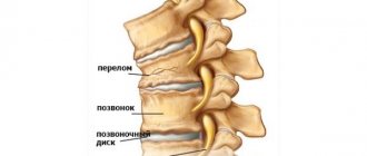
It is important to distinguish between complete dislocation and subluxation of the atlas vertebra. When a dislocation occurs, there is a complete loss of contact between the surfaces of the joints. In this case, the person is not able to rotate his head at all. A dislocation is usually characterized by severe pain, and when the joint is displaced, a characteristic click is heard. Dislocation is also not rotational: the vertebrae are not displaced along the spinal axis, but in any way. This almost always results in disability or death.
But much more often with injuries and impacts, it is rotational subluxation of the atlas that occurs.
With rotational subluxation, the vertebra is not completely displaced (by a few millimeters) and precisely relative to the spinal axis, and the atlantoaxial and atlanto-occipital joints are blocked by tense muscles that will hold the vertebrae in a new state. The head turns in all directions, but the amplitude of rotation is limited. Pain with rotational displacement is not always observed.
It is important to note that congenital (not mechanical origin) pathology of the atlas position also occurs, but in this case correction is possible.
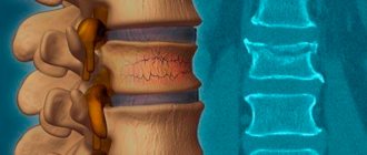
In the picture: rotational subluxation of the atlas (first cervical vertebra), view from the back.
The picture shows how the vertebra moves. The dotted red line shows the position of the atlas when it is displaced (some people may have the pathology all their lives and not know it), and the green dotted line indicates the normal natural position.
Symptoms
Doctors have identified the main symptoms of subluxation of the vertebrae of the cervical segment:
- pain;
- the patient's head is in a forced position;
- muscle tone increases;
- swelling occurs in the damaged area.
When the spinal cord is pinched, neurological disorders occur.
The nature of pain during subluxation of the cervical vertebrae is different; it can be aching, pulling, and annoying. Painful sensations appear during the dislocation or a little later.
It is much more difficult to detect subluxation in a newborn than in an adult patient. With pathology, the child often cries, sleeps poorly at night, is underweight, etc. However, it is difficult for parents to understand that this is due to pathology of the cervical segment of the spine. Specific signs of subluxation in children include:
- tilting the head in the direction opposite to the displacement;
- severe pain in the back of the head and neck;
- neck deformity;
- limitation of head mobility, etc.
Subluxation, in which the spinal cord is damaged, is accompanied by dizziness, hearing, vision disorders, etc.
When the spinal cord is damaged, the following symptoms appear:
- vertigo;
- sleep disorders;
- headache;
- convulsions;
- pain in the jaw, shoulders, or back (upper part);
- hearing disorders (temporary or permanent);
- numbness, “crawling” sensation;
- limited mobility and decreased strength in the arms or legs.
In addition, cramps in the arm on the affected side may occur.
Rotational subluxation of the 1st cervical vertebra is manifested by the following symptoms:
- pain in the upper neck;
- with a right-sided lesion, the head is turned to the left, and with a left-sided lesion, the head is turned to the right;
- dizziness when trying to turn your head in the direction of the lesion, and sometimes loss of consciousness.
The main sign of the pathology is still a nagging pain, which intensifies with movement; it can spread to the jaw.
Symptoms of subluxation of the 2nd cervical vertebra:
- pain in the affected area of the neck;
- difficulty swallowing;
- possible swelling of the tongue.
Subluxation of the 3rd vertebra shows the same signs.
Injury to the remaining cervical vertebrae is manifested by the following symptoms:
- pain that extends to the shoulders;
- flatulence;
- discomfort behind the sternum.
Congenital pathologies in the first months after birth are asymptomatic. But as you grow older, the load on the spine increases, and then the first symptoms begin to appear. A little later, the clinical picture is supplemented by headaches, deterioration of memory, attention, increased fatigue, etc.
[node:field_similarlink]
Types and causes of Atlanta injuries
Displacement or skew of the atlas most often occurs in newborns due to the actions of obstetricians during the birth process or due to incorrect presentation of the fetus. Subluxation is sometimes characterized by limited neck mobility or torticollis.
During childbirth, the baby experiences an incredible external mechanical load for its small size. The cervical spine suffers the most as a result of high pressure, especially if the birth process is forced to be stimulated using obstetric forceps and other methods.
At one time in the Soviet Union there was a practice of “preserving the perineum of a woman in labor.” The so-called “humane” method was aimed at reducing the rate at which the fetus comes out, which was supposed to reduce the recovery period of the mother and help avoid injury to the perineum. In fact, the midwife simply pressed the baby’s head in the opposite direction with her palm, increasing the already high load on the baby’s neck.
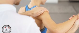
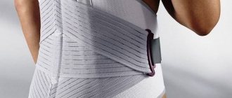
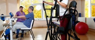
Patient rehabilitation
After reduction, a corset is put on the neck to fix the cervical spine. The duration of wearing it can be up to 3 months, depending on what the doctor says. After removing the support agent, rehabilitation begins. It includes special exercises compiled by a doctor, physiotherapy procedures (electrophoresis, ultrasound, magnetic therapy), massage and acupuncture. Together they speed up the recovery of the damaged area.
Subluxation of the cervical vertebra, even if mild, requires observation by a doctor. Displaced vertebrae put pressure on nerves and blood vessels, which without proper attention leads to negative consequences for the nervous and circulatory systems. Moreover, you cannot reduce an incomplete dislocation yourself without a competent specialist. Timely treatment, an experienced doctor and a well-chosen course of rehabilitation are the main components of a quick recovery.
Damage to the cervical spine - specialist recommendations
The myth about safe caesarean section
In the photo: caesarean section is one of the common causes of subluxation of the atlas.
This is important for parents to know!
There is an opinion that a caesarean section relieves a woman in labor from painful labor and almost completely relieves the load on the cervical spine. In pursuit of bonuses and other privileges, some doctors advertise this method as the safest. This is very far from the truth! Caesarean section, like any other surgical intervention, should be done only if there are objective indications for it.
A Caesarean section is a method of delivering a baby through abdominal surgery, in which the newborn is removed through an incision in the uterus. The main problem in such childbirth is bleeding. To reduce it, the surgeon makes an incision as small as possible (so that only the arm goes through) and pulls the baby out by the neck with his fingers. To pull the fetus out through a small incision, a lot of force has to be applied, and the main part of this load in most cases falls on the child’s fragile neck. Due to the fact that the newborn is actually pulled by the head with great force, this method cannot be considered safe: it often leads to displacement of C1.
Also, during a caesarean section, there is a sharp change in intrauterine pressure to atmospheric pressure, which can lead to another type of birth injury - barotrauma.
Minor birth rotational subluxation (incomplete dislocation) of C1 is the most common type of birth injury of the cervical spine. The key word here is “minor.” This fact receives relatively little attention in medicine. Firstly, a slight displacement of C1 does not cause sharp and irreversible pathological reactions in the musculoskeletal system, and secondly, this phenomenon is so common that it is considered almost normal.
There is even a theory according to which the body itself compensates for the C1 shift with different bone thicknesses and leg lengths and seems to reduce the resulting imbalance to nothing. Whether this is good or bad, and what the consequences of such compensation are, one can guess even without a medical education.
Unfortunately, neither natural childbirth nor cesarean section are safe and do not avoid high stress on the newborn’s neck, and therefore do not exclude the possibility of birth injuries in infants.
Classification
In this section we will consider only the classification of rotational subluxation. There are 4 types of rotational subluxation:
- Type A - stable rotational subluxation. It is a simple rotational displacement without damage to the transverse ligament, with the axis tooth being considered as the center of rotation.
- Type B - manifests itself as an anterior displacement of C1 to C2 by a distance of 3-5 mm with an incomplete transverse ligament with one lateral mass, which allows maintaining the function of axial rotation. There is potential for non-permanent subluxation with this type of injury.
- Type C - this type is characterized by anterior displacement of the atlas by a distance exceeding 5 mm. In this case, the lateral masses of the atlas are either subluxated or damaged, thereby fixing the resulting dislocation, significantly destabilizing the atlantoaxial joint.
- Type D is an extremely rare type of rotational subluxation characterized by posterior displacement of C1 to C2, often associated with a deficient/broken axis tooth.
The types of rotational subluxations at the atlantoaxial joint are shown in the figure below.
Causes of C1 subluxation in adults
In the photo: trauma to the cervical spine during an accident is one of the common causes of subluxation of the atlas.
Displacement of the cervical spine and any other vertebrae in adulthood usually occurs due to strong impacts and injuries. The most dangerous injuries are to the upper cervical region.
The most common cases of spinal injuries due to road accidents (when the head makes a whipping movement backwards or forwards from a push), as well as as a result of falls on the head from a height of more than 50 cm. In accidents, other consequences are possible, for example, antelisthesis - displacement of a vertebra forward relative to other vertebrae .
Needless to say, the recovery period after such incidents takes time: from a month to a year, and sometimes the damage to health turns out to be irreparable, up to lifelong disability and even death.
Injury sustained during recreational and athletic activities due to hitting the head on a hard surface also sometimes leads to subluxation.
Pathogenesis
Due to the impossibility of describing in a short article the mechanism of development of subluxations in various joints, as an example we will consider only the pathogenesis of subluxation of the atlas . The mechanism of development in relation to the occipital condyles/second vertebra depends on the cause that caused it. So, subluxation, caused by the action of a traumatic force, when the neck sharply bends/unbends during car accidents, develops as a result of damage to the ligamentous apparatus. Rotational/anterior subluxations of the atlas occur both under the influence of tensile forces and simultaneously during rapid labor/forceps/caesarean section, leading to tear/sprain of ligaments and subluxations of the vertebrae. The pathogenesis of uncoordinated sharp lateral movements of the head/neck in individuals with low muscle tone/congenital hypermobility is caused by the opening of the articular surfaces of the axis-atlas and pinching of the articular capsule, which develops as a result of spasm and pain in the group of adjacent cervical muscles.
With the development of inflammation in the lateral atlantoaxial joint due to the spread of infection from the nasopharynx to it, the pathogenesis of rotational subluxation of the atlas is caused by the accumulation of inflammatory fluid ( Grisel's disease ). In systemic diseases that cause connective tissue damage, the pathogenesis of subluxation of the atlas is caused by weakness of the neck muscles/extension of the ligamentous apparatus. With inflammation of the synovial membrane of the atlantoaxial joints, which develops against the background of rheumatoid arthritis , the mechanism of subluxation of the atlas develops due to weakening of the transverse ligament, which fixes the atlas to the odontoid process of the 2nd cervical vertebra.
Symptoms and consequences of atlas subluxation in children
Even minor trauma to the cervical spine and displacement of the cervical vertebrae lead to a narrowing of cerebral blood flow and compression of the vertebral artery and important blood vessels. The consequences of this are increased intracranial pressure, headaches, migraines, impaired growth and the development of diseases of the musculoskeletal system, and delayed motor development. Such problems are observed even in children 6, 8, 10 years old. Attempts to treat concomitant diseases and prescribing exercise therapy (physical therapy) do not eliminate the problem.
Children who have suffered a birth injury to the neck often experience poor posture and scoliosis. Due to poor blood supply to the brain, the central nervous system suffers, and this affects the character, psyche of the child, and his physical development.
Not all consequences of birth trauma go away with age; some remain for life and only get worse over time.
List of sources
- Lutsik A. A., Ratkin I. K., Nikitin M. N. Craniovertebral injuries and diseases. - Novosibirsk, 1998. - 552 p.
- Selivanov V.P., Nikitin M.N. Diagnosis and treatment of dislocations of the cervical vertebrae. M., 1971.
- Kazakevich I.E. Unilateral subluxations and dislocations of the cervical vertebrae // “Orthopedics, traumatology and prosthetics”. 1986, no. 1, pp. 31-34.
- Tyazhelkov A.B., Zhila N.G., Belyaev Yu.A. About dislocations of cervical vertebrae in children // “Orthopedics, traumatology and prosthetics.” 1983, no. 7, pp. 58-60.
- Bondarenko N.S., Kazitsky V.M., Dovgan V.P. Dislocations and subluxations of the atlas in children and adolescents // Orthopedics, traumatology and orthopedics, 1988, No. 2, pp. 51-55.
General consequences of C1 subluxation in children and adults
In the picture: subluxation of the atlas vertebra negatively affects the entire musculoskeletal system.
Rotation does not manifest itself immediately, but over time it will inevitably make itself felt.
Regardless of the reasons, C1 misalignment has a serious impact on the functioning of the musculoskeletal system, ligaments and tendons, the cardiovascular system, and all body systems, causing diseases and disorders such as radiculitis, cervical osteochondrosis, all kinds of pinching, pain in the neck, back, lumbar pain, protrusion and intervertebral hernia and even joint pain (due to the spinal column being in an incorrect position for a long time and uneven distribution of the load in the limbs).
First, pain caused by subluxation may be associated with muscle overload, then pain appears due to protrusions, and damage (trauma) to the intervertebral discs may occur due to their incorrect position relative to each other. Often the result of a subluxation is scoliosis, pain in the tailbone or pain in the entire back. The back of the neck is tense all the time. Often, curvature of the spinal column and protrusion of the vertebrae due to the displaced atlas in the cervical region are visible to the naked eye.
Old subluxation always affects the functioning of the blood vessels of the head. Mechanical compression of the arteries sooner or later leads to a deterioration in the blood supply to the brain. VBI develops (vertebrobasilar insufficiency, vertebrobasilar syndrome, vertebrobasilar arterial system syndrome). This manifests itself in neurosensory disorders, weakened vision and hearing, dizziness, headaches, and the appearance of “spots” before the eyes during sudden movements.
When the vessels supplying the brain are pinched, the risk of stroke, malfunction of the central nervous system significantly increases, blood pressure rises, weather dependence appears, there is often tinnitus, numbness in the hands, runny nose and nasal congestion, difficulty breathing and even asthma. Also noted are rapid fatigue, poor memory, poor adaptation to increased stress, and unstable gastrointestinal function.
The medulla oblongata is located in the spinal canal. Subluxation of C1 can directly or indirectly affect its functioning, and this will lead to changes in intracranial pressure, the level of brain activity and malfunction of internal organs. Also at the craniovertebral junction (the area where the head meets the neck) are many blood vessels that supply the brain, including the vertebral artery. When they are squeezed, blood circulation in the cranial zone worsens, nerve endings and membranes of the medulla oblongata are pinched.
As a result, headaches of varying intensity and frequency occur, disturbances in the functioning of the cardiovascular system, VSD (vegetative-vascular dystonia), the habit of hunching, prolonged muscle tension, and it is difficult and/or painful to turn the head left and right. There is a need to constantly lower your head, heavy legs. There is also damage to the organs of vision and hearing, changes in bite and many other symptoms and pathologies that, at first glance, are difficult to attribute to the syndrome of rotational subluxation of the atlas.
Serious pathologies with C1 subluxation
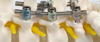
In young children (including infants), due to subluxation of the first cervical vertebra, a number of diseases sometimes develop, including cerebral palsy and paralysis, which leads to disability. Sometimes, due to road accidents and injuries, retrolisthesis occurs - the simultaneous displacement of several vertebrae relative to the position of the spinal column. In such a situation, step-by-step recommendations for restoring the health of the spine will be given by an atlas specialist from our center. You may first have to undergo surgery, conservative treatment of the neck, and wear a Shants orthopedic collar for some time.
Preventive measures
To prevent subluxation of the cervical vertebrae, you need to avoid increased stress on this area. Therefore, strength sports should be abandoned if a person does not follow safety precautions. For the same reason, it is forbidden to sit in an incorrect position for a long time. Parents must hold the child correctly, support his head, and ensure that his back is level. Older children at risk should avoid carrying heavy objects and sitting at a desk for long periods of time.
Patients need to maintain moderate physical activity, try to avoid injuries, observe working conditions, eat right, and get proper rest.
Doctors recommend performing special exercises that will help strengthen the muscles of the neck and upper back. If performed regularly, the risk of subluxation will decrease.
How to detect if the atlas is dislocated
Since all people could be exposed to birth trauma, every person needs a preventive examination to detect displacement. Some patients are diagnosed with signs of old diseases, the main cause of which is a displaced atlas. Therefore, the sooner the diagnosis is made and the atlas is reduced, the better for the person affected by the displacement of the vertebra.
When it is possible to eliminate the pathology, rapid sanogenesis (self-healing) of the body begins and the functioning of all its systems begins. But this does not mean that a victim of displacement can be cured of all the symptoms of existing diseases only by editing C1.
Forecast
Most atlas dislocations/subluxations have a favorable outcome. An unfavorable prognosis occurs with instability of the articulation, which cannot be eliminated, or with vertical displacement of the axis tooth towards the occipital foramen. In this case, there is a risk of damage to the medulla oblongata and surgical intervention is necessary. Patients develop myelopathy . With timely jaw adjustment and proper rehabilitation, the prognosis is favorable. Sometimes there are repeated subluxations and joint stiffness .
How to diagnose subluxation of the upper vertebra?
Diagnosis of C1 displacement at the Atlas-Standard clinic is made on the basis of:
- Kinesiological tests (muscle tone and the relationship between their tension and the functioning of internal organs are assessed). Using this method, you can detect whether there are malfunctions in the craniovertebral zone (cervical spine), and most importantly, the tests help to see the consequences of these malfunctions.
- X-rays, CT (computed tomography) and MRI (magnetic resonance imaging) demonstrate statistical changes in the vertebra. They are done to control the condition of the spinal column.
MRI picture: subluxation of the first cervical vertebra (atlas).
What specific methods will be selected for correcting rotational subluxation will be clarified during a consultation with a specialist. Note that hardware research methods can display the structure of the joint, but cannot demonstrate the consequences of displacement. An example would be a paralyzed limb where the cause of paralysis is a subluxation of C1. This is why special kinesiological tests are needed.
The same tests are performed after correction of the C1 misalignment to ensure that there is no misalignment and the position of the vertebrae allows the body to function normally.
Treatment
If a subluxation of the neck is suspected, the patient should be given first aid and then transported to the hospital. Further treatment of the pathology is carried out in a hospital.
If a cervical vertebra is subluxated, the victim’s neck must be secured.
If you notice symptoms of displacement (pain, forced position of the head, swelling), then fix the victim’s head and neck to avoid further deformation of the spine. As a rule, a special collar or splint is used for this. If there are no orthopedic devices, then carefully place the patient on a hard board, secure his head with bandages or rags, while turning over or raising his head is prohibited. Then the patient is transported to a medical facility, where he will receive qualified care.
You can use a homemade collar to secure your neck. It is made from several balls of cotton wool, which are tightly wrapped in gauze. You need to set it up very carefully, you need to make sure that the device securely fixes the neck, but does not impair breathing.
Carefully. Do not try to reduce a subluxation yourself, even if it seems to you that the victim needs it. Such manipulation can only be carried out by a doctor in a hospital setting.
Reduction must be carried out as soon as possible, otherwise swelling will appear in the damaged area. Then it will be more difficult to return the vertebrae to their normal position.
To reduce a subluxated cervical vertebra, a Gleason loop is used. The procedure goes according to this plan:
- The victim is placed on the couch, and a low cushion is placed under the shoulders.
- A Glisson loop is placed on the head and secured with clasps under the chin.
- The doctor takes the loop and does a pull, and then turns his head. You can also gradually realign the vertebra using a small load.
- During realignment, you can hear a characteristic crunch, which indicates that the vertebra has fallen into place. After this, the patient’s pain decreases, neck mobility improves, and relief occurs.
After the procedure is completed, the doctor prescribes a repeat x-ray. The study will help confirm that the normal position of the vertebra has been restored.
After subluxation of the cervical vertebra, the ligaments are damaged, so there is a risk of relapse. To avoid this, the patient should wear a Shants collar, which will relieve stress from the cervical spine and ensure its rapid recovery. The duration of use of the orthopedic device is from 2 weeks to 3 months. While wearing it, it is forbidden to perform even minimal head movements.
Next, you need to be treated using conservative methods:
- taking medications;
- physiotherapy;
- massage;
- Exercise therapy.
To relieve back muscle tension, the patient is prescribed Tolperisone. Preparations based on B vitamins normalize blood circulation and stimulate metabolic processes in nerve structures that are damaged during subluxation. To improve microcirculation, the patient is prescribed Pentoxifylline.
Complex therapy is complemented by massage. Sessions are indicated from the first days after injury, they are carried out in a gentle manner. After a course of procedures, muscle tone is normalized, blood flow and tissue trophism in the damaged area are improved.
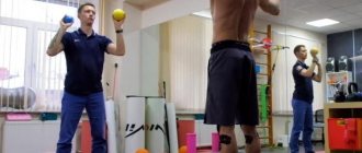
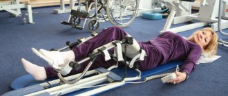
Therapeutic exercises are indicated almost immediately after realignment of the vertebrae. At first, the patient trains only the shoulders and upper arms, and after removing the neck brace, the complex is supplemented with exercises for the neck. Physical education is indicated until complete recovery; it helps strengthen muscles and relieve stress on the spine.
Sports activities are alternated with physiotherapeutic procedures. Most often during rehabilitation, electrophoresis with anesthetics (novocaine, lidocaine), ultrasound, ultra-high frequency therapy, and thermal procedures are prescribed.
No ads 3
Unique diagnosis of atlas position
In almost all patients suffering from various diseases of the spinal column and spinal cord, seemingly not directly related to subluxation (headaches, migraines, cervical osteochondrosis, dizziness, back pain, lumbar pain, etc.), dislocation (rotation) is detected during diagnosis ) first cervical vertebra. Sometimes people have difficulty turning their heads since childhood: this is also a consequence of vertebral displacement. Correcting the situation (subluxation) simultaneously solves many health problems.
How subluxation is eliminated using the Swiss technique of adjusting the atlas
Setting C1 to a normal state is achieved through vibration effects on the muscles of the body that stabilize and support the vertebra, which have memory.
When vibrating at a certain frequency, which is produced by a special device developed in Switzerland, the muscles relax. The atlas falls into place, and the muscles are fixed in a new, correct position. First, a free initial examination is carried out and a diagnosis is made. The procedure is performed by a doctor specially trained in the nuances of the technique. You can read more about the method on the main page of our official website.
After undergoing the procedure, the patient is prescribed a special program to restore the functions of the musculoskeletal system, nervous and cardiovascular systems. Vitamin complexes are also prescribed and preventive recommendations are given depending on the initial condition and the presence of specific diseases.
General information
A dislocation is a displacement of the articular surfaces of two bones relative to each other, different from the anatomical norm, which is accompanied by a pronounced violation of the configuration of the joint and its function in the form of limited mobility.
Its variety is the condition of subluxation, in which there is a relatively slight displacement of the articular surfaces relative to each other while maintaining the point of contact and, in some cases, there is the possibility of spontaneous reduction. In most cases, subluxations are acquired, while congenital subluxations are relatively rare. Acquired subluxations are formed in the presence of risk factors such as:
- Traumatic effects (sharp jerks, falls, prolonged hanging, impacts) caused by injury or high physical exertion.
- Various diseases of the musculoskeletal system ( osteomyelitis , dysplasia /joint instability, poliomyelitis , osteochondrosis , etc.).
The most susceptible to subluxation are joints with a large range of motion, which have maximum mobility in various planes. Therefore, subluxation most often occurs in the joints of the upper/lower extremities (ankle joints, toes, subluxation of the patella), spine, jaw, dislocation of the upper extremities (shoulder and elbow joint).
Subluxations can be combined with rupture/sprain of ligaments and muscles, rupture of the joint capsule, damage to blood vessels and nerve fibers. Let's take a more complete look at the subluxation of the cervical vertebra (the so-called subluxation of the atlas), and also briefly touch on the most common subluxations of the shoulder, clavicle and joints of the upper (elbow) and lower limb - subluxation of the knee (patella), hip joint and ankle.
Subluxation of the cervical vertebra (subluxation of the atlas)
The craniovertebral region is the transition point of the spinal column into the skull and includes the upper cervical vertebrae (atlas, axis), as well as the basal part of the occipital bone. This vertebra performs an extremely important function, holding the cranium with its contents to itself.
The atlas is a slightly flattened ring-shaped bone formation, which is essentially a bone meniscus, a kind of washer between the lateral masses of the axis and the condyles of the occipital bone, providing the transition from flexion-extension to rotational movements. The wedge-shaped lateral masses of the atlas are connected by the anterior/posterior arches into a single ring-shaped formation and are connected by a powerful transverse ligament, which provides additional stability to the bone ring (Fig. below).
The atlas connects from above with the help of tiny joints to the occipital bones, and from below to the axis (second cervical vertebra). The weight of the head is transmitted through the condyles of the occipital bone to the lateral masses of C1 and is further transmitted to the upper articular facets of the axis, where it is distributed predominantly to the anterior structures (up to 80%) and to a much lesser extent to the posterior structures of C2.
The lateral masses of the atlas/body of the axis stabilize axial loads; shear loads - anterior arch of the atlas, tooth/lower articular processes of the axis; rotational, flexion-extension and dynamic shear load - ligamentous apparatus: the transverse ligament prevents anterior displacement of the atlas; The pterygoid ligaments stabilize axial rotation of the head, during which there are no bone restrictions on movement in the joints. The C1-C2 stabilization ligaments are ligaments of tension, which provide secondary stability in the joint. The primary/secondary stabilizers for the atlantoaxial and atlanto-occipital joints are different. When the function of different ligaments is impaired, the stability of the joint is affected differently.
One type of pathology of the craniovertebral segment is atlantoaxial instability, manifested by subluxation. There are three main types of atlantoaxial instability: distraction , flexion-extension and rotation , which can be isolated or complex in various combinations. Instability in the atlantoaxial joint is caused by damage to the structures of the craniocervical junction, mainly damage to the transverse ligament.
The most common occurrence in practice is rotational subluxation of the C1 cervical vertebra (Fig. below), which is a pathological asymmetrical displacement to the right/left of the articular surfaces of the ring-shaped C1 vertebra relative to the axis of the C2 vertebra (left-sided/right-sided subluxation).
Rotational subluxation of the atlas is a common injury of the cervical spine, accounting for about 30% of the total pathology of the cervical spine. Subluxations of the C1/C2 vertebrae are most often caused by isolated trauma. Rotational subluxations of the atlas are detected much less frequently in adults than in children.
A relatively rare occurrence is subluxation according to Kovacs (habitual subluxation), which manifests itself in the form of instability of one/several segments of the spine: when the head is tilted, the process of the underlying vertebra slips and layers on the posterior-inferior corner of the body of the overlying vertebra, and when the neck is extended, it returns to its original position. position.
In case of rotational atlantoaxial subluxations, spinal cord injuries are extremely rare, and compression/damage to the vertebral artery is practically never encountered.
The most common manifestation is dysesthesia (unpleasant sensations) caused by damage to the roots of the C2 vertebra. As a rule, after careful reduction of the C1-C2 vertebrae, restoration of all impaired spinal functions is observed.
Subluxation of the hip joint
Subluxation of the hip joint is a condition in which the surface of the articular head of the femur is displaced partially outward and upward in relation to the acetabulum, without leaving the limbus while maintaining contact between the elements of the joint. Due to the stretching of the capsule/teres ligament, as well as the upward displacement of the limbus, the support function is weakened, which causes the possibility of upward/partial displacement of the femoral head (Fig. below).
This pathology is based on joint dysplasia , caused by insufficiency of connective tissue, the development of which is based on mutations in different combinations of various genes and adverse environmental influences. Anatomical manifestations are underdevelopment of the joint: hypoplasia /flattening of the acetabulum, slow development of the bursal-ligamentous apparatus of the joint/head, which contributes to increased mobility of the joint.
Subluxation of the knee joint
The most common pathology of this joint is external subluxation of the patella (synonymous with lateral patellar hyperpressure syndrome ), which is most often caused by high position of the patella, ligament weakness, hypoplasia of the lateral femoral condyle, valgus curvature of the knee , outward rotation of the femur/inward rotation of the tibia, and less commonly - congenital insufficiency/improper formation of the femoral-patellar joint. This is a common pathology of the patellofemoral joint, especially among children, adolescents and athletes (Fig. below).
It develops against the background of an imbalance between the lateral/medial stabilizers of the patella, congruent relationships between the articular surfaces of the patella and femoral condyles. This pathology is characterized by a redistribution of specific pressure in different zones of the articular surfaces, which contributes to overload of its lateral parts.
Since with long-term subluxation of the patella, the cartilage that covers the condyles/patella begins to suffer, there is a high risk of developing patellofemoral arthrosis . In addition, with long-term subluxation of the patella, synovitis (fluid accumulation in the joint) may develop, which will manifest itself as swelling of the knee joint.
Subluxation of the ankle joint
Subluxations of the talus are among the most common cases of intra-articular injuries of the ankle joint.
This pathology is most typical for a period of life with high activity (20-45 years). Depending on the nature of the translational movement of the talus (along, around) the longitudinal axis of the tibia/transmalleolar axis, 12 variants of subluxations are distinguished: anterior/posterior, upper/lower subluxations, internal/external, plantar/dorsal, supination/pronation, etc.
At the same time, each type of dislocation is characterized by its own mechanism of injury, typical anatomical changes in bones, ligaments, muscles, x-ray picture and specific treatment.
Subluxation of the ankle joint often occurs when the foot is twisted, loss of balance/fall, the foot touching a physical barrier, jumping, in the presence of ankle hypermobility, systemic connective tissue diseases, etc.
Subluxation of the shoulder joint
The shoulder joint is one of the joints that has the maximum range of motion in various planes.
Shoulder subluxation is a standing condition in which displacement of the humerus from the glenoid cavity of the scapula (forward/backward) occurs during movements associated with external rotation/abduction of the arm during active sports or awkward movements in everyday life. The shoulder joint is characterized by frequent development of articular instability, which is caused by degradation of the articular cartilage of the humeral head and the formation of a Hill-Sachs defect . As a rule, the patient eliminates dislocations with a certain movement independently.
Jaw subluxation
Caused by weakening of the temporomandibular joint ligaments. It is characterized by partial exit of the articular process of the jaw from the articular cavity, accompanied by pain/inability to close the mouth. The development of dislocations of the lower jaw is caused mainly by contraction of the muscles that open the mouth during various physiological acts during which the articular heads protrude forward and, stretching the capsules, partially emerge from the articular sockets (with excessive opening of the mouth, yawning, laughing, vomiting, less often with a blow to the chin, falling ). They are more common in females, which is due to the smaller size of the articular tubercle/depth of the articular fossa and the relatively weak ligamentous apparatus.
Depending on the cause, the following subluxations are distinguished:
- traumatic, caused by forced forced movement in a joint (excessive opening of the mouth, blow, falling on the chin, yawning, screaming, dental procedures);
- habitual, the development of which is based on stretching of the articular capsule/insufficient height of the articular tubercle, found mainly in older people, with rheumatism, deforming arthrosis of the lower jaw joint, gout, epilepsy.
The difference between the Swiss method and traditional methods of treatment
Since the craniovertebral (upper cervical) zone of the spine is a “risk zone” - the most dangerous for exposure, doctors and osteopaths usually try to use conservative treatment: gentle traction and a system of physical exercises to strengthen the muscles, as well as manual therapy, and so on. However, these osteopathic methods in the vast majority of cases do not provide a complete return of C1 to its natural position.
Correction of C1 using the Swiss technique is based on a discovery in the field of physiology that allows you to overcome muscle memory. If the effect is carried out not with the help of a device that vibrates for a sufficiently long time, but with the help of manual therapy, then the result from realigning the vertebra will not last long at best: sooner or later instability of the first cervical vertebra will appear, and the atlas will again take an incorrect position in the atlantoaxial joint In the worst case, there will be no result at all, or even greater harm will be caused to the musculoskeletal system and the entire body.
If you want to learn more about how the reduction of the atlas vertebra is carried out and what the effect of the method is, then you can read and watch detailed reviews and video reviews about it.
How to make an appointment with an atlas specialist
Leave a request for an appointment with a doctor using a special form.
A specialist will contact you as soon as possible. If you live in Moscow or St. Petersburg, you can agree on a visit time that is convenient for you. If you are in another city in Russia, doctors from the Atlas-Standard clinic will be able to travel to your city according to the schedule established on the website (updated every week).
Promotions and discounts are regularly offered to residents of certain cities. Check with the clinic specialists for information.