Symptoms of cervical osteochondrosis
Symptoms of cervical osteochondrosis are usually divided into radicular and reflex.
Radicular symptoms of cervical osteochondrosis
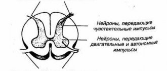
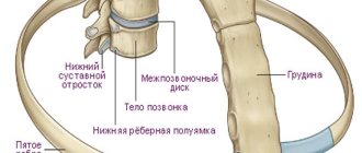
There are many nerves coming out of the spine. They are called spinal nerves. Each such nerve gradually branches and follows a specific area of the body with clearly defined boundaries. This area is called the zone of segmental innervation. Each vertebra, disc, nerve and zone are numbered, strictly corresponding to each other. If a nerve is exposed, then symptoms will appear in the zone of segmental innervation corresponding to a given nerve, and not just anywhere - in an arbitrary place.
Radicular symptoms of cervical osteochondrosis include:
- Decreased or lost reflexes;
- Muscle weakness;
- Impaired sensitivity;
- Radicular pain.
Not all areas of the cervical spine are equally susceptible to pathology. The most mobile segments are most often affected: C3–C4, C4–C5, C5–C6 and C6–C7. According to the principle - “More movements - more wear.”
Innervation zones of the cervical segments
Osteochondrosis C3–C4
- indicates that the 3rd and 4th cervical vertebrae and the disc between them are affected. In this case, the 4th spinal nerve, which goes to the neck, is affected. The main signs are decreased sensitivity along the entire circumference of the neck. There is discomfort, and sometimes pain, in this area.
Osteochondrosis C4–C5
- affects the 5th spinal nerve. Pain from the neck extends along the shoulder girdle to the upper front of the shoulder. Sensitivity decreases along the outer surface of the shoulder. Numbness occurs in the same area. The deltoid muscle definitely weakens.
Osteochondrosis C5–C6
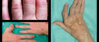
- the most common diagnosis. In this case, the 6th spinal nerve is affected. This manifests itself as pain from the biceps to the thumb and index finger along the outer surface of the arm. Sensitivity decreases, numbness or “goose bumps” appear in the same area. And biceps weakness inevitably occurs.
Osteochondrosis C6–C7
- affects the 7th spinal nerve. Pain occurs in the triceps area and at the back of the arm. Sensitivity decreases in the area of the middle and ring fingers. Numbness and “pins and needles” may appear in the same area. Definitely triceps weakness.
Reflex symptoms of cervical osteochondrosis
Pain in the neck, back of the head, and collar area—more often in the morning. Difficulty in movement, crunch in the neck. Neck muscle tension. Constant aching pain. Headache. Dizziness. Intracranial pressure. Acute pain. Weakness in the arms, numbness, pins and needles and goosebumps. Pain in the shoulders, sometimes radiating under the shoulder blade. Burning between the shoulder blades. A burning sensation in the heart area resembles angina pectoris. Pain in the left side of the chest can sometimes radiate to the left arm. Numbness of the hands or fingers. “Lump” in the area of the seventh cervical vertebra. Poor dental condition. Transient visual disturbances, temporary darkening or “floaters” in the eyes. Decreased vision. Noise or ringing in the ears. Hearing impairment. Nausea, sometimes leading to vomiting. Pressure surges. Fainting or pre-syncope. Loss of consciousness. Feeling of a lump in the throat, problems with swallowing. Sore throat. Weakening or hoarseness of the voice. Feeling short of air. Sleep disturbances, frequent insomnia. Snoring is a consequence of neck muscle tension. Feeling like you didn't get enough sleep. It is difficult to move, especially in the morning. Coordination of movements is impaired - this affects the gait. General weakness, weakness. Irritability. Fast fatiguability.
Symptoms of cervical osteochondrosis:
- depend on the stage of osteochondrosis
- worsens when tilting and turning the head
- more often appear after 35–45 years
- in women they occur approximately 3 times more often than in men
You, of course, noticed that the radicular symptoms are defined quite clearly, while the reflex symptoms are very vague and non-specific. And as you know, everything that does not have clear definitions serves as a convenient cover for professional helplessness. This applies to both reflex symptoms and such a favorite concept among doctors as “age-related changes.” Surely many of you are familiar with the situation when the doctor explained the problem as “reflex” or “age-related” processes. Most people at such moments rightly believe that the doctor simply cannot figure out what is happening and is trying to veil his incompetence in the fog of these “magic words.”
At one time there was a popular phrase: “Every accident has a name, surname and position.” Every disease has its own unique symptoms. And it is the doctor’s duty to know them clearly. And then there will be no need to cast a fog and blame cervical osteochondrosis for everything. Now you understand how important it is to find an experienced and knowledgeable doctor. Both the correct diagnosis and treatment results will depend on this.
When choosing a clinic, the main thing is to get to an experienced and knowledgeable doctor.
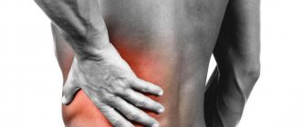
Lumbar radiculopathy
Radiculitis is usually associated with radicular syndrome localized in the lumbosacral spine. Lumbar radiculopathy clinic:
- acute onset;
- intense pain in the lumbar spine;
- the pain intensifies in a sitting position, especially in a soft chair, bending, turning, coughing and sneezing, pressing on the jugular veins;
- pain is reduced in a position on the side with a bent leg, at rest;
- triggers of pain in the buttocks, calf muscle, thigh;
- the body is slightly bent (forced position);
- tension of the paravertebral muscles in the lumbar region;
- history of lumbar pain;
- severe limitation of movements (bending forward);
- functional scoliosis (curvature to the side);
- pain can radiate along the back of the thigh, groin, back of the lower leg, buttock;
- paresthesia or numbness along the back surface of the lower limb, including the foot;
- restriction of movements in the hip joint;
- impaired functioning of the feet (rising on the toes, flexing the ankle joint;
- wasting of the muscles of the pelvic girdle and free lower limb [1,6].
Rice. 7. Lumbar radiculopathy
Treatment of cervical osteochondrosis
As you understand, osteochondrosis is a real “tangle” of symptoms, which, by unraveling, the doctor will relieve you of pain and torment. But it is not possible to eliminate changes in the vertebrae and discs. Therefore, the words “treatment of osteochondrosis” must be understood correctly. If you are interested in eliminating pain and other suffering, then yes, it is quite possible. And if you conduct an academic discussion on the topic of returning the vertebrae and discs to their original appearance, “like a newborn child,” then no, the past cannot be returned. You need to be realistic, and then you won’t fall for scammers.
- Don't fall for scammers!
- It is impossible to return the vertebrae and discs to their original appearance!
What method of treatment is considered the main one?
Gentle manual therapy is the main type of treatment for cervical osteochondrosis. It’s like an antibiotic for pneumonia—you can’t do without it. The remaining types - massage, medications, physiotherapy and exercise therapy - are auxiliary.
How does gentle manual therapy work?
The nutrition of the discs is directly related to the muscles surrounding the cervical vertebrae. In addition, the neck muscles themselves are one of the constituent causes of pain in osteochondrosis of the cervical spine. Gentle manual therapy is a special method that allows you to return muscles to their natural physiology, eliminate spasms, muscle tension and improve nutrition of the discs.
Intervertebral discs are the only part of the body that does not have blood vessels and is nourished by the proper functioning of the muscles.
In addition, when performing treatment using hands, the chiropractor:
- will relieve the load from the affected vertebrae and discs and distribute it correctly;
- will relax the muscles and help them return to normal.
Thereby:
- relieves the patient of clamps;
- improve disk power supply;
- will restore the motor functions of the body;
- normalizes blood circulation.
Manual influence mobilizes the internal forces of the body and triggers self-healing mechanisms. The treatment is absolutely safe.
Classification
The classification of radicular syndrome takes into account the topography of the pinched nerves (cervical, thoracic, lumbar, etc.).
Depending on the nature of the lesion, there are three main types of radiculopathy:
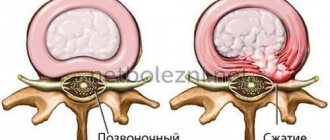
- discogenic - the most common type, occurs with disc hernias, with a long course there is a high risk of paralysis;
- vertebrogenic - pinched nerve in stenotic foraminal openings, requires urgent medical attention;
- mixed - the nerve is pinched due to deformation of the intervertebral disc and osteophytes on the vertebral bodies.
Prevention of cervical osteochondrosis
To avoid relapses, create comfortable conditions for sleeping and working. Watch your weight and eat right. Keep up your physical activity. But the main thing is not to neglect your health and not to skimp on it. Don't let things take their course. After recovery, try to do at least one maintenance session of gentle manual therapy once every three to six months - this will reduce risk factors. Don’t forget, advanced osteochondrosis leads to complications - protrusion and disc herniation. Remember: your health comes first!
Advanced osteochondrosis leads to complications - protrusion and disc herniation.
Compression lesions of the cauda equina
The cauda equina is an anatomical formation in the lumbosacral region, corresponding to the end of the spinal cord and the exit of the axon bundle (they form the cauda equina).
Clinical picture:
- severe pain syndrome;
- foot paresis;
- hypotrophy of the gluteal and calf muscles, foot muscles;
- decreased reflexes;
- decreased sensitivity in the “rider's pants” area;
- delayed urinary passage followed by incontinence;
- encapresis (fecal incontinence) and enuresis (urinary incontinence);
- paresthesia in the pelvic area;
- paresis of the lower extremities [1,6].
Rice. 8. Cauda equina compression syndrome
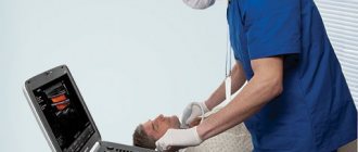
Shock wave therapy in the treatment of radicular syndrome
Extracorporeal shock wave therapy (ESWT) for intense pain syndromes has a convincing clinical effect. For therapeutic purposes, a focused acoustic wave with a frequency of 16-25 Hz is used, which improves tissue trophism, reduces muscle tone, and stops the transmission of impulses from nociceptors to the brain [4].
A change in the permeability of cell membranes of the nociceptive system promotes the supply of endorphins, and active neoangiogenesis improves blood supply and resolves inflammatory edema. In fact, the anti-inflammatory effect of ESWT also relieves pain [4,8]. Biochemical changes are activated due to the production of prostaglandin, interleukin and cyclooxygenase [13]. The cavitation effect helps to inhibit the breakdown of inflammatory mediators (free radicals, nitric oxide, endothelial HSFR), which provides an anti-inflammatory effect [9].
Rice. 9. ESWT procedure
Contraindications to the use of ESWT:
- purulent inflammation;
- tumors in the affected area;
- coagulopathies;
- mental disorders;
- pregnancy;
- artificial pacemaker [4,8].
The method allows for relatively quick pain relief. After the first session, patients note subjective improvement.
ESWT as a method of pain relief: review of scientific literature
Among the indications for the use of extracorporeal shock wave therapy, the leading place is occupied by pain, with muscle-joint pain being the most sensitive to the effects. L.M. Shteh et al described the effects of ESWT on pain trigger points. Of the 86 patients, at the end of the course, consisting of five weekly procedures, 80% noted significant improvement [14].
A massive study of 650 units demonstrated pain relief and increased spinal mobility in myofascial pain syndromes in 96% of cases [2].
Zuev et al described the use of ESWT in combination with osteopathy in 120 patients with myofascial pain syndrome. The method allowed maintaining the clinical effect throughout the year in 77.1%, which exceeds the same indicator when using isolated ESWT by 24.3%, and when using isolated osteopathy – by 14.9% [7].
E.V. worked with the same category of patients. Kostenko. She prescribed shock wave therapy to patients in combination with Tizanidine. Of the 110 patients, in 90% of cases the intensity of the pain syndrome regressed to 3.3 ± 0.9 points on the VAS scale, muscle tone normalized, and range of motion in the spine expanded. The author performed from three to eight procedures using a stable-labile technique with a frequency of 4-6 Hz, a power of 0.037–0.399 mJ/mm2, and the daily dose of Serdalut was 8 mg [8].
Chistov and Serov influenced trigger zones (or trigger points - TT) of chronic pain syndrome, and the diagnosis of TT is optimally performed using ESWT. The device allows you to treat several pain and trigger zones at different levels, since its therapeutic depth in the form of a “candle flame” reaches 125 mm. In this case, the first procedure ensures the extinction of the pain syndrome by 20%, then, after a slight exacerbation, the pain is completely relieved within a week after the course [1,13].
Thus, shock wave therapy, used alone or in combination with drug or non-pharmacological treatment, can relieve pain as the main manifestation of radicular syndrome
ESWT in the treatment of piriformis muscle syndrome: a review of the scientific literature
Piriformis syndrome complicates the course of lumbar vertebrogenic radiculopathy, manifesting itself with inflammation, swelling, reflex hypertonicity of the piriformis muscle and pain. As a result of compression of the sciatic nerve, urinary dysfunction occurs.
A team of authors from the Ural State University of Physical Culture and SONAR LLC presented a description of 128 patients with piriformis syndrome who received shock wave therapy from the STORZ MEDICAL Duolith SD device on the buttocks and paravertebrally. As a result of three procedures, not only subjective and clinical improvement was noted, but also an improvement in laboratory parameters (increased antioxidant activity and decreased lipoperoxides) [9,11].
Min Kyun Sohn and colleagues described a convincing antispastic effect of ESWT for gastrocnemius syndrome in patients who suffered stroke in a small sample of 20 units. The described results are extrapolated to the use of shock wave therapy for gastrocnemius syndrome, which develops with lumbar radiculopathy [15].
Sumnaya D.B. et al. presented the physiological rationale for the complex alternating use of ESWT and laser therapy (LLLT) with a semiconductor laser with a wavelength of 0.89 μm in combination with exercise therapy and manual correction in patients with piriformis muscle syndrome back in 2021. It was possible to achieve not only pain relief, but also normalization of muscle tone [11].
Rice. 10. Diagram of piriformis syndrome