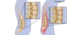
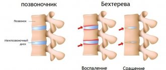
Ankylosing spondylitis (the pathology is named after the famous Russian physician V.M. Bekhterev, who first described it in 1892) is a progressive chronic inflammatory disease that primarily affects the spine, sacroiliac joints, and paravertebral soft tissues. As a result of this disease, irreversible changes appear in the structures of the musculoskeletal system, such as fusion of articular surfaces, leading to the development of ankylosis.
The basis for the occurrence of BD is the manifestation of aggressiveness of the immune system against the cells of one’s own joints and ligaments (an inadequate immune response occurs, as it were). In this case, the body's immune system mistakenly perceives some of its tissues as foreign, and this is precisely the cause of immune aggression.
The prevalence of ankylosing spondylitis is relatively low, approximately 4–5 people per 1000. Men are affected 5–7 times more often than women. The disease begins at a young or middle active age (16–40 years).
Most importantly, spondyloarthritis is NOT EQUAL to ankylosing spondylitis!
Spondyloarthritis is a group of inflammatory diseases
characterized by damage to the spine and/or joints. They are grouped according to the mechanisms of their development and similar clinical manifestations. This group is divided into two subgroups - axial and peripheral spondyloarthritis. The difference is that the first affects only the sacroiliac joints and/or spine, while the second can also affect the joints of the upper and lower extremities.
In addition, a subgroup of axial spondylitis is divided into ankylosing spondylitis (this is ankylosing spondylitis) and non-radiographic axial spondylitis. The difference between them is the presence/absence of damage to the sacroiliac joints on radiography. X-ray changes occur very slowly, sometimes it takes 7-8 years for signs of “fusion” to appear, so there is still heated debate about whether these two forms are stages of the same process, or different diseases from the same group.
For a better understanding, below we will talk about the group of axial spondyloarthritis (AS).
2. Clinical picture and complications of ankylosing spondylitis
The onset of the disease does not have characteristic symptoms. Mild aching pain and stiffness after a night's sleep in the lower back and buttocks can be perceived by a person as a consequence of physical activity. Symptoms go away with physical activity. Over the course of several months, the soreness may become permanent. At rest (at night), the pain can be so unbearable that it forces a person to get up and move in the middle of the night in order to get relief.
Sometimes the disease initially manifests itself as pain in the chest, which is especially noticeable during coughing and sneezing.
Pain syndrome in the limbs, hip and shoulder joints at the initial stage is less common and is asymmetrical in nature.
Generalization of the process can cause fever, weight loss, weakness, and nocturnal hyperhidrosis.
The vertebral localization of the first foci of inflammation is typical for adult patients. In the puberty period, ankylosing spondylitis most often has the character of peripheral arthritis and enthesopathies.
The gradual loss of motor function of the spine leads to its complete immobility. In this case, the back remains completely flat or, conversely, takes on a curved kyphotic shape.
Due to damage to the costovertebral and sternocostal joints, breathing functions largely fall on the diaphragm, which causes a transition to the “abdominal” type of breathing. The most dangerous complication of ASA, spinal fracture, occurs quite often. Less common is the development of pulmonary and cardiac disorders, which are characteristic only of the later stages of ankylosing spondylitis.
In general, ASA rarely leads to death, but in most cases it causes disability, characterized by complete loss of ability to work.
Visit our Rheumatology page
Causes of the disease
The exact cause of the disease is unknown.
Spondyloarthritis, unlike rheumatoid arthritis, is less of an autoimmune disease and more of a hyperinflammatory disease. They rarely cause systemic inflammatory reactions, but affect certain organs and tissues - joints, skin, intestines, eyes. One of the key factors in the development of AS is the presence of the HLA-B27 antigen. Despite the 40-year history of its study, the role of the antigen remains not entirely clear. According to one theory, the process that triggers inflammation may be microbes in which some of the genes are so similar to the genes of the patient - a carrier of HLA B-27 that the human body “confuses” them and begins to work against its own.
However, when the disease has already manifested itself, do not look for these mysterious microbes - most likely the body has long ago gotten rid of them, and the inflammation will continue.
Another theory is based on the possibility of an abnormal structure of HLA-B27, which itself activates a full-blown immune response. Any factor (trauma, infection) can be a factor—a “trigger”—to activate the “improper functioning” of this antigen. Be that as it may, this notorious HLA B-27 can be detected in 95% of patients with AS.
And then a cascade of reactions unfolds: T-lymphocytes, the cells of the immune system, are sequentially activated and the formation of protein molecules, “inflammatory mediators” (cytokines), increases. They lead to changes characteristic of the entire group of spondyloarthritis and AS in particular. Unlike rheumatoid arthritis, where the main mechanism of joint destruction is the formation of erosions—“eaten away”—of cartilage and bone destruction, in AS the inflammation process ends with bone proliferation—the formation of new “excess” bone tissue. The result is ankylosis (fusion) of the joints and ossification (ossification) of the spinal ligaments, resulting in immobility.
Men are affected more often, with onset occurring before the age of 45 years. It is possible to develop AS in childhood with a transition to adulthood, which is usually quite severe.
Causes and mechanisms of development of ankylosing spondylitis
The etiology of BD has not yet been fully studied. But in recent years, it has been established that there is a genetic predisposition to this disease, caused by the presence of the HLA-B27 antigen, which is activated mainly in men after puberty. In this regard, their bodies begin to produce specific antibodies, the action of which is aimed at their own connective tissue elements involved in the formation of the ligamentous and articular apparatus. At the same time, inflammatory reactions are formed in the latter, leading to the resorption (resorption) of cartilage with its replacement with fibrous (scar-like) tissue, followed by the deposition of calcium salts in it and subsequently the proliferation of bone tissue. As a result of this, first there is stiffness in the joints, then there is a limitation of movements in them and, finally, ankylosis develops, characterized by fusion of the articulating articular surfaces.
Initially, inflammation is most often observed in the sacroiliac joints, then in the intervertebral joints (connecting the vertebral bodies), and, gradually spreading higher and higher, is already localized in the costovertebral sections of the chest. Much less often, spondyloarthritis simultaneously occurs in large joint structures of the body - shoulder, knee, hip, and very rarely - in small joints of the feet or hands.
As the pathology progresses, the above areas of the human musculoskeletal system are “switched off” from movement, the increased or unusual load compensatory “falls” on the adjacent sections, which contributes to the appearance of degenerative changes in them (osteoporosis, osteochondrosis, spondylosis, spondyloarthritis, synovitis, myositis) . This certainly affects the well-being of patients and their physical activity.
So, for example, if ankylosis occurs in the costovertebral joints of the chest, then the range of its movements during breathing gradually decreases until it almost completely disappears, and then patients breathe only due to excursions of the diaphragm. This leads to shortness of breath at the slightest exertion and the formation of respiratory failure. One third of patients may experience, to one degree or another, damage to other structures of the body: for example, the development of myocarditis, valvular heart defects or aortitis (if the cardiovascular system is involved in the process), sometimes disturbances in the functioning of the organs of vision (uveitis, iridocyclitis, episcleritis), urinary tract (nephritis, cystitis), etc.
Symptoms
The most typical symptom of the disease is pain in the lower back (in the area of the sacrum and on the sides of it) of an inflammatory nature, i.e.
occurring early in the morning or after standing still. The pain can be subtle, like discomfort or a feeling of heaviness, often accompanied by morning stiffness (stiffness), and can radiate to the buttocks, but very often the pain forces the patient to wake up at 4-5 o’clock in the morning.
Symptoms usually progress very slowly (years or even decades), and patients are often observed with a diagnosis of osteochondrosis. The doctor or patient should be alert to the persistent nature of the pain, young age (up to 45 years) and lack of improvement after rest.
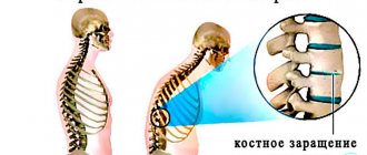
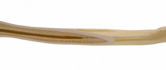
After affecting the sacroiliac joints, AS next affects the vertebrae. Usually the process goes “bottom up”, but less often there may be a different order. In this case, inflammatory changes in the vertebral bodies are first observed, and then intervertebral “bone bridges”—syndesmophytes—develop. Together with calcification of the spinal ligaments, this creates a characteristic “bamboo stick” picture on the x-ray, and during an external examination, the doctor observes the “petitioner’s pose”: the vertebral curves are smoothed, the patient’s head is tilted forward, lateral movements in the spine are possible only by turning the entire torso. The rheumatologist can see such a patient “from afar”, but, unfortunately, at this stage the treatment options are extremely limited. Therefore, modern diagnostic criteria are aimed at the earliest possible diagnosis of AS.
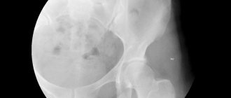
The “classical” instrumental study is an X-ray of the pelvis, which reveals bilateral sacroiliitis - inflammation of the sacroiliac joints. But symptoms on X-rays appear no earlier than after 4-5 years, so for early diagnosis, as the key to effective treatment, the MRI method is used. If sacroiliitis is detected on MRI, but not on radiography, it is called non-radiological. The main symptom of sacroiliitis is swelling of the bone marrow in the area adjacent to the sacroiliac joints.
The laboratory criterion for AS is the HLA-B27 antigen. Remember that, unlike MRI, this is not a sign of a disease, but a marker of PREDISPOSITION to it. Having HLA B-27 is not a death sentence! In general, in the population, HLA B-27 can be found in 8-15% of the population, and only every twentieth of its carriers will have any manifestations associated with this gene.
Other traditional inflammatory changes - an increase in ESR and C-reactive protein - can be observed in only 30% of patients with AS, so normal readings of these tests do not exclude or confirm the presence of the disease.
Unfortunately, the presence of HLA B-27 can be associated not only with AS, but also with damage to other organs. The classic reminder for a rheumatologist is “musculoskeletal system – skin – eyes – intestines.” And how this mosaic will develop in which of the patients is unknown. And since this gene is inherited, one family member may have skin manifestations, while another, for example, may have a combination of AS and intestinal lesions.
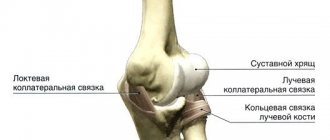
In addition to damage to the spine, pathology of the musculoskeletal system associated with HLA B-27 can manifest itself as so-called “enthesitis” - inflammation in the area where the tendon attaches to the bone. The favorite localization in this case is pain in the heel area (on the side or side of the foot) at the insertion of the heel ligament or Achilles tendon.
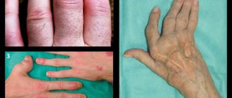
Another manifestation of the musculoskeletal system may be dactylitis (translated as “inflammation of the finger”), although only the tendons become inflamed, not the joints. In this case, the finger takes on the appearance of a sausage. Inflammation of the joints itself - arthritis: often asymmetrical with greater involvement of the lower extremities, is also a symptom that will direct the doctor to search for HLA B-27. By the way, if the “mosaic” in a particular patient develops in such a way that only the peripheral joints are inflamed without involving the spine, then the disease will acquire the name “peripheral spondyloarthritis.”
Sometimes the manifestation of the disease is anterior uveitis - inflammation of the anterior chamber of the eye. Symptoms: severe pain and redness of the eye, lacrimation, blurred vision (blurred vision), photophobia. A combination with AS may be intestinal damage, accompanied by abdominal pain, weak or loose stools, which may contain mucus or blood. A colonoscopy may diagnose Crohn's disease or ulcerative colitis. Another target organ for HLA B-27 may be the skin, so the doctor will certainly ask about the presence of psoriasis.
Thus, a combination of the patient’s complaints and medical history, conducting specific tests, and analyzing instrumental and laboratory data allows the doctor to diagnose AS and begin therapy.
Main clinical manifestations
One of the most common complaints of patients is pain in the lower back and buttocks, most often making itself felt at night and at rest, accompanied by morning stiffness, but subsiding after slight physical activity. Gradually, the pain “rises” higher, over time it is felt in the back and interscapular region, and later in the neck. Such people are characterized by a “petitioner pose,” which is associated with ankylosis of the interarticular processes of the cervical and upper thoracic spine, leading to the formation of kyphosis of varying degrees of severity.- In this case, shortness of breath and the inability to take a full deep breath through the entire chest may occur. This significantly reduces the quality of life of patients, as well as their ability to work and leads to disability.
- Heart pain, tachycardia or arrhythmia also often occur, which may be a symptom of myocarditis, valve damage or conduction disturbances (for example, atrioventricular block).
- In some cases, with pronounced changes in the spinal column, compression of the spinal cord roots may be observed, which is not only accompanied by severe pain or, conversely, the development of paresthesia (decreased skin sensitivity), but can also lead to disruption of the pelvic organs.
- Sometimes there may be pain in the places where the tendons attach to the bones (for example, to the spinous processes of the spine), the plantar fascia, the Achilles tendon to the calcaneal tubercle, the tendons of the oblique abdominal muscles to the iliac crests, etc.
- When the organs of vision are affected, patients may complain of pain in the eyes, intolerance to bright light, rapid fatigue of the eye muscles, and even increasing loss of vision.
The prognosis of the disease is currently relatively favorable, thanks to modern methods of drug therapy. However, the pathology tends to gradually progress, and 25-40 years after its onset, a decrease in mobility is possible, followed by loss of function of the intervertebral and sacroiliac joint structures, often leading to disability. This is also most relevant when at least one hip joint or the cervical spine is involved in the process (this leads to compression of the spinal cord with the likelihood of developing subluxation between the first and second cervical vertebrae). Kidney damage is less common; adequate, timely administration of non-steroidal anti-inflammatory drugs helps reduce the likelihood of nephropathy and other manifestations of ankylosing spondylitis.
Treatment
The patient needs to obtain as much information as possible from the doctor and strictly follow clinical recommendations.
Despite the fact that the list of groups of drugs that affect the course of the disease is small, it is quite possible to avoid ankylosis of the spine and the development of extra-articular complications from other organs and systems. A very important role is played by daily gymnastics, which is indicated for absolutely everyone, regardless of the activity of the disease and the development of ankylosis. Its goal is to slow down the progression, prevent and treat deformities, and improve overall well-being. The main exercises are stretching the spine and strengthening the paravertebral muscles. If we evaluate the arsenal of drugs proven against AS (Attention! Now we are talking only about the spine), then there are two main classes of drugs.
First of all, these are non-steroidal anti-inflammatory drugs (NSAIDs). They should be prescribed to the patient immediately after diagnosis, regardless of the stage of the disease, should be taken for a long time and without interruptions, and can not only reduce pain, but also slow down the progression of AS.
In other words, the fusion of vertebrae with each other occurs four times slower compared to those patients who used NSAIDs only “on demand”. The selection of NSAIDs is carried out by the attending physician, taking into account many factors, including the patient’s concomitant diseases, the characteristics of the drug’s purpose and its possible side effects.
Genetic engineering therapy
– these are TNF-α blockers (infliximab, golimumab, adalimumab, certolizumab pegol, etanercept) and antibodies to interleukin-17 (secukinumab, netakimab). Effective at any stage of AS development (but at an early stage more than at a late stage) both for reducing activity and for preventing deformities. As a rule, they are prescribed if the effect of NSAIDs is insufficient. Taking into account the availability of several drugs from the group of genetic engineering therapy, the doctor has the opportunity to “switch” the patient to another drug if the first one is ineffective.
In the event that extra-articular lesions of the eyes, intestines or skin occur, the doctor’s arsenal in terms of effective therapy expands even further.
Severe complications arise either when the patient presents late or when he ignores the recommended treatment.
Nowadays, therapy allows us to avoid the rapid and pronounced progression of ankylosing spondylitis and make the classic “supplicant pose” a thing of history.