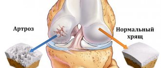
Deforming osteoarthritis is one of the most common joint pathologies. About 80% of all rheumatic diseases are caused by this disease, which is based on cartilage damage. As a rule, the disease develops when the articular cartilage is exposed to excessive stress and can no longer cope with it. Why does this happen and is it possible to resist this process?
Cartilage is a fragile substance: it must be protected
general information
Arthrosis is a chronic, long-term process that affects not only the joints. As it progresses, dystrophic and degenerative changes and auxiliary apparatus occur. In the process, the patient faces inflammation of the cartilage and bone tissue, joint capsule and periarticular bursa, as well as the muscles, ligaments and subcutaneous tissue in contact with them.
Regardless of the location, the pathological process follows a single pattern. First, in the thickness of the tissue, the balance between the processes of growth and destruction of cartilage is disturbed, and the balance shifts in favor of dystrophy and reverse development (degeneration). At this time, invisible changes occur in the microstructure of the cartilage, which leads to its thinning and cracking.
As the disease progresses, the joint loses its elasticity and becomes more dense. This reduces its ability to absorb shock; the rate of tissue damage is constantly increasing due to vibration and microtrauma during movements. The thinning of the cartilage layer provokes the active growth of bone structures, as a result of which spines and protrusions appear on the smooth surface of the joint - osteoarthritis develops. Movement becomes increasingly limited and painful. Spasms of the muscles surrounding the affected area develop, which aggravates the pain and deforms the limb.
Make an appointment
Degrees (stages) of development of osteoarthritis
Osteoarthritis of the joints (arthrosis) is a complex disease that is difficult to diagnose in the initial stages of development. Slow processes of cartilage tissue degeneration become noticeable only when the surfaces of the joints become unprotected and experience friction, causing pain.
Today in medicine it is customary to distinguish 3 stages (degrees) of disease development:
- 1st degree – characterized by immediate pain in the joint, without causing anxiety in the patient. It is almost impossible to independently diagnose the disease at this stage of development, since the rhythm of a person’s life does not change, and hidden disorders do not affect comfort.
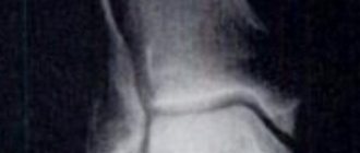
X-ray examination can help determine grade 1 osteoarthritis of the joint;
- Grade 2 – severe pain and limitation of movement force the patient to seek qualified help. Painful manifestations become more frequent and begin to disturb not only during movement, but also at rest.
At this stage, the affected limb loses up to 50% of its usual mobility, which is associated with swelling of the muscle tissue. The lifestyle changes slightly, all actions are doable with minor difficulties.
The main method for diagnosing stage 2 osteoarthritis of the joint is x-ray examination;
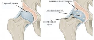
- Grade 3 - easily detected based on the occurrence of a serious problem with the joints. The cartilage tissue is completely destroyed, the joint space is absent. Negative lesions have spread to the joint tissue, which makes it impossible to treat the disease with medication.
As a result of the lack of treatment for arthrosis, a complete loss of the ability to move the limb and, as a result, disability is likely.
Stages of the disease
Arthrosis of the joints develops gradually and in the process goes through three successive stages that determine the severity of the disease:
- Stage 1: the pathology is not detected on X-ray or ultrasound, but the destruction processes have already started; the composition of the joint fluid changes, as a result of which the tissues receive fewer nutrients and become more sensitive; increased stress on the affected area causes inflammation (arthritis) and pain;
- Stage 2 is characterized by active destruction of cartilage tissue, and bone spines and growths appear along the edges of the articular platform (the area of contact of surfaces); at this time, the pain becomes habitual, and the inflammatory processes become stronger and weaker; spasms of the muscles associated with the joint are periodically observed;
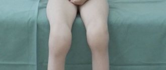
- Stage 3: areas of destruction affect almost the entire surface of the cartilage, the articular area is deformed, the affected limb deviates from its axis; range of motion decreases and ligaments weaken and become short.
Some experts also distinguish stage IV of the development of arthrosis. It is characterized by almost complete immobility of the joint.
How to carry out the procedure
- In an operating room, fat is removed from the patient’s thighs, buttocks, and abdomen through liposuction.
- Using a special centrifugation technology, it is purified from impurities of blood, anesthetic and other substances.
- The result is autofat - pure living cell mass suitable for colonization. It contains mesenchial cells that can activate the growth of new blood vessels and accelerate regeneration.
- The area of skin where the problem joint is located is numbed with lidocaine.
- Under ultrasound control of the joints, autologous fat is injected into the joint. Ultrasound increases the safety of injection, so it is no coincidence that it is also used for intra-articular injections of synovial fluid prostheses into large joints.
- For 1-2 days after the manipulation, the patient is advised to avoid physical activity, however, the joint cannot be completely immobilized. Movement speeds up healing.
In arthrosis, autofat activates platelet growth and joint regeneration
Kinds
Depending on the cause of the disease, primary and secondary arthrosis are distinguished. In the first case, the pathology occurs independently against the background of the complex influence of predisposing factors. The secondary form is a consequence of other diseases and is divided into the following groups:
- joint damage resulting from metabolic disorders or endocrine diseases (gout, diabetes mellitus, acromegaly, hyperparathyroidism);
- destruction associated with congenital pathologies (Paget's disease, congenital hip dislocation, scoliosis, hemophilia, etc.);
- post-traumatic arthrosis, which arose against the background of fractures, cracks, necrotic processes or surgical operations, as well as due to the characteristics of the profession.
The most popular classification of osteoarthritis is depending on the location of the pathological process:
- gonarthrosis: damage to the knee, one of the varieties of which is palletofemoral arthrosis - destruction of the joint between the femur and the patella);
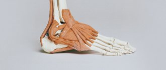
- arthrosis of the ankle joint: occurs against the background of heavy load and frequent injuries;
- arthrosis of the foot joints: the big toe most often suffers at the junction with the foot; the lesion develops against the background of gout or hallux valgus;
- shoulder arthrosis is characterized by damage to the shoulder and often occurs at a young age against the background of increased physical activity (loaders, athletes, construction workers);
- coxarthrosis: damage to the hip joint; can be both unilateral and bilateral and is one of the common causes of disability in people over 50 years of age;
- vertebral arthrosis: destruction of the cartilage discs between the vertebrae, most often affecting the cervical and lumbar spine;
- arthrosis of the joints of the hand: the joints of the fingers are most often affected; women in menopause are especially susceptible to pathology;
- arthrosis of the temporomandibular joint: it is quite rare, most often against the background of chronic inflammation due to malocclusion or improper prosthetics;
- Arthrosis of the elbow joint: a rare form of the disease, most often associated with injuries to this area.
Reasons for development
The main factor in the development of arthrosis is the discrepancy between the load experienced and the ability of the joint to withstand this load. Acute or chronic, this process inevitably leads to tissue destruction.
The list of reasons that increase the risk of developing arthrosis of any localization includes:
- heredity;
- endocrine pathology (diabetes mellitus);
- injuries of the articular apparatus: bruises, dislocations, fractures or cracks of bones inside the joint capsule, complete or partial ruptures of ligaments, penetrating wounds;
- regular increased load on the joints associated with the profession);
- obesity;
- hypothermia;
- previous inflammatory diseases of the joints: acute arthritis, tuberculosis, etc.;
- blood diseases in which bleeding into the joint often occurs (hemophilia);
- sudden changes in hormonal levels (pregnancy, menopause);
- local circulatory disorders due to atherosclerosis, varicose veins, thrombophlebitis, etc.;
- autoimmune diseases (rheumatoid arthritis, systemic lupus erythematosus, etc.);
- connective tissue dysplasia (congenital pathology, accompanied, among other things, by excessive joint mobility);
- congenital pathologies of the musculoskeletal system (flat feet, dysplasia or congenital dislocation of the hip joint, etc.);
- age over 45-50 years (increased risks are associated with a decrease in collagen synthesis);
- osteoporosis (bone loss);
- chronic intoxication of the body (including salts of heavy metals, drugs, alcohol);
- surgical interventions on joints.
What can happen if left untreated for osteoarthritis?
If the disease is ignored and untimely seeking professional medical help, morning swelling and joint pain can lead to serious diseases of the musculoskeletal system.
Osteoarthritis of the joints (arthrosis) is a destructive and deforming disease that irreversibly brings a person closer to disability. As the disease progresses, it reaches certain stages when the use of the functional capabilities of the musculoskeletal system becomes impossible.
In advanced cases of arthrosis, it is necessary to resort to drastic methods of treatment, which ultimately leads to the installation of a prosthesis and forced adaptation to a special rhythm of life.
Symptoms
Symptoms of arthrosis practically do not depend on its cause and location, since changes in the joints follow the same scenario. The disease develops gradually and begins to manifest itself when the cartilage is quite seriously damaged.
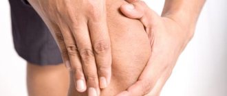
One of the first signs of trouble is a crunching sound in the problem area when moving. It most often occurs when the knee or shoulder is affected. At the same time, a person may feel a slight decrease in mobility after prolonged inactivity, for example, in the morning.
When asked what symptoms appear with arthrosis, most patients name pain first. At first insignificant and weak, it gradually gains strength, preventing normal movement. Depending on the stage and location of the pathology, a person may feel:
- starting pain: occurs during the first movements after prolonged inactivity of the joint and is associated with the formation of a thin film of destroyed tissue on the surface of the cartilage; after starting work, the film moves and the discomfort disappears;
- pain during prolonged physical activity (standing, walking, running, etc.): appears due to a decrease in the shock-absorbing properties of the joint;
- weather pain: provoked by low temperature, humidity, changes in atmospheric pressure;
- night pain: associated with venous stagnation and increased blood pressure inside the bones;
- joint blockade: sharp, severe pain associated with pinching of a piece of cartilage or bone located in the joint cavity.
As arthrosis develops, the symptoms become more noticeable, the patient notes the following signs:
- increase in morning stiffness;
- intensification and increase in duration of pain;
- decreased mobility;
- joint deformation due to bone growths;
- deformation of bones and surrounding tissues: the process is clearly visible on the limbs and fingers, which become noticeably curved.
When inflammation occurs, the affected area swells, turns red and becomes hot to the touch. Pressing on it causes a sharp increase in pain.
Contraindications to physiotherapy
Physiotherapy for arthritis and arthrosis , like any other type of treatment, has its contraindications. Limitations will depend on the physical treatment method, however, general problems can be identified when this type of therapy is not recommended:
- oncological diseases;
- pregnancy;
- presence of a pacemaker;
- pathologies of the thyroid gland;
- epilepsy;
- severe viral infections;
- heat;
- taking photosensitizing drugs;
- coronary heart disease (may require observation by a cardiologist during physical treatment).
Tests and diagnostics
An orthopedic surgeon diagnoses arthrosis. He conducts a detailed interview with the patient to identify complaints and medical history. The doctor asks in detail about the time of the first signs and the speed of their development, previous injuries and illnesses, and the presence of similar problems in relatives.
A general blood test can reveal the inflammatory process that often accompanies arthrosis.
The main diagnostic method is radiography. The following signs are clearly visualized in the image:
- narrowing of the joint space;
- change in the contours of contacting bones;
- impaired bone structure in the affected area;
- bone growths (osteophytes);
- curvature of the axis of a limb or finger;
- subluxation of the joint.
For a more detailed diagnosis, the following may be prescribed:
- computed tomography (CT);
- magnetic resonance imaging (MRI);
- Ultrasound of the joint;
- arthroscopy (internal examination of the joint cavity using a camera inserted through a small puncture);
- scintigraphy (assessment of the condition of bones and metabolism in them using the introduction of radiopharmaceuticals).
If a secondary nature of the disease is suspected, appropriate tests and consultations with specialists are prescribed.
Treatment of joint arthrosis
The choice of treatment for joint arthrosis depends on the cause of the disease, its stage and symptoms. In the arsenal of doctors there are:
- medications;
- non-drug treatment;
- surgical techniques.
In addition, the patient is required to strictly follow a diet and adjust their lifestyle to minimize further damage to the joints.
Drug treatment
Prescribing medications for arthrosis has two main goals:
- relieving pain and inflammation;
- restoration of cartilage tissue or, at least, stopping further degeneration.
To alleviate the patient's condition, various types of drugs are used:
- non-steroidal anti-inflammatory drugs: ibuprofen, ketorolac, diclofenac and their analogues in the form of tablets, injections, ointments or suppositories; they relieve pain and inflammation well;
- hormones (corticosteroids): indicated for severe pain and, most often, injected directly into the joint cavity;
- other analgesics, for example, antispasmodic (mydocalm): help reduce pain by relaxing muscles;
It is important to remember: all types of painkillers are used only to alleviate the patient’s condition. They do not affect the condition of cartilage, but when taken for a long time, they accelerate its destruction and cause serious side effects.
The main drugs for joint restoration today are chondroprotectors. They help saturate the cartilage with nutrients, stop degeneration and initiate cell growth processes. The most common remedy in this group is glucosamine and its analogues. They have an effect only at the early and middle stages of the disease and with regular long-term use.
Drugs that improve microcirculation in tissues and antienzyme agents help enhance the effect of chondroprotectors. The former provide a good supply of oxygen and nutrients to the affected area, while the latter slow down the processes of tissue destruction.
The doctor selects specific medications, their dosage and regimen.
Non-drug treatment
Non-drug treatment includes the following methods:
- physiotherapy: shock wave therapy: destroys bone growths and stimulates blood circulation due to ultrasound;
- automated electromyostimulation: exposure to electrical impulses to stimulate muscle contraction;
- ultraphonophoresis: exposure to ultrasound combined with the use of medications;
- ozone therapy: introduction of a special gas mixture into the joint capsule;
Surgery
Most often, the help of a surgeon is required in severe stages of the disease. Depending on the localization of the pathological process and the degree of damage, the following may be prescribed:
- puncture: puncture of the joint with removal of part of the fluid and, according to indications, administration of drugs;
- corrective osteotomy: removal of part of the bone followed by fixation at a different angle to relieve the load on the joint;
- endoprosthetics: replacement of a damaged joint with a prosthesis; used in extremely advanced cases.
Make an appointment
Is it worth treating arthrosis using this method?
Injecting fat cells to reduce pain and increase joint mobility is a new method. It has proven itself well in the treatment of diseases of tendons and ligaments, but with degenerative changes in cartilage tissue, such as occur with arthrosis, there is no significant improvement to speak of. In addition, difficulties will arise at the stage of searching for a clinic: not all medical institutions provide such a service, not to mention its high price.
Today, orthopedists are more inclined to another method of treating arthrosis - synovial fluid prostheses. They are also not able to completely cure the disease, but have significant advantages over the injection of autologous fat into the joint:
- The action of synovial fluid prostheses is quite understandable and logical, unlike fat cells. They simply mechanically push the rubbing surfaces apart and make up for the lack of lubrication. Due to this, improvement occurs.
- Noltrex and other synthetic drugs last a long time - up to 1.5-2 years.
- Improvements often occur after the first administration.
- The manipulation does not require expensive preparatory procedures, as with the introduction of fat cells.
- It is carried out in many medical institutions and medical offices.
- There will also be no difficulties in purchasing a liquid prosthesis.
If we were talking about a complete cure, then it would be worth paying attention to innovative, expensive techniques. However, upon closer examination, it turns out that this is only one of the methods of pain relief for arthrosis. You can achieve this result for a long period with much less effort and financial investment thanks to intra-articular injections of liquid endoprostheses.
Arthrosis in children
Arthrosis is considered a disease of older people, but it can also occur in children. The most common causes of pathology are:
- congenital pathology of connective tissue;
- severe injuries;
- heredity;
- metabolic disorders and functioning of the endocrine glands;
- orthopedic disorders (flat feet, scoliosis, etc.);
- overweight.
Children's arthrosis is rarely accompanied by severe symptoms: the pain is aching, and there is virtually no stiffness or limitation of function. Degenerative changes are detected on x-rays, MRI and ultrasound. The treatment process uses the same means as for adults. Maximum attention is paid to exercise therapy and physiotherapy, since they are especially effective at a young age. Without treatment, the disease sooner or later goes into an advanced stage with complete loss of mobility.
Diet
Diet is one of the most important factors in the treatment of arthrosis. If you are overweight, you need to reduce it to reduce the stress on your joints. In this case, a balanced diet with a calorie deficit is prescribed. Regardless of body mass index, doctors recommend completely avoiding:
- fast carbohydrates (sugar, desserts, flour);
- alcoholic drinks;
- spices;
- legumes;
- strong tea and coffee;
- excessively fatty and spicy foods.
Canned food and offal are not excluded, but are significantly limited, as is salt. An ideal diet for osteoarthritis includes:
- lean meats;
- fish and seafood;
- eggs;
- dairy products;
- flaxseed and olive vegetable oils;
- vegetables and fruits, a large amount of greens;
- cereals in moderation, durum wheat pasta;
- products with a high content of collagen (jellied meat, jellied meat, jelly).
What is the effectiveness of the technique for arthrosis?
The research results confirmed that the technique is most effective in the treatment of arthrosis of the shoulder and knee. In 80-90% of cases, after the procedure, patients experience improvement, pain decreases and mobility increases. For coxarthrosis, the method is less effective: only 60-70% of patients report improvements. However, in any case, we are not talking about a complete cure: this is only one of the options to influence the symptoms.
Improvements occur approximately 6-8 weeks after the course of therapy
Prevention
Arthrosis is easier to prevent than to treat. To maintain healthy joints for many years, it is recommended:
- to live an active lifestyle;
- do exercises regularly and visit the pool;
- eat right, consume enough omega-3 and collagen;
- do not exceed the BMI norm;
- wear comfortable shoes.
If the disease is diagnosed at an early stage, it is recommended to undergo regular spa treatment, as well as to exclude occupational risk factors: prolonged standing, heavy lifting, vibration.
What's the difficulty?
At first glance, the procedure looks attractive due to its high efficiency. But in reality it is not so simple. The method is still very new and little tested, it is used in single clinics, and is expensive. In addition, in order to get the desired results, the patient is offered to undergo osteoarthritis treatment according to the full protocol, which includes other expensive procedures:
- Stage 1. Ozone therapy - as preparation of the joint for future healing.
- Stage 2. Direct injection of fat cells.
- Stage 3. Administration of autologous blood plasma (PRP therapy) two weeks after the manipulation. The procedure is aimed at maintaining the activity of the transplanted fat cells. It is also recommended to repeat it after another 2 months.
Injection of fat cells is a complex and expensive treatment option for arthrosis
Treatment at the Energy of Health clinic
Medical orthopedists invite everyone to check the condition of the joints and, if necessary, begin treatment. We use modern effective techniques:
- full-fledged drug therapy in accordance with indications;
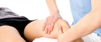
- physiotherapy, exercise therapy and massage;
- diagnostic and therapeutic punctures of the joint with the introduction of a synovial fluid substitute or medications;
- drug blockades for complete pain relief and other techniques.
You can view the full list of services on the website or by phone.
Advantages of the clinic
“Health Energy” is a multidisciplinary medical center equipped in accordance with modern standards. We offer patients:
- consultations with experienced doctors;
- examination using high-quality diagnostic equipment;
- individual approach to treatment selection;
- regular monitoring and performance monitoring.
The joints of our body can withstand enormous loads, but there are situations when they need help. Don’t let arthrosis change your life, sign up for a consultation at Health Energy.