Osteoarthritis of the hands begins when the wear of cartilage exceeds its regeneration. The rate at which cartilage tissue is produced depends on genetics, general health, lifestyle and environmental factors. But even despite regeneration, it tends to wear out - to a large extent, this is facilitated by monotonous loads on the small joints of the hand (working at a computer, machine, online games, writing text by hand, cutting food, wringing laundry, etc.). Degenerative processes in the articular surfaces over time lead to deterioration in mobility and even pain in the hands. Such complaints are observed in 10-15% of all humanity, especially in people over 45 years of age. And this problem is getting younger every year, increasingly affecting patients aged 25-30 years.
Let's look at what osteoarthritis of the hands is, the symptoms and treatment of the disease, the general prognosis, as well as methods for its prevention.
Osteoarthritis of the hands: what is it?
Osteoarthritis of the interphalangeal joints of the hands refers to a whole range of pathological conditions in which the joints of the fingers suffer. The dystrophic process leads to thinning and cracking of the cartilage, gradual destruction of connective tissue and, in later stages, to deformation of the joint. At the same time, the load on the ligaments and muscles of the hands increases - they are in chronic overstrain, which is expressed by pain, swelling, a reduction in the range of motion, as well as rapid fatigue and decreased performance. The shock-absorbing function of the joint capsule deteriorates, the cartilage ceases to retain the amount of moisture that it needs to maintain elasticity. In an effort to prevent further destruction of the joints, the body triggers the production of osteophytes - spine-like and other bone growths that limit mobility and injure adjacent tissues. This leads to chronic pain and inflammation. The metacarpophalangeal joint is poorly involved or not involved at all (unless there are generalized joint diseases or wrist injuries). In the early stages, the process is considered conditionally reversible, but in the absence of treatment, the changes become permanent, and the condition of patients can only be alleviated and maintained.
Osteoarthritis of the hands without treatment quickly spreads to all tissues of the hand - bone, cartilage and muscle. Undesirable, physically noticeable friction appears in the joints, their destabilization is observed, and the capillary supply of the hands deteriorates significantly. Fine motor skills are steadily deteriorating. For patients, performing the simplest household and professional duties becomes painful, grasping movements cause discomfort, handwriting changes, and irritability appears. In the early stages, irregularities and damage to the joint capsule, clearly visible on x-rays, appear. The last stages are characterized by loss of ability to work and complete disappearance of cartilage.
Not only medicines
Medicines eliminate symptoms, reduce pain and inflammation, but alone they will not cope with their task soon. Physiotherapeutic techniques are very important in the treatment of osteoarthritis of the fingers. Recommended procedures:
- electrophoresis : it avoids complications and allows the patient to carry out procedures at home (after detailed consultations and doctor’s recommendations). It becomes possible not only to improve metabolism and blood circulation in the affected joint, but also to deliver a mass of medications to it - from novocaine, zinc and sulfur to dimexide with analgin;
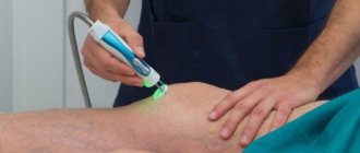
- laser - its effects can only be obtained in a physiotherapy room. But the device relieves reflex muscle spasms, normalizes the permeability of cell membranes, stimulates tissue respiration and metabolism. The only limitation is that this technique is not applicable during the relapse period; you will have to wait for a stable remission;
- cryotherapy. Cold applications to the affected joint become a real salvation when treatment of osteoarthritis of the fingers is limited to exacerbation of the disease. Their use stops the development of irritation in the joint capsule, relieves swelling, and facilitates treatment after the relapse ends.
UHF and magnetotherapy also give good results; methods provided by traditional medicine are effective. But in the latter case, coordination with the attending physician is required: “healer” approaches may conflict with the main course of treatment.
Causes of osteoarthritis of the hands
Experts distinguish several types of carpal arthrosis, among which the most common are professional (associated with everyday increased load on the joints and unfavorable working conditions), traumatic, autoimmune (as a consequence of rheumatoid arthritis) and others. The disease can be secondary and occur due to:
- diabetes mellitus and other metabolic and hormonal disorders;
- previous operations, fractures, dislocations, sprains;
- problems with peripheral arteries and other diseases that impair blood supply to the hands (cervical, thoracic osteochondrosis and others);
- a number of infectious diseases (for example, sickle anemia);
- frequent stress;
- severe or persistent hypothermia.
In addition to the genetic predisposition, which is usually observed in the patient’s relatives, the development of the disease is facilitated by a sedentary lifestyle, excessive stress (sports and household), and excess weight. If the patient already suffers from gout or arthritis, inflammatory diseases of the hand, the risk of arthrosis increases in this case every year.
Deforming osteoarthritis of the hands has a gender “preference”—women suffering from low estrogen levels due to hormonal imbalance or menopause are especially susceptible to it.
Age-related changes also provoke a degenerative process. The depletion of cartilage tissue and its gradual drying out is associated with reduced production of collagen and proteoglycan - these substances are responsible for the construction of cartilage cells and the preservation of nutrients in them.
Manifestations of polyosteoarthrosis
Classic treatment of arthrosis is not always suitable for this diagnosis, since the disease has specific manifestations:
- with generalized arthrosis, three or more joints are affected, most often symmetrical (knee, hip, interphalangeal), Heberden's nodes are noticeable;
- with lesions of the intervertebral joints, pain occurs in different parts of the spine, but secondary symptoms are also possible - dizziness, blurred vision, migraine, indicating compression of the branches of the vertebral artery;
- if polyosteoarthrosis is accompanied by spondylosis of the lumbar and cervical regions with the growth of osteophytes and degenerative changes in cartilage tissue, there is numbness of the limbs, “intermittent” claudication, tinnitus and other unpleasant symptoms;
- sometimes multiple osteoarthritis develops along with periarthritis or tenosynovitis - tissue inflammation, swelling of tendons and capsules.
All this rich symptomatology complicates not only the diagnosis of the disease, but also its treatment.
Polyosteoarthrosis can be accompanied by the most unexpected symptoms
Signs of deforming osteoarthritis of the hands
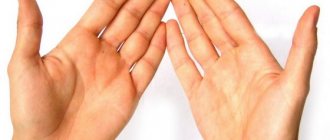
Carpal osteoarthritis begins unnoticed; only X-ray examination can reliably identify it before the onset of constant pain. Patients usually attribute short-term aches and slight discomfort in the fingers to fatigue - and therefore only every 5th person consults a doctor. Symptoms manifest themselves most clearly during climate change, hypothermia, stress, physical activity or lack of sleep. Gradually, the time intervals between episodes shorten and pain becomes an unfortunate norm of life for most patients. A characteristic crunching sensation appears in the fingers, the joints swell, making the hands appear knotty. Then stiffness increases (especially in the morning), mobility is limited, and deformation of the hand is observed. The movements become tight, bending the fingers resembles an exercise on a wrist expander. Cysts may form. The feeling of constant load corresponds to a real pathological process - small joints begin to bend, taking on an unusual shape, and the burden of decompensation falls on the muscles.
As a rule, the first joints to succumb to disease are the joints of the fingers located behind the nails (the last phalanx of the fingers), as well as the joints of the thumbs. Painless but noticeable thickenings appear on them - the so-called. Bouchard's nodes and Heberden's nodes - which are easy to notice on the back of the hand. Often the skin on them turns red and becomes hot.
Types of polyosteoarthrosis
Depending on the visual condition of the joints, the disease can take the following forms:
- knotless;
- nodular (with thickenings on small joints - Bouchard's or Heberden's nodes).
Depending on the nature of the course, the disease is classified as follows:
- low-symptomatic form - typical for young patients, rarely accompanied by severe pain, instead of which there are cramps in the calves and nodules on the upper phalanges of the fingers;
- manifest form.
In its manifest form, the disease progresses slowly or quickly. In the first case, a person may not pay attention to the symptoms for more than five years, after which moderate pain appears - due to the weather, when moving, “starting pain”. Rapidly progressive multiple osteoarthritis is typical for young people. Rapid damage to several joint groups occurs with limitation of movement, muscle atrophy and neurological complications.
In old age, multiple osteoarthritis does not progress as quickly as in young people.
Stages of osteoarthritis of small joints of the hands
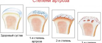
As a rule, treatment of osteoarthritis of small joints of the hands is delayed due to the slow progression of the disease. Symptoms increase gradually, which makes it difficult for the patient to objectively perceive their intensification. However, experts distinguish 3-4 stages of the disease, which make it possible to adjust therapy and make predictions about the course of the process with considerable reliability.
Osteoarthritis of the interphalangeal joints of the hands of the 1st degree is characterized by mild, barely noticeable pain, joint mobility is preserved and the patient may not suffer any restrictions in daily activities. The tissue around the affected joints begins to harden.
Osteoarthritis of the interphalangeal joints of the hands, grade 2, involves the presence of constant, but not too severe pain. The range of motion in the joint is reduced, and osteophytes, clearly visible on x-rays, appear, which prevent full movement. Towards the end of the stage, the fingers may become thinner because the muscles responsible for normal hand activity are no longer used and begin to atrophy. Against the background of this process, the joint nodes become especially noticeable and begin to crunch audibly when bending the fingers due to the narrowing of the lumen in the joint.
Osteoarthritis of the interphalangeal joints of the hands of the 3rd degree is expressed by severe pain and extreme difficulty in flexion movements. The number of osteophytes increases, almost completely limiting the mobility of the joint. The photographs show an extremely small amount of cartilage tissue. The atrophy of the muscles responsible for fine motor skills of the fingers is completed. Bone density increases, severe deformation and signs of osteosclerotic changes are noticeable.
The following are used as agents that alleviate the course of polyosteoarthrosis:
- physiotherapeutic procedures - to relieve swelling, stimulate blood circulation and metabolism, accelerate regeneration;
- manual therapy - to reduce pain;
- acupuncture – to restore mobility;
- other rehabilitation measures (physical therapy, massage, orthopedic treatment, etc.).
Part of the treatment is a proper diet and giving up bad habits.
With a timely approach, even such a complex disease as polyosteoarthritis can be kept under control. The disease begins with pain in the spine and hands, and is manifested by deformation of one of the large joints - the knee or hip. Therefore, you should not ignore any pain symptoms or attribute them day after day to physical fatigue. It is impossible to completely cure arthrosis, and when it comes to multiple lesions, the future is at stake. Be vigilant and attentive, first of all, to yourself - then the disease will not have a chance!
Which doctor treats osteoarthritis of the hands?
If symptoms are detected, you should consult a doctor to determine the stage and, if possible, the cause of the disease. You can contact a therapist, orthopedist or rheumatologist - they can conduct a visual examination or immediately refer you to a radiologist to confirm the diagnosis. Based on the collected medical history, the specialist prescribes an individual treatment plan. It depends on the degree of narrowing of the joint space, the presence of osteophytes and their number, the presence of deformation or osteosclerosis. It is also important to rule out other similar diseases - for example, rheumatoid arthritis, which is characterized by longer morning stiffness and so on. It is impossible to determine these criteria at home, so an examination by a specialized doctor is more mandatory than desirable. If osteoarthritis of the hands is secondary, tests or treatment of primary diseases may be required.
How to treat osteoarthritis of the hands
The treatment program for carpal osteoarthritis usually includes conservative methods, but sometimes the situation requires surgical intervention to eliminate deformities and osteophytes. In severe cases, intra-articular injections of glucocorticosteroids are also prescribed (no more than 3 courses per year) - they relieve pain and inflammation, improve mobility, but do not prevent the development of the disease and do not build up cartilage tissue. The main goal of therapy is to slow down destructive processes and relieve clinical manifestations that worsen the quality of life (so-called symptomatic treatment).
Treatment of osteoarthritis of the hands with drugs is carried out in several directions:
- pain relief (analgesics);
- relief of inflammation (NSAIDs) with systemic and local drugs;
- cartilage regeneration (chondroprotectors, sometimes in combination with risedronic acid);
- restoration of the elasticity of cartilage and replenishment of nutrients in it (taking rehydration, micro-, macronutrient and vitamin preparations, including calcium and vitamin D);
- according to indications - medical prosthetics using hyaluronic acid injections.
Rest is also indicated for patients. To redistribute the load and prevent joint deformation, it is recommended to wear orthoses.
Physiotherapeutic procedures include phonophoresis, electrophoresis, magnetic and shock wave therapy, radio wave and laser exposure, mud wraps, paraffin baths, massage (including self-massage - to relieve spasms, strengthen muscles and improve tissue nutrition), hand exercises with use of auxiliary means. It is recommended to use an ergonomic keyboard and avoid inconvenient working tools.
Diagnostics
Diagnosis of osteoarthritis is based on clinical and radiological examination data. Clinical studies have examined the ability to measure autoantibodies and synovial fluid markers as indicators of OA [4]. None of these indicators have proven reliable for diagnosing and monitoring OA.
As a rule, indicators of the acute phase of inflammation in OA are within normal limits. In the case of erosive arthritis, an increase in ESR is possible. In the synovial fluid, leukocytes are detected in quantities below 2000 per μl, with a predominance of mononuclear cells.
The method of choice in diagnosing OA is radiographic examination [5]. Some features of articular cartilage and soft tissue that are not visible on radiographs can be visualized using MRI. However, in most patients with OA, MRI is not necessary.
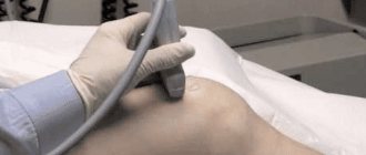
Ultrasound has no role in the routine clinical assessment of a patient with OA. It can be used as a tool for monitoring cartilage degeneration, as well as for intra-articular injections.
Arthrocentesis can help rule out inflammatory arthritis, infection, or crystalline arthropathy associated with deposition of microcrystals of varying composition.
The diagnostic results determine which methods will be used to treat osteoarthritis.
Osteoarthritis of the joints of the hands: treatment determines the prognosis
Timely diagnosis can prevent the development of the disease even if it is caused by genetic and age-related factors. For example, a “non-functioning” gene that is responsible for the production of type 2 collagen can be compensated by taking chondroprotectors - such as Artracam - which contain it in easily digestible forms.
If the disease is advanced to the last stage, the prognosis is unfavorable. Unlike osteochondrosis of large joints, when the interphalangeal joints are affected, endoprosthetics are rarely performed. Even the installation of an implant usually does not guarantee 100% restoration of functionality. Other operations - arthrodesis (fusion) and reconstruction of joints are also characterized by increased complexity.
The best prognosis is observed with the use of complex medication and physiotherapy for grade 1 osteoarthritis of the hand and grade 2 osteoarthritis of the hand joints. Lifelong use of over-the-counter chondroprotectors triggers regenerative processes in bone and cartilage tissue - this prevents further destruction of cartilage and, subject to other medical prescriptions, even helps to achieve lasting improvements.
You may be interested in:
Osteochondrosis of the lumbar region Osteoarthrosis of the shoulder joint Osteoarthrosis of the elbow joint Osteoarthrosis of the ankle joint Reactive arthritis - a disease of the young
Main symptoms of the disease
Considering the clinical picture of the disease, we can distinguish three main symptoms of joint osteoarthritis:
- painful sensations of a mechanical nature, which have varying degrees of severity and are localized in the area of the affected joint;
- increased volume of joints, swelling;
- crepitus (crunching sound when moving a joint).
In addition, osteoarthritis is often accompanied by diseases such as varicose veins (dilated veins) of the lower extremities, thrombophleitis (inflammation of the vein wall and the formation of blood clots).
