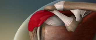
Anatomy of the ankle joint
The ankle connects the bones of the foot to the bones of the lower leg. The ankles are the distal portions of the tibia and fibula.
The structure of the ankle is quite complex: it is formed by the talus and tibia bones, which are connected by cartilage and muscle tissue. Each joint has an extensive network of blood vessels and nerves, which provide tissue trophism and coordinated movement of the limb.
In the event of an injury, all this is disrupted and, if not treated correctly, is never fully restored.
Clinic and diagnostics
If a person breaks his ankle, there is a sharp and acute pain. Looking at the damaged area, you may see swelling, blue discoloration, and in some cases, skin damage.
If the injury site is touched, the patient will feel a sharp, shooting pain. Joint movement becomes unnatural or almost completely absent. Professional diagnosis includes an initial survey, visual examination and fluoroscopy. For a clearer understanding of the clinical picture, pictures are taken from the end, back and front sides.
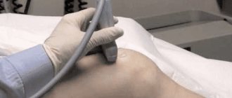
Additionally, ultrasound, sonography and arthroscopy may be prescribed. After all diagnostic measures, the specialist prescribes comprehensive treatment.
Causes and symptoms of an ankle fracture
The main causes of ankle fractures are direct (impact) and indirect (twisting of the lower limb) injuries.
Indirect injuries are more common. They can occur when falling during icy conditions, sports training, roller skating and snowboarding, slipping on a wet floor, etc.
Causes of direct ankle injuries can include car accidents, something heavy falling on your foot, etc. An ankle fracture is accompanied by:
- Pain syndrome;
- Swelling of soft tissues;
- Hematomas;
- Impaired joint function.
How does a fracture occur?
Ankle fractures, more commonly known as ankle fractures, occur when one or more of the bones that make up the ankle joint are broken. Although an ankle fracture is an injury seen at almost any age, it is more common among older adults and athletes who are physically active.
When the bones are damaged, the ankle becomes unstable. When adding ligament damage, the injury can become a very serious problem, and complications after an ankle fracture are possible.
An ankle fracture can be caused by a sudden, accidental movement, such as twisting the ankle during sports or activity. Car accidents can also cause ankle fractures, especially after hard impacts.
A person can also trip, fall, or twist an ankle, which can cause an ankle fracture. Depending on age and previous health conditions, this problem can develop into a much more serious injury if left untreatedSource: Ankle Injury Analysis. Taylashev M.M., Salatin P.P., Sobolev V.V., Pozikov V.V., Kolesnikov A.S. Acta Biomedica Scientifica, 2008. p. 144-145.
First aid for a fracture of the outer ankle
If the ankle is broken, the victim must be taken to the emergency room. It's better to call an ambulance. When this is not possible, you will have to organize a stretcher from available materials and deliver the victim to the nearest medical facility.
Before the doctors arrive, the patient needs to be provided with pre-medical care:
- Free the leg from traumatic factors, of course, if this is possible without additional damage to the limb.
- Elevate the affected leg. To do this, you can make a roller out of clothes.
- If bleeding occurs, apply ice or something cold, if possible - a hemostatic tourniquet, which should be loosened for 20 seconds, every 20 minutes.
- If the integrity of the skin is damaged, under no circumstances should you try to set bone fragments.
- If the pain is severe, give the victim an analgesic.
- When there is no specialized splint for immobilization, and the patient needs to be taken to a medical facility independently, you need to apply a splint from improvised means.
Gently bend the injured limb at the knee joint and position the foot so that the heel is at a right angle to the shin. Apply an improvised splint and secure it with a bandage, belt, belt, etc.
Rehabilitation
The entire rehabilitation period can be roughly divided into three stages, which we will return to a little later. The main thing is to follow simple rules - keep your leg higher than your body (blood flow improves), eat right, follow all the recommendations of the traumatologist.
First stage of rehabilitation
The total length of the period under review does not exceed 14 days. Seven days after injury, gradual development of the limb and light physical activity begins. For patients who have undergone surgery, the time period increases slightly (you can move your leg only after 21 days).
Therapy includes UHF, laser heating and ultraviolet exposure. If the patient's condition is normal, walking on crutches and massages are allowed.
Second
The victim stops using crutches and tries to walk on his own. The main goal of the second recovery period is to normalize the functioning of the ankle joints. The doctor attributes a number of special exercises and water procedures (your weight in water is less, and, therefore, the load).
After some time, the patient begins exercises on an exercise bike and similar rehabilitation techniques. In order to relearn how to walk correctly, instep supports (devices for adjusting the amplitude of movements) are used.
Physiotherapy is represented by magnetic and laser irradiation and segmental massage. A satisfactory state of health allows you to prescribe paraffin treatment (warming procedures) and electrical stimulation. If there are metal fasteners in the bone, ultrasound procedures are prohibited, since the influence of sound waves can cause heating of the structure and its deformation.
Third
The final stage returns the person to normal life. You can develop muscles on treadmills; you don’t need to run, just walk quickly. Do not forget to unload the joints, use an elastic bandage for this. As practice has shown, underwater massage is very effective. Additionally, laser and ultrasound therapy is performed. You can put full weight on your leg only 70 days after discharge from the hospital.
Treatment of an ankle fracture
An ankle fracture is treated conservatively and surgically.
Conservative treatment methods are used in the following cases:
- Closing fracture without displacement;
- Displaced fracture, but immediate closed reduction is possible;
- Minor damage to ankle ligaments;
- The operation cannot be performed (risk of anesthesia for serious diseases, patient refusal, some pathologies of the cardiovascular system and central nervous system, decompensated diabetes mellitus).
Stages of conservative treatment of an ankle fracture:
- Closed reduction (reposition) of the bone.
- Application of a plaster cast.
- Removing the plaster.
Indications for surgical therapy:
- Open fracture;
- Ineffectiveness of closed reduction (reduction);
- Old injuries;
- Damage to both limbs;
- Rupture of the ligamentous apparatus of the ankle joint;
- Damage to the ankle with simultaneous fracture of the tibia and fibula, rupture of ligaments and outward subluxation of the foot.
Possible complications
After treatment, it is important to follow your surgeon's instructions. Failure to do so can lead to infection, deformity, arthritis, and chronic pain. Source: Mistakes and Complications in the Treatment of Compound Ankle Fractures. Salikhov R.Z., Pankov I.O., Plakseychuk Yu.A., Soloviev V.V. Practical medicine, 2014. p. 128-131.
Article sources:
- Errors and complications in the treatment of complex fractures of the ankle joint. Salikhov R.Z., Pankov I.O., Plakseychuk Yu.A., Soloviev V.V. Practical medicine, 2014. p. 128-131
- Analysis of ankle joint injuries. Taylashev M.M., Salatin P.P., Sobolev V.V., Pozikov V.V., Kolesnikov A.S. Acta Biomedica Scientifica, 2008. p. 144-145
- Comprehensive diagnosis of ankle joint injuries. Kim L.I., Dyachkova G.V. Genius of Orthopedics, 2013. p.20-24
- Fractures of the ankle joint and treatment methods. Cherednik A.A., More Gautam Saherbao, Al-Fakih Abdulaziz. Bulletin of Medical Internet Conferences, 2014. p.432
- Therapeutic tactics for intra-articular fractures of the ankle joint (literature review). Zedgenidze I.V., Tishkov N.V. Acta Biomedica Scientifica, 2013. pp. 178-182
| Name of service (price list incomplete) | Price, rub.) | In installments* |
* You can read more about the conditions here - Treatment on credit or in installments.
First aid
Self-treatment of a fracture at home is unacceptable. Trying to change the position of the leg or realign a bone can aggravate the condition and make the person disabled. At the first signs of the disease, it is recommended to immediately consult a traumatologist. Before the ambulance arrives, you can provide first aid to the person:
- To prevent severe swelling, apply ice to the affected area. This must be done carefully and always over clothing. A medical ice pack is applied for 25 minutes. After this, you need to take a break for 15 minutes and apply a cold compress again. If you keep ice continuously, a side effect will occur - tissue hypothermia.
- Give the person pain medication. To relieve pain, the patient can be given tablets from the group of non-steroidal anti-inflammatory drugs. These include Ibuprofen, Nurofen, Ketanov or Nise. Before giving the patient a pill, read the list of contraindications and ask the patient if he has one of the listed diseases. Consider the presence of allergic reactions and intolerance to components.
- Pay attention to the symptoms that occur as a result of the fracture. If you are providing first aid to a person with a chronic illness, ask about their condition. If necessary, give him medications that the patient must take regularly.
What is a trimalleolar fracture?
What components does such a fracture consist of?
- Fracture of the outer malleolus (fibula).
- Fracture of the inner malleolus (tibia).
- Fracture of the posterior edge of the tibia (tibia).
All three of these anatomical structures participate in the formation of the articulatio talocruralis, thus, when fragments are displaced, the relationship of the articular surfaces is disrupted. In the future, if this displacement is not eliminated, severe deforming osteoarthritis of this joint occurs.
As a result of such a fracture, a subluxation of the foot may occur, which must be eliminated. It is also possible to combine this injury with a rupture of the tibiofibular syndesmosis; then a divergence of the “fork” of the articulatio talocruralis will be observed.
Symptoms
The clinical picture largely depends on the nature of the damage. Doctors identify a number of common symptoms by which pathology can be recognized:
- Deformation of the ankle joint, due to which the foot noticeably changes its shape.
- Paleness of the skin in the affected area.
- Severe pain when touching or walking.
- Numbness of the foot or entire limb (noted when nerve fibers are damaged or blood vessels are damaged).
- Tension of the skin over the area where the fracture is located. When the fragments are displaced, the skin is injured and may even tear.
- Decreased or loss of motor abilities. In some cases, the patient cannot even move his fingers.
- Formation of hematomas - accumulations of blood in places where blood vessels rupture. If left untreated, the symptom worsens, causing the hematoma to affect the toes.
Patients suffering from a trimalleolar fracture complain of severe swelling. The leg becomes deformed and swells. This occurs due to injury to the ligaments and joint capsule. Synovial fluid and blood accumulate in the affected area, which leads to the formation of edema.
Even with the listed symptoms, the pathology can be mistaken for a sprain. To clarify the diagnosis, the patient is sent for an X-ray examination.
Bone reduction
If a dislocation is diagnosed during an x-ray examination, the patient undergoes reduction. The procedure involves returning the bones to the place from which they were displaced. Reposition is prescribed to patients in cases where displacement has occurred by more than a third of the bone diameter.
Reposition is divided into closed and open. In the first case, we are talking about a traditional method of treatment without surgery. Open reduction is a surgical procedure.
Closed reduction
Closed reduction is prescribed in cases where the skin remains intact and completely hides the fracture site. For minor or transverse fractures, the surgeon and his assistant perform the procedure manually. This process is carried out as follows:
- The initial process of reduction is called traction. It involves stretching a limb. The task of the trauma surgeon is to align the axis of the broken bone. To do this, the doctor and his assistant move the bone fragments around the axis until the required distance is formed between them.
- While the doctor works with the fragments, his assistant holds the limb in the joint above the area in which the fracture is located. This is necessary in order to create the effect of stretching the limb.
- Once the doctor achieves diastasis (the required distance between bone fragments), the next step is to rotate the limb along the axis. This is done by palpation (palpation). By palpating, the trauma surgeon compares the fragments and achieves maximum correspondence between the fragments and the bone, due to which the displacement is completely eliminated.
Risk groups and mechanism of injury
This fracture often occurs as a result of an accident.
Who is at risk for this injury?
This includes the following categories of people:
- Motorcyclists.
- Unbelted car passengers.
- Patients whose professional activities involve working at heights.
- Aged people.
- Patients suffering from osteoporosis.
- Patients involved in speed skating, alpine skiing, and skateboarding.
What is the mechanism of injury?
The answer to this question could be:
- Excessive flexion/extension of the joint
- Rolling the foot outwards or inwards
- Ankle twist
- May be a consequence of high-energy injuries (road accidents, catatrauma - fall from height)
Exercises in the first stage of treatment
Therapeutic exercises should be started long before the cast is removed (it is better to check the timing with your doctor). However, at this stage, exercises should not put stress on the sore leg. They are prescribed to restore blood circulation in an immobile limb, since good blood supply to tissues promotes their restoration and avoids swelling. Let's look at a few exercises (perform them while standing on your healthy leg and holding onto a wall or the back of a chair):
- As you inhale, move your injured leg to the side. As you exhale, make a cross movement in front of your healthy leg. Repeat the exercise for 30 seconds.
- Swing your casted leg to the side. Try to take it as high as possible and hold it in this position for a few seconds. Lower your leg. Repeat the exercise 10-15 times.
- Raise your knee until the thigh of your affected leg is parallel to the floor. Hold this for a few seconds and then lower it. Repeat the exercise for 30 seconds.
- Take your sore leg back, trying to lift it as high as possible, but without arching your lower back. Hold the limb in this position for a couple of seconds, then lower it. Repeat this exercise for about 30 seconds.
The considered gymnastics will seem very difficult to you at first. Therefore, avoid excessive loads and exercise every other day. Once you get used to the exercises, you will do them every day without feeling tired or painful.