- Treatment of arthrosis of the knee joint: Goals of autologous chondrocyte transplantation
- Who is suitable for this type of treatment for knee arthrosis?
- Methods for restoring articular cartilage
- What type of doctor performs autologous cartilage cell transplantation?
- How successful will the treatment of arthrosis be with chondrocyte transplantation?
- What happens before surgery?
- How is arthroscopic chondrocyte transplantation performed?
- What type of anesthesia does the patient receive during surgical treatment of knee arthrosis?
- Will my knee hurt after surgery?
- Conditions of accommodation at the Gelenk Clinic
- What should you pay attention to after surgery?
- How much does it cost to treat knee arthrosis with autologous chondrocyte transplantation?
- Is this method right for me?
- How can a foreign patient make an appointment and the operation itself?
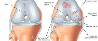
Painful wear and tear of the articular cartilage of the knee joint is explained by several factors: arthritis, arthrosis, injuries with deformations of bones or cartilaginous structures, metabolic disorders, as well as gout or hemochromatosis.
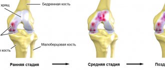
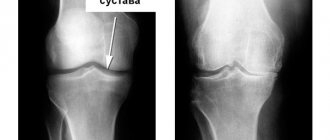
© bilderzwerg @ fotolia Arthrosis (wear and tear) of articular cartilage is the most well-known joint disease. One of the most common forms of the disease is arthrosis of the knee joint - a pathology that causes chronic pain, as well as limited movement of the patient. Over the course of several years, the sliding surface of the articular cartilage gradually wears out, and therefore slowly loses its original properties. Since cartilage tissue lacks nerve endings (nociceptors), knee injuries become noticeable only when the defect reaches the bone layer located under the articular cartilage. Autologous chondrocyte transplantation, or in other words cartilage cell transplantation, is an innovative surgical treatment for knee arthrosis that aims to correct cartilage deformities using the patient's own cartilage cells. Successful treatment of knee arthrosis using this technique is carried out by only a few knee specialists in Germany. Dr. Baum is the first surgeon in the world to perform arthroscopic articular cartilage transplantation. In addition, he is one of the developers of this method of treating knee arthrosis, who regularly trains new specialists.
Who is suitable for this type of treatment for knee arthrosis?
The method of cartilage tissue transplantation as a treatment for arthrosis of the knee joint is not suitable for every patient. At a progressive stage of the disease, this technique will no longer bring the desired result. In such cases, the surgeons at Gelenk Klinki in Freiburg will recommend the patient joint-preserving partial replacement or total knee replacement. Successful autologous chondrocyte transplantation depends on the following factors:
- knee stability, intact cruciate and collateral ligaments
- straight axis of the joint without deformities (e.g. hallux valgus or varus deformity of the legs)
- absence of articular mouse (tissue fragments formed due to defects in articular cartilage)
- at least partial preservation of the menisci
- cartilage damage on only one of the two articular surfaces
The usual indications for cartilage tissue (chondrocyte) transplantation are local injuries to the articular cartilage in the knee.
What are the ideal conditions to perform autologous chondrocyte transplantation?
- Patient age from 15 to 55 years
- Defect size up to 10 cm2 in the presence of healthy areas of cartilage
- Stability of the knee joint, undisturbed ligament structure
- No excessive stress on the knee, particularly due to excess weight
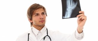
As a rule, this technique is suitable for people under the age of 55. However, the decisive factor in this case is not the biological, but the calendar age of the patient. Sometimes, if all the necessary indications are present, cartilage tissue transplantation is also performed for patients over 65. In order to accurately say whether this surgical intervention is right for you, the specialists of our orthopedic clinic need X-rays, as well as an MRI of the knee. Often, only during the operation the surgeon makes a decision regarding the final treatment method: sometimes partial endoprosthetics is enough for the patient, and sometimes, to restore mobility, it is necessary to perform a cartilage tissue transplant.
If doctors can determine that autologous chondrocyte transplantation does not make sense, you will be offered methods of treating arthrosis, such as partial prosthetics using the HEMICAP patellofemoral prosthesis, Repicci prosthetics or complete endoprosthetics.
Chondroplasty of the knee joint: who will it help?
Injuries, excessive physical activity, metabolic disorders, heredity and other reasons can seriously affect the condition of the cartilage in the knee joint. Chondroplasty can help identify cartilage damage and defects in this area. We asked Alexey Yuryevich Krasovsky, a traumatologist-orthopedist of the highest category at the Expert Clinic Irkutsk, to tell us more about this type of surgical intervention.
— Alexey Yurievich, what is chondroplasty of the knee joint?
— This is a plastic surgery performed to replace cartilage defects in the joint. It can be performed either using autologous tissue (the patient’s own tissue) or using a bipolymer film.
Chondroplasty helps patients who have a defect in the load-bearing surface of the cartilage, arthrosis of the second or third degree, but at the same time the range of motion is preserved.
— What types of knee chondroplasty are there? What is their difference?
— Chondroplasty at the present stage is carried out endoscopically, i.e. using arthroscopy. There are generally three main types of arthroscopic chondroplasty of the knee.
1) Abrasive chondroplasty of the knee joint - what is it? This is when, during arthroscopic surgery, pathological areas of cartilage are removed that are mobile, create a conflict in the joint and further destroy the articular cartilage. During this operation, both pathological foci are removed and the edge of the cartilage is stabilized, which helps prevent its further destruction.
The next stage is the so-called microfracture, or tunnelization of cartilage defect areas. The surgeon creates perforations using a needle with a diameter of 1.8 - 2 mm or a special tool (awl for microfracturing). In this case, mesenchymal cells emerge from the bone marrow. These are red bone marrow cells that fill defects and promote the growth of fibrocartilage.
The second point is the use of artificial collagen membranes in abrasive chondroplasty. A collagen membrane is applied to the microfractured surface of the cartilage (in the tunneling zones). It is glued using fibrin glue or sewn with a special suture. This provides ideal conditions for obtaining stem cells and subsequent restoration of cartilage. That is, mesenchymal cells emerge from the tunneling (or microfractured) holes and cartilage begins to grow under this collagen membrane.
2) The next type is mosaic chondroplasty of the knee joint. Its essence is as follows. Using special technology, through a small incision (or additional endoscopic ports), a column of cartilage is taken from the non-load-bearing area along with a section of bone, and at the same time the same section is taken in the area where there is a defect. After that they change places. That is, the osteochondral column from the non-load-bearing area, where the cartilage is of satisfactory thickness, is transferred to the area where there is a defect. And the column from the defect zone is transferred to the unloaded zone. The resulting tissues are transplanted in the form of a mosaic into the damaged area. As a result, the articular surfaces are restored, and the person gets rid of pain.
3) There is also a technique that combines mosaic chondroplasty and collagen membrane. There are dissertations confirming that the use of combined techniques gives greater effect. However, long-term results indicate that the use of both abrasive chondroplasty and mosaic chondroplasty separately gives quite good results. According to the international standard, mosaic chondroplasty should be used for deep cartilage defects.
Arthroscopic mosaic chondroplasty of the knee joint can be performed either purely arthroscopically or combined with a small, minimally invasive approach.
— Do you need special preparation for chondroplasty of the knee joint?
— Preparation, as a rule, consists in the fact that it is necessary to eliminate the inflammatory process, that is, remove synovitis. Initially, it is possible to carry out sanitation arthroscopy or anti-inflammatory therapy, achieve weight loss for the patient, and teach him how to walk correctly using crutches to unload the joint.
In my opinion, in the postoperative period it is necessary to use special types of orthoses that allow you to unload certain areas of the loaded surface of the knee joint. The type of orthosis depends on the location of the lesion (in particular, the medial, i.e., internal, or lateral, i.e., external, part of the knee). Therefore, when choosing, you need to focus on the recommendations of your doctor.
— How long does rehabilitation last? What is it?
— The duration of rehabilitation after chondroplasty of the knee joint is approximately 6 to 8 weeks. If the patient wears an orthosis, then normal physical activity is allowed after 6 weeks. If a person is engaged in physical labor, then, of course, it is necessary to change his lifestyle. Heavy physical activity, jumping and impact are prohibited.
If we used the so-called film chondroplasty, i.e. a special artificial film, then we need to start developing the joint no earlier than in a week. If the patient has undergone another type of chondroplasty, then, in principle, movement is allowed immediately, but axial load is prohibited for the first three weeks.
The basic principles of treatment in the postoperative period are that the patient can move the limb as often as he needs and in full, but axial load is excluded for up to 6 weeks. That is, as much movement as possible, but at the same time prohibiting axial load. Chondroprotective therapy is also used, and in the late postoperative period, hyaluronic acid preparations, plasma lifting or PRP technology (injection of patient’s blood plasma enriched with activated platelets into the damaged joint) can be used.
— Who is contraindicated for knee chondroplasty?
— First of all, the procedure is contraindicated in case of purulent or acute inflammatory processes in the area of the knee joint (synovitis), or violation of the integrity of the skin.
Chondroplasty is also prohibited in case of gunshot osteomyelitis, severe joint contractures, when there will be no effect with this surgical procedure.
The patient's mnestic-intellectual decline is also a contraindication. That is, when the patient is not determined and does not understand that in the postoperative period he will need to adhere to a certain rehabilitation plan, and he will not follow all the recommendations. It is better for such patients to refrain from surgery.
Interviewed by Marina Volovik
The editors recommend:
What to do if your knees hurt? “I fell, I woke up...” What is a meniscus tear?
For reference:
Krasovsky Alexey Yurievich
In 1995, he graduated from the Faculty of Medicine of Irkutsk State Medical University. 1996–98 – residency in the specialty “Traumatology and Orthopedics”. He has been involved in joint arthroscopy for more than 15 years. For 12 years he was the chief traumatologist in Irkutsk. Currently, he is a traumatologist-orthopedist of the highest category at the Expert Clinic, Irkutsk. Receives at the address: st. Kozhova, 9a.
Methods for restoring articular cartilage
After extracting a small sample of autogenous cartilage tissue, the resulting chondrocytes are cultured in a special laboratory under sterile conditions. © Istockphoto.com
Step 1: Taking a sample of cartilage tissue (biopsy) from the knee joint
At the beginning of arthroscopic surgery (arthroscopy), Gelenk Clinic surgeons remove a small sample of autogenous cartilage tissue. In this case, a biopsy sample, the size of a grain of rice, is taken from a healthy, least loaded area of the knee. Then the resulting chondrocytes (cartilage cells) are isolated in a special, highly specialized laboratory, after which they are cultured (grown) under sterile conditions.
The operation is performed on an outpatient basis and lasts approximately 30 minutes. The next day, the patient is sent for a blood test, during which 120 to 150 ml are removed from the vein. blood, from which serum (the liquid element of blood without formed parts) is obtained in the laboratory.
Step 2: Cell culture in the laboratory
The biopsy sample, about the size of a grain of rice, is delivered to the laboratory along with a blood test. The chondrocytes are then separated from the tissue sample and expanded under sterile conditions in the patient's own blood plasma. This method completely eliminates contact with foreign proteins or blood components.
At the end of the entire process, which usually lasts from 6 to 8 weeks, small spherical cell aggregates are formed containing hundreds of thousands of cartilaginous tissues capable of reproduction. In order to maintain quality, cells are delivered to the clinic within several hours in special refrigerated containers.
Mosaic chondroplasty technique
The patient's position is on the back with the leg bent at 120°. An x-ray with a reference mark helps ensure that the knee is flexed enough to access the bone donor site.
Next, arthroscopic access is made to the femoral condyle with the damaged area of cartilage. The area is cleaned with a curette and the edges are excised to obtain a smooth, healthy contour. Then a probe with marks is inserted - it is used to measure the exact size of the defect. If the defect is too large or located very far away, the operation is performed using arthrotomy (open approach).
The next step is to make a tissue incision and gain access to the area of the bone from which the graft will be taken. The cross principle is used: if a defect is restored on the medial condyle, then the graft is taken from the lateral surface of the lateral one and vice versa. The tissue is taken with a tubular chisel. It is very important to maintain exact perpendicularity with respect to the surface of the cartilage at the moment of “driving in” the chisel.
The number of cylindrical osteochondral fragments harvested varies from three to five and depends on their size: the larger the graft, the fewer are required. Usually 3 grafts with a diameter of 10-11 mm or 5-6 with a diameter of 5-7 mm are taken.
After obtaining the required number of osteochondral fragments, the first “landing socket” is drilled on the defective area of the cartilage. This is also done with a tubular chisel, but its diameter is 1 mm smaller than that used to extract grafts. The length of the landing hole is measured, the length of the graft is adjusted to it, which is carefully fixed to the place intended for it using the press-fit method. The procedure is repeated the required number of times. In this case, the holes are drilled in such a way that they are not parallel and fan out from the hypothetical center of curvature of the condyle, this allows the curvature of the articular surface to be restored as accurately as possible.
What type of doctor performs autologous cartilage cell transplantation?
A very important element for the employees of the orthopedic medical center Gelenk Klinik in Freiburg is the close connection between doctors and patients. This means that your attending physician will take care of you from the day you take your medical history until the operation itself. Your knee specialist will provide you with appropriate care during the postoperative period. This way, you will have a contact person who understands your situation and can answer all your questions at any time. Experts who treat knee arthrosis with autologous chondrocyte transplantation are Dr. Peter Baum, Prof. Dr. Sven Ostermaer and Privatdozent Dr. med. Dr. Bastian Markwas..
How successful will the treatment of arthrosis be with chondrocyte transplantation?
The technique of autologous chondrocyte transplantation has been known for 25 years. Various scientific studies have shown that cell tissue transplantation leads to significant improvements in the cartilage surface of the knee joint. After surgery, the graft is difficult to distinguish from real articular cartilage. Such treatment of arthrosis helps reduce pain and also restores the patient’s previous mobility.
Dr. Baum is one of the world's top specialists in this field and performs autologous chondrocyte transplantation at the Gelenk Clinic at the highest level. Dr. Baum was the first orthopedic surgeon to perform this procedure arthroscopically, which means it is gentle and has almost no scars. Prof. Dr. Ostermaier and PD Dr. Markwas are leaders in the study of knee arthrosis treatments and have published numerous scientific publications on the topic.
Historical aspects of mosaic plastic surgery of the knee joint
First, procedures were developed for transplanting large osteochondral grafts obtained from the patella, the medial and lateral surfaces of the condyles, and the intercondylar groove. These methods were too invasive and did not allow obtaining sufficiently congruent grafts, and also often led to disruption of the biomechanics of the joint.
Therefore, it has been proposed to use multiple cylindrical osteochondral grafts. Successful surgery using this technique was demonstrated by Matsusue et al. in 1993. During the operation, a 15 mm defect in the femoral condyle, caused by a rupture of the anterior cruciate ligament, was successfully closed. This technique is called mosaic cartilage plastic surgery (“Mosaicplasy”).
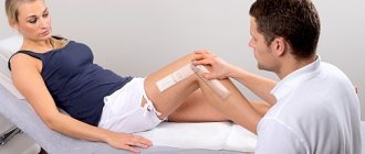
What happens before surgery?
First, a comprehensive clinical and imaging examination is carried out, which, in addition to a history and physical examination of the patient, includes x-rays under stress, as well as magnetic resonance imaging (MRI).
This will make it easier for the doctor to determine the extent of damage to the articular cartilage. If all the examinations completed show that the above treatment for arthrosis is suitable for the patient, the attending physician conducts an explanatory conversation with him, during which he explains the course of the operation, and also talks about possible complications after the intervention. After this, an anesthesiologist talks with the patient, who once again checks the patient’s health status. As a rule, the operation is performed the next day after the consent of the surgeon and anesthesiologist.
How is arthroscopic chondrocyte transplantation performed?
Transplantation of articular cartilage of the knee joint: after culturing the cells in a special laboratory, implantation of chondrocytes is carried out using a minimally invasive technique.
© dissoid, Fotolia Transplantation of cartilage cells obtained in the laboratory is always carried out arthroscopically, that is, in a minimally invasive manner. To insert the necessary instruments into the knee, the surgeon only needs minimal incisions.
At the beginning of the operation, the knee joint is cleaned of damaged cartilage tissue, after which grown cartilage balls (chondrospheres) are placed on the damaged area of the knee. Thus, the chondrospheres immediately adhere to the bone and after a few weeks correct the defect by forming connective tissue of the cartilaginous type.
After contact of cartilage cells with the prepared site of the cartilage defect, connecting molecules (adhesive proteins) begin to firmly adhere to the bone within 10 minutes. In this way, the chondrocytes grow into the damaged part of the knee until it is completely filled. If there is no need for correction of the cruciate ligaments or meniscus, surgical treatment of arthrosis of the knee joint by autologous chondrocyte transplantation at the Gelenk Clinic lasts approximately 30-60 minutes.
In order for the surgical treatment of knee arthrosis using the above mentioned method to be successful, it is necessary to identify and, if necessary, treat the problems (for example, deformation pathologies, rupture of the cruciate ligament, meniscus injuries) that caused the injury to the articular cartilage. That is why the patient needs to take into account long-term postoperative treatment, as well as the need to take care of their health.
Mosaic plastic surgery - questions on biomechanics and histology
There are several questions regarding mosaic plastic surgery of the knee joint:
What is the minimum size of a cartilage defect that can be closed with mosaicplasty?
Studies on cadavers show that the maximum pressure at the periphery of the defect is observed when its size is more than 10 mm. Therefore, this threshold value is used to determine indications for surgery.
Does mosaicplasty reduce the load on cartilage?
It was found that destruction of cartilage over an area of 16 mm (about 2 cm2) leads to an increase in peripheral tension by 92%, which further increases the destruction of cartilage and contributes to increased pain. When three 8-mm grafts are transplanted, peripheral tension increases by only 35%
Where is the best place to get transplants?
The optimal location for the fence is the areas with the least load, sufficient thickness and curvature of the surface similar to the recipient area.
Studies have shown that the lateral and lateral surfaces of the femoral condyles experience the least load in the knee joint. It is from these areas that transplants are taken.
Does the curvature of the grafts matter?
Restoring the curvature of the joint as close as possible to the original one is very important for even distribution of the load. Therefore, it is very important to select the curvature of the grafts.
Is the thickness of the cartilage on the donor fragment important?
The thickness of cartilage directly depends on the load and varies in different parts of the joint. Naturally, in donor areas the cartilage is somewhat thinner (on average 1.8 mm), and in areas requiring restoration it is thicker - up to 2.5 mm. However, this difference does not cause big problems.
Arthroscopy or arthrotomy - which is better when harvesting a transplant?
In a study by Keeling et al. it was shown that grafts obtained by arthroscopy had a discrepancy of up to 1 mm in 69% of cases, and by arthrotomy - in 57%. It was noted that arthroscopy is more difficult when harvesting the graft from the lateral surface of the condyles, and it also increases the risk of marginal fractures.
What determines the stability of the graft?
Animal studies have established the following facts:
- grafts with a diameter of 11 mm and a length of 15 and 20 mm have better vertical stability;
- ideal adjustment of the graft length to the depth of the “planting socket” ensures a 2-3 times increase in stability;
- grafts with press-fit fixation are more stable (this effect is achieved due to the fact that the transplanted osteochondral fragment itself is slightly wider than the hole for implantation).
What happens if the curvature of the graft does not match the curvature of the area being restored?
In a study on sheep, it was shown that a curvature mismatch of up to 1 mm is acceptable and the cartilage is preserved; if the discrepancy reaches 2 mm or more, then aseptic necrosis of the graft occurs and it gradually resolves.
Does the viability of graft cartilage depend on the force during its fixation?
When the pressure force during fixation is up to 10 MPa, the chondrocytes do not suffer, but if pressure is applied with a force above 15 MPa, cartilage damage occurs. Therefore, it is recommended to insert the graft with several gentle pressures rather than intense ones.
What type of anesthesia does the patient receive during surgical treatment of knee arthrosis?
As a rule, the operation is performed under general anesthesia. Sometimes, in order to avoid the consequences of complete anesthesia, surgeons perform the intervention under spinal anesthesia, which is a type of local anesthesia. During this manipulation, an anesthetic is injected into the spinal canal of the lumbar spine, which allows the patient to remain fully conscious. The specialists at the orthopedic clinic Gelenk Klinik in Freiburg have many years of experience in both techniques. What type of anesthesia is most suitable in your individual case is discussed before the operation itself, taking into account all the tests and your state of health.
Will my knee hurt after surgery?
Surgical treatment of arthrosis of the knee joint, like other interventions, is associated with some pain. The high professionalism of our surgeons allows us to reduce the risk of pain to a minimum. As a rule, before surgery, a drug blockade of the damaged knee joint is performed, which relieves pain in the knee for about 30 hours. After this, the worst wave of pain is over and treatment can be carried out using conventional medications. Our goal is maximum pain relief for the patient.
Mosaic chondroplasty of the patella and tibial articular surface
As we know, the knee joint has two more articular surfaces - the inner side of the patella and the articular surface of the tibia. They can also become damaged and require “repair”.
In the case of the patella, mosaic chondroplasty is not difficult. It is always performed in an open manner, as it requires dislocation of the patella. The source of grafts is usually the intercondylar groove, and the osteochondral fragments themselves are thinner - no more than 12 mm (for operations on the femoral epicondyles, their length is 15-18).
Carrying out mosaic chondroplasty of the articular surface of the tibia of the knee joint is technically quite difficult. This requires a guide ligamentoplasty of the anterior cruciate ligament - this provides access to the operation site. The sources of transplants are still the same.
Conditions of accommodation at the Gelenk Clinic
Private room at the Gelenk Clinic in Gundelfingen in Germany
As a rule, during an inpatient stay at the clinic you are in a separate room with a shower and toilet. In addition, we provide you with towels, a robe and slippers. You can also use the minibar and safe. Each room has a window.
You only need to bring your own medications, comfortable clothes and nightwear. After surgical treatment of arthrosis of the knee joint, we guarantee 24-hour care by qualified staff and experienced physiotherapists. Generally, the length of hospital stay after surgery is three days. Your family members can stay in a hotel, which is located a few steps from the clinic. We will be happy to take care of your hotel room reservation.
What should you pay attention to after surgery?
Immediately after surgery, the knee should be cooled and kept elevated. After approximately 12 days, the stitches are removed. After this you can take a shower again.
In order to avoid complications after surgical treatment of arthrosis of the knee joint, we recommend not placing special stress on the knee. During this time, you will be given sick leave and crutches with elbow support. Prevention of thrombosis during periods of inability to perform full exercise is mandatory. At this stage, physiotherapeutic treatment is a very important element in order to counteract the loss of muscle mass and preserve the function of the knee joint.
The return flight can be planned no less than 10 days later. However, we recommend flying home only after two weeks.
Physiotherapy is carried out to preserve the functions of the knee joint and strengthen the knee muscles.. © Elnur, Fotolia
Recommendations after arthroscopy (1st operation)
- Inpatient treatment: 2 days
- Recommended time of stay in the clinic: 7 days
- When is a return flight possible: 10 days after surgery
- When can you shower: after 5 days
- Recommended duration of sick leave: 2 weeks
- When the stitches are removed: after 5 days
- When can you drive again: after 2 weeks
Recommendations for autologous chondrocyte transplantation or elimination of the cause of articular cartilage damage (2nd operation)
- Inpatient treatment: 3 days
- When to stop using elbow crutches: after 6 weeks
- Gradual restoration of normal physical activity on the operated leg: after 6-8 weeks
- Recommended time of stay in the clinic: 10 days
- When is a return flight possible: 10 days after surgery
- Recommended return flight: after 2 weeks
- When can you shower: after 7 days
- Recommended duration of sick leave: 6 weeks
- When the stitches are removed: after 7-12 days
- When is it safe to drive again: 6-12 weeks
What are the causes of destruction of cartilage tissue?
The main reason for the destruction of cartilage is a decrease in the quality of synovial fluid - a natural lubricant that protects the joint from excess friction and provides its nutrition. After all, there are no blood vessels in cartilage, but only “pores” that are saturated with joint lubricant. This means that degenerative processes in cartilage can be triggered by:
- insufficient production of joint lubrication
(even with a physiologically normal volume of lubrication there may not be enough due to increased loads on the joint); - lack of nutrients
in the synovial fluid (for example, due to a poor diet or poor tissue nutrition); - lack of conditions for the circulation of joint fluid
(sedentary lifestyle, immobilization of a limb after injury) - after all, in order for it to wash the joint, it must be alternately in a state of loading and rest; - insufficient consumption of clean drinking water
and, as a result, chronic dehydration - cartilage, which is 70-80% water, loses its elasticity and begins to crack.
In case of injury, excessive stress without rest, or, less commonly, a systemic disease (metabolic or autoimmune), the nutrition of the periarticular tissues is disrupted. Due to inflammation, nutrients cease to flow normally into the synovial fluid, causing its production and quality to suffer, and the restoration of cartilage tissue slows down or stops. The cartilage is “starving”. When a limb is immobilized after an injury, there is no circulation of synovial fluid at all. Because of this, cartilage tissue loses its shock-absorbing properties and begins to deteriorate and become thinner. That is why, after an injury, it is important to correctly dose the load, receive proper anti-inflammatory treatment, drugs for the restoration of cartilage tissue and supportive therapy.
In addition to poor diet, physical inactivity and injury, cartilage destruction can be caused by:
- infectious diseases, especially chronic ones;
- endocrine disorders;
- chronic stress and lack of sleep;
- difficult working conditions;
- congenital or acquired anomalies of the musculoskeletal system;
- hypothermia or overheating;
- age-related changes;
- overweight;
- bad habits;
- genetic prerequisites.
How much does it cost to treat knee arthrosis with autologous chondrocyte transplantation?
In addition to the cost of the surgical treatment itself, treatment of knee arthrosis with autologous chondrocyte transplantation also involves diagnostics, doctor appointments, and additional resources such as elbow-supported crutches. Thus, the amount can range from 1,500 to 2,000 euros. If you require physiotherapeutic treatment after your stay at the Gelenk Clinic, we will be happy to help you and provide you with a preliminary cost estimate.
Information regarding the cost of hotel accommodation, as well as subsequent treatment at the rehabilitation clinic, can be found on the website of the clinic itself.
How can a foreign patient make an appointment and the operation itself?
In order to assess the condition of the knee joint, it is necessary to provide the results of imaging diagnostics - X-rays, as well as MRI. After viewing the sent images on our website, within 1-2 business days you will receive all the necessary information, a treatment proposal, as well as a cost estimate for surgical treatment of arthrosis of the knee joint.
Foreign patients can make an appointment with a Gelenk Clinic specialist in a short time frame that suits their plans. We will be happy to assist you with obtaining a visa after the advance payment indicated in the cost estimate has been received into our account. If your visa application is refused, we will refund your advance payment in full.
For foreign patients, we try to reduce the time interval between the preliminary examination and surgical treatment of arthrosis of the knee joint to a minimum.
This way you will not need to come to the clinic several times. During your outpatient and inpatient stay at the Gelenk Clinic in Freiburg, our multilingual patient management staff (English, Russian, Spanish, Portuguese) will answer all your questions. In addition, we provide a translator (for example, into Arabic), which is paid for by the patient himself. Also, we will be happy to help you organize a transfer, find a hotel and tell you how to spend your free time in Germany for you and your relatives. Send request
How to start the restoration of cartilage tissue in joints?
If treatment is started in a timely manner, conservative methods of restoring cartilage tissue of joints are used. These include:
- treatment with chondroprotectors;
- massage (classical therapeutic, hydromassage, lymphatic drainage, vacuum, self-massage);
- physiotherapy;
- taking vitamins (at least 2 times a year, in the autumn-spring period), homeopathic remedies and blood microcirculation correctors;
- swimming;
- physiotherapy;
- injection therapy (intra-articular administration of hyaluronic acid, platelet-rich plasma, Orthokine);
- use of orthoses (orthopedic bandages, corsets, insoles, as well as orthopedic furniture, backpacks, etc.).
It is also important to eliminate habits that increase its destruction:
- addiction to tobacco and alcohol;
- excessive consumption of coffee (especially decaffeinated);
- overeating or eating 1-2 times a day;
- lack of sleep;
- penchant for sweet and salty foods;
- self-treatment of infectious diseases and injuries;
- excessive exercise;
- the habit of dressing inappropriately for the weather.
If the cartilage is not subject to natural restoration, in addition to taking chondroprotectors to restore cartilage tissue, surgical treatment is recommended - for example, arthroscopic debridement (cleaning and polishing of cartilage), arthroscopic microfracture (creating holes for blood flow with growth factors), osteochondral transplantation (transplantation of cartilage of a patient from an unloaded area of the joint), transplantation of cultures of chondrocytes grown in the laboratory from a fragment of healthy cartilage, and others.