Home » Diseases » Treatment of the musculoskeletal system » Spinal spondylitis » Tuberculous spondylitis
Tuberculous spondylitis is a specific disease of an infectious nature that develops as a result of penetration of Koch's bacillus into one or more segments of the spine. This pathology, accounting for about 30–40% of the total number of cases of bone tuberculosis, can affect any part of the spinal column and lead to the formation of severe neurological disorders and deformation of the skeletal axis.
What it is?
Tuberculous spondylitis is the name given to pathological changes occurring in the spine . They are caused by an infectious pathogen. For this reason, this disease is classified as a chronic infectious pathology.
The development of the disease occurs due to infection with the tuberculosis bacillus , known as Koch's bacillus. The name of the disease - Pott's disease - was given in honor of the name of the doctor who was the first to describe its clinical signs back at the end of the 18th century. The disease is most often chronic. In most cases, patients complain of intense spasms in the back and problems with the functioning of the affected vertebrae.
Picture of the course of the disease
The development of the disease begins with the penetration of Koch's bacillus into the structure of the vertebrae . Most often it comes from the bloodstream, but can also spread through the lymph or by contact, due to which it moves from the primary site of inflammation located in the lung tissue.
In appearance it may look smooth or curved. Its length can reach 10 microns, and its width is 04.06 microns. After hitting the spine, the stick actively multiplies. This period sometimes takes up to two years, after which the patient may first notice clinical signs.
The result of the proliferation of the tuberculosis bacillus is complete destruction or deformation of the vertebrae , as well as the intervertebral discs adjacent to it. Then a closed fibrous membrane forms around the purulent exudate (abscess).
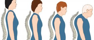
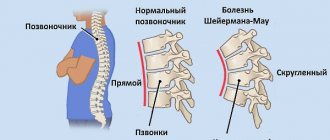
Tuberculous spondylitis provokes deformation and destruction of the vertebrae
After this, the infection moves to neighboring vertebrae, which it also gradually destroys. These processes lead to the formation of compression fractures and deformation of the spine. Children may begin to develop a hump.
Degrees and classification
According to the nature of localization, the following types of tuberculous spondylitis are distinguished::
- cervical;
- chest;
- lumbosacral.
This pathology can occur in any of these segments.
Prevalence
Tuberculous spondylitis is most often observed among the population of developing countries , which include Central and South America, some African countries and Asian countries, primarily India, China, Nepal, and Indonesia. It is in countries where the standard of living is very low that the incidence of this disease is highest.
In most cases, the disease affects children and adolescents . Among adults, pathology occurs in very rare cases. At the same time, the child’s body is characterized by increased sensitivity to bacteria, the destructive effects of which can affect up to four vertebrae simultaneously. The area of damage in an adult organism is significantly smaller, as is the speed of spread of the pathology.
Video: “Clinical picture of tuberculous spondylitis”
Causes of tuberculous spondylitis
The main and only cause of tuberculous spondylitis, as clearly evidenced by the name of the disease, is Mycobacterium tuberculosis, named after its discoverer Koch's bacillus. In most clinical cases, the pathogen, together with the blood flow from the primary inflammatory focus located in the lung tissues, is carried into the spinal motion segments.
Risk factors that can provoke the development of an infectious-inflammatory process include:
- decrease in the body's immune forces;
- unfavorable socio-economic conditions (for example, living in areas where infection is widespread);
- malnutrition;
- childhood and adolescence;
- immunosuppressive state (immune suppression) after organ transplantation;
- long-term use of hormonal drugs;
- chronic internal diseases;
- malignant tumors.
Risk factors, causes
The greatest predisposition to the development of this pathology is noted among the following categories of people:
- people caught in the epidemic zone;
- those who have chronic diseases of internal organs and systems;
- population living in unfavorable conditions, especially if there are sanitary and hygienic violations;
- children and adolescents;
- patients suffering from endocrine system disorders;
- people who have undergone organ implantation surgery;
- patients undergoing hormonal therapy;
- people who have been diagnosed with a malignant neoplasm.
Consequences
If the necessary therapeutic measures are not applied to the identified disease, the following complications may occur::
The main complication of Pott's disease is disruption of the functioning of the spine; problems with the motor function of the spine;- paralysis of the lower or upper limbs;
- bladder dysfunction;
- pathologies in the functioning of the large intestine;
- problems with reproductive function;
- tuberculous meningitis;
- breakthrough of the abscess and its penetration into the lumen of the spinal canal, leading to epiema (a complication that represents the accumulation of pus in any body cavity or inside organs).
Due to the risk of disease outcome, it is important to start therapy as early as possible to avoid the development of serious complications. If proper treatment of the disease is not carried out or started too late, there is a high risk of death .
Symptoms of tuberculous spondylitis
Tuberculous spondylitis can develop either several months or several years after the initial infection. The first symptoms of the disease include dull pain in the back, intensifying towards the end of the day and receding after a night's sleep or a long daytime rest. As the pathological process develops, the patient begins to develop muscle contracture (a state of involuntary tension in the long back muscles). As a result, the spine at the site of the lesion stops bending.
Further, the above symptoms are accompanied by pain, which occurs even with a slight load (when pressing on the shoulders or head). Often the pain intensifies when sneezing, coughing, jumping, or going down stairs. In case of damage to the cervical spine, difficulties arise when tilting and turning the head; many patients support their chin with their hands while sitting.
Subsequently, as the deformation of the spinal cord develops, symptoms of brain compression arise, the function of the organs of the genitourinary system is disrupted, and the so-called. drip abscesses (accumulations of pus in the immediate vicinity or at a distant distance from the tuberculosis focus), which do not cause acute inflammatory changes. In particularly severe cases, loss of sensation and motor function can lead to complete paralysis of the upper and lower extremities.
If you notice the first symptoms of trouble, you should immediately contact a specialist. The disease can only be correctly diagnosed in a medical facility.
Symptoms and diagnostic methods
Did you know that...
Next fact
Symptoms of the pathology include the following::
- lung damage, which can be detected by a persistent cough accompanied by copious sputum, hemoptysis, shortness of breath, and chest pain;
- feeling of general weakness and malaise;
- increased fatigue;
- loss of performance, weakening of attention;
- nausea;
- headaches;
- a slight increase in body temperature to 37.0-37.5 degrees.
Such disturbances can be observed over a long period from 2-3 months to two years . Subsequently, if left untreated, the infection begins to spread to the spine.
During this period, patients complain of the following negative symptoms : pain in the spine; movement disorders; curvature of the spinal column.
| If the cervical spine , you may experience: | Patients suffering from thoracic note: | People who have been affected in the lumbosacral region notice: |
|
|
|
In advanced forms of damage, the formation of the Pott triad occurs:
- the appearance of abscesses;
- the appearance of a hump - curvature in the thoracic area;
- paralysis of limbs.
If you suspect the development of this disease, patients should seek professional medical help as soon as possible.
First of all, the doctor usually prescribes the following diagnostic procedures:
- general urine and blood tests, as well as glucose levels;
- biochemical study (bilirubin, protein content, level of transaminases, alkaline phosphatase, urea, creatinine);
- study of protein fractions using the proteinogram method;
- rheumatological test;
- study on the composition of blood serum.
Instrumental methods are also used : x-ray; CT scan; Magnetic resonance imaging; ultrasonography.
Consequences of tuberculous spondylitis on MRI
Patients are recommended to be examined by the following doctors : therapist; neurologist; traumatologist; rheumatologist; phthisiatrician.
Video: “Diagnostics of tuberculous spondylitis”
Also read with this article:
- You can read about the diagnosis of spinal spondylodiscitis on the page
- Symptoms and treatment of spinal osteomyelitis
- You will find information about the symptoms and treatment of spinal myelitis here
- To explore the causes and risk factors for spinal discitis, click here
- You can find out when an epidural abscess of the spine occurs in the following article.
Spondylitis - what kind of disease is it?
Spondylitis refers to chronic inflammatory diseases of the spinal column. Spondylosis is a form of spondylopathy, during which destruction of the vertebral body occurs, and this is fraught with multiple deformities. Spondylitis is rare in the population and is associated with a bacterial or autoimmune complication. The prevalence does not exceed 0.6% of cases in the world. The disease can occur at any age and its course does not depend on gender. Spondylitis can be specific, associated with other chronic diseases, and nonspecific, which is an independent malfunction in the body.
Treatment
Therapy is carried out exclusively in specially equipped TB hospitals.
Provided that treatment is started on time, therapy can be very successful, especially in the early stages.
A long full course of therapy is protracted and can last from 6 months to several years.
A person with tuberculous spondylitis is considered contagious. To avoid infecting others, he should be placed in a TB department. He needs strict bed rest, long-term antibiotic therapy and exercise restrictions. .
Drugs
The following types of treatment are usually prescribed from medications::
- etiotropic therapy , in which several anti-tuberculosis drugs are used at once (Rifampicin, Ethambutol, Isoniazid, Streptomycin). The course of treatment can be 2-6 months;
- symptomatic therapy , which uses corticosteroids, muscle relaxants, non-steroidal drugs and B vitamins.
Surgery
Surgery can be used in cases where the effect of conservative therapy is not observed , and drainage of abscesses is required, and progression of neurological disorders is observed.
The operation may be scheduled after the acute phase has subsided in the first year of the disease. The operation consists of removing necrotic tissue. In this case, the vertebrae are replaced with prostheses and compression is eliminated. The spinal column must also be strengthened with plates and screws. An abscessectomy may be performed.
Exercise therapy and massage
Patients who have undergone surgery require long-term rehabilitation, during which wearing a plaster corset, physical therapy and physiotherapy are recommended.
Treatment at home
Patients suffering from this disease can benefit greatly from sanatorium treatment. It is important to properly organize your work and rest schedule, avoid excessive stress, spend more time in the fresh air, and stop smoking and drinking alcohol.
Pott's disease is treated in a hospital. Therefore, the use of folk remedies acts as an additional therapy. You need to carefully plan your diet. Meals should be taken at least 5 times a day. The diet should consist of high-calorie foods enriched with vitamins.
It is useful to drink the following drinks:
- freshly squeezed carrot and celery juice 100 ml before meals once a day;
- brewed rosehip;
- infusion of St. John's wort. It can be prepared by brewing 10 g of herbs with boiling water and leaving for a third of an hour. Then strain. Drink up to three times a day, a tablespoon before meals.
For this pathology, taking a mixture of walnuts and honey can be very effective. To prepare it, mix 3 tbsp. chopped nuts and one tablespoon of honey. Take twice a day, morning and evening.
Prevention
To avoid the development of complications with this disease, it is necessary:
- timely identification of its first signs and prescription of competent therapy;
- undergoing an annual examination, including fluorography;
- careful attention to personal hygiene;
- improving the quality of living conditions;
- educational work in the field of sanitary and hygienic standards.
Spinal tuberculosis
Mycobacterium tuberculosis (MBT) is a facultative intracellular parasite. Mycobacterium tuberculosis (MBT) belongs to the family of bacteria Micobacteriacae, order Actinomycetalis, genus Mycobacterium. The genus Mycobacterium has over 100 species, most of which are saprophytic microorganisms widely distributed in the environment.
Etymologically, the word “mycobacterium” comes from the Greek words myces - mushroom and bacterium, bactron - stick, twig. The "mushroom" component of the name comes from the tendency of these microorganisms to form filamentous and branching mold-like forms.
From the standpoint of clinical medicine, Mycobacterium tuberculosis, discovered by the German scientist Robert Koch, is the most important type of actinomycetes, which are united in a complex that includes M. tuberculosis (MBT); M. bovis and its variant BCG (Bacillus Calmette-Guerin); M. africanum and M. microti. This group of mycobacteria is distinguished by pronounced genetic similarity.
M. microti is considered non-pathogenic to humans, but causes a disease in mice resembling tuberculosis. BCG culture is not pathogenic for humans. Mycobacterium tuberculosis (MBT) is the cause of human tuberculosis in up to 95% of cases, depending on the area of residence. However, M. bovis and M. africanum cause a disease in humans that is clinically no different from classical tuberculosis.
Mycobacteria not included in the M. Tuberculosis complex can cause mycobacteriosis. Such mycobacteria are combined into complexes, the most important of which are: M. avium, M. fortinatum and M. terrae, M. leprae, M. ulcerance.
The materials presented below on tuberculosis are relevant only to the disease caused by M. tuberculosis (MBT) - Koch bacteria (KB), typus humanus.
The natural reservoir of mycobacterium tuberculosis is humans, domestic and wild animals, and birds.
MBTs are externally thin, curved sticks that are resistant to acids, alkalis and drying. The outer shell of the bacterium contains complex waxes and glycolipids.
MBT can multiply both in macrophages and outside cells.
MBT reproduce relatively slowly. Reproduction occurs mainly by simple cell division. On enriched nutrient media, MBT multiply with a doubling period of 18 to 24 hours. For growth in culture of Mycobacterium tuberculosis obtained in clinical conditions, it takes 4 to 6 weeks.
The genetic structure of MBT has been established. The nucleotide sequence of MBT can be found in international data banks. The nucleotide sequence of MBT (strain H37Rv) is 4,411,529 bp
MBTs do not have independent movement. Temperature limits for growth are between 29 and 42 °C (optimal - 37-38 °C). MBT are resistant to physical and chemical agents ; they remain viable at very low temperatures, and can withstand increases up to 80 °C for 5 minutes.
In the external environment, Mycobacterium tuberculosis is quite stable. It can survive in water for up to 150 days. Dried mycobacteria cause tuberculosis in guinea pigs after 1-1.5 years, lyophilized and frozen are viable for up to 30 years.
With intense sun irradiation and high ambient temperatures, the viability of MBT decreases sharply; on the contrary, in darkness and dampness their survival rate is very significant. Outside a living organism, they remain viable for many months, especially in dark, damp rooms.
MBTs are detected by a unique staining property (acid fastness), which distinguishes them from many other infectious agents. Ziehl and Neelsen in 1883 developed a special contrast method for staining MBT, based on the property of acid resistance. Unlike non-acid-fast bacteria, tuberculous mycobacteria are stained red, do not discolor when exposed to an acid solution, and are clearly visible against a blue background under microscopy. The Ziehl-Neelsen method is still one of the main methods for staining MBT during microscopy. More sensitive than the acid-fast staining method is MBT auramine staining followed by fluorescence microscopy.
The lipid fraction of the outer shell of MBT is associated with the resistance of tuberculosis pathogens to acids, alkalis and alcohols.
Variability of MBT morphology. The morphology and size of the office are not constant; it depends on the age of the cells and especially on the living conditions and composition of the nutrient medium.
Cord factor. Lipids in the surface wall of mycobacteria determine its virulence and ability to form clusters of bacteria in the culture in the form of braids (cord factor).
The cord factor was already mentioned by Koch in his initial message regarding MBT. Initially, the cord factor was associated with the virulence of MBT. The ability to form braids is observed among other mycobacteria that have low or no virulence. The cord factor, as it was later established, is associated with an unusual biological substance trehalose 6,6-dimyco-late, which is highly virulent.
L-shape. One of the important types of MBT variability is the formation of L-forms. L-forms are characterized by a reduced level of metabolism and weakened virulence. Remaining viable, they can remain in the body for a long time and induce anti-tuberculosis immunity.
L-forms are distinguished by pronounced functional and morphological changes. It was found that the transformation of MBT into L-forms increases with prolonged exposure to antibacterial therapy and other factors that disrupt their growth and reproduction, and the formation of the cell membrane.
It has been established that the sputum of “abacillary” patients with destructive forms of tuberculosis may contain L-forms of MBT, which, under appropriate conditions, can reverse (modify) into a rod-shaped variant, thereby causing reactivation of the tuberculosis process. Consequently, abacillation of the cavities of such patients does not yet mean their sterilization with respect to MBT.
MBT by its nature is insensitive to many antibiotics. This property is primarily due to the fact that the highly hydrophobic cell surface serves as a kind of physical barrier to therapeutic agents and antibiotics. The main reason for resistance is encoded in the structure of the genome of the tuberculosis bacillus.
At the same time, MBT can develop resistance (resistance) to anti-tuberculosis drugs. In recent years, simultaneous drug resistance of MBT to several drugs has significantly reduced the effectiveness of tuberculosis treatment.
As a result, modern healthcare deals not just with the dangerous pathogen of tuberculosis, but with a whole set of its strains that are resistant to various drugs. In practice, to organize effective treatment of tuberculosis, it is important not only to detect MBT, but also to simultaneously determine their resistance, and quickly enough - within two to three days - in order to prescribe effective chemotherapy in a timely manner.
At the end of the 80s. In the last century, a method has emerged that significantly reduces the time of such analysis. New diagnostics are based on selective amplification of nucleic acids (DNA or RNA) in vitro using polymerase chain reaction (PCR).
The PCR method has great potential and underlies precise DNA diagnostics, which allows one to identify any MBT strain and determine the root cause of a particular drug resistance.
Laboratory studies have shown that the emergence of resistance in M. tuberculosis is associated with nucleotide substitutions (mutations) in genes encoding various enzymes that directly interact with drugs.
The resistance of some MBT strains to isoniazid is associated with mutations in the katG gene, leading to the replacement of some amino acids in the enzymes catalase and peroxidase.
MBT insensitivity to streptomycin is associated with a missense mutation in the rpsL gene, encoding the S12 mitochondrial protein, or with nucleotide substitutions in the rrs gene, encoding 16S RNA.
Source of infection. The main source of MBT is a person with tuberculosis who spreads MBT (bacillus shedding) .
The focus of tuberculosis infection becomes dangerous in cases where patients suffer from an open form of tuberculosis, i.e. secrete mycobacterium tuberculosis. Of particular importance when infected with tuberculosis is direct, long-term and close contact of a healthy person with the bacilli shedding agent. Infection can most often occur in a family, place of residence, or group where there is a patient with tuberculosis who secretes mycobacteria. The danger of dispersal of the infectious principle is eliminated if the bacilli-releasing agent is promptly identified and isolated.
The occurrence and course of infection depend not only on the virulence of the pathogen, but also on the state of resistance and reactivity of the macroorganism.
Of great importance is the place of penetration of MBT into the body, where primary contact with the microbe is made (entry gate of infection). The following routes of transmission of tuberculosis are distinguished: 1) airborne; 2) nutritional (through the digestive tract); 3) contact; 4) intrauterine infection with tuberculosis.
Airborne transmission of tuberculosis Mycobacterium tuberculosis enters the air through droplets when a patient with active tuberculosis coughs, talks, or sneezes. When inhaled, these contaminated droplets enter the lungs of a healthy person. This method of infection is called airborne infection.
Depending on the strength of the cough impulses and the size of the droplets, MVTs spread in the air over different distances: when coughing - up to 2 m, when sneezing - up to 9 m. On average, sputum particles disperse over a distance of 1 m right in front of the patient.
Droplets of tuberculosis sputum that settle on the floor dry out and turn into dust particles. The tuberculous mycobacteria found in them remain viable in the dust for some time. It has been established that by the 18th day 1% of living bacteria remains in dried sputum. When there is strong air movement, sweeping the floor, or people moving, dust particles containing mycobacterium tuberculosis rise into the air, enter the lungs and cause infection.
Alimentary route of infection through the digestive tract Special experiments on animals show that the nutritional route requires a significantly larger amount of mycobacteria than aerogenic infection. If one or two mycobacteria are enough when inhaled, then hundreds of microbes are required for infection through food.
The ways of spread of tuberculous mycobacteria in the human body during alimentary infection with a tuberculosis culture are demonstratively shown by sectional materials published in connection with the trial in Lübeck. By mistake, 252 infants were given a tuberculosis culture (Kiel strain) instead of BCG during per os vaccination. As a result of infection, 68 children died from tuberculosis, 131 children fell ill and 53 remained healthy.
During the autopsy of the corpses of 20 deceased children, it was found that in most cases the process was localized in the abdominal organs.
The entry point for infection was the digestive organs.
One of the features of this route of infection in young children is the frequent involvement of mesenteric lymph nodes by tuberculosis.
It must be borne in mind that the penetration of tuberculous mycobacteria into the intestines can also occur when patients with pulmonary tuberculosis swallow their own bacillary sputum, which is confirmed by using the flotation method of gastric lavage water.
Contact transmission of tuberculosis Cases of infection through the conjunctiva of the eyes of small children and adults have been described; in this case, acute conjunctivitis and inflammation of the lacrimal sac are sometimes detected.
Infection of tuberculosis through the skin is rare. Cases of tuberculosis in milkmaids due to the penetration of MBT through damaged skin of the hands from cows with tuberculosis have been described.
Intrauterine infection with tuberculosis The possibility of infection of the fetus with tuberculosis during intrauterine life has been established in the section by cases of tuberculosis in children who died in the first days after birth. Infection occurs either when the placenta is affected by tuberculosis, or when the damaged placenta becomes infected during childbirth by a tuberculous mother. This route of infection with tuberculosis is extremely rare.
Immunity Morphological and biochemical components of a microbial cell cause various reactions in the body.
The main biochemical components of MBT are proteins, carbohydrates, and lipids. Proteins (tuberculoproteins) are the main carriers of the antigenic properties of MBT.
Tuberculin is one of the tuberculoproteins widely used in practice to detect MBT infection.
Delayed-type hypersensitivity (DSHT) Substances that make up the outer shell of the office induce a specific tissue inflammatory reaction of the macroorganism and the formation of granuloma. At the same time, delayed-type hypersensitivity (DHT), determined by the reaction to tuberculin tests, and weak antibody formation appear. Mainly, HCT is used to characterize the type IV immune response (the presence of induration that developed after 48 hours at the site of intradermal tuberculin administration) in individuals infected with MTB. However, HCT is associated with an immune response to factors that damage tissue.
Relationship between the immune response and pathogenesis Local and generalized tuberculosis damage in the body is determined by the protective reactions produced by the body's immune system against MTB. When describing this complex process, we will limit ourselves to a simple listing of the events that occur from the moment of the initial penetration of MBT into the alveoli until the results of the natural struggle between the macroorganism and the MBT. This process determines the fate of at least a third of the world's population that is infected with Mycobacterium tuberculosis.
The development cycle of tuberculosis from infection of the body with Mycobacterium tuberculosis to clinical manifestations of the disease and the spread of MTB in the environment can be divided into 5 stages.
Stages 1. Spread of infection (infection). 2. Onset of infection, proliferation and dissemination in the infected organism. 3. Development of the body's immune response. 4. Caseation (development of caseous necrosis) and accelerated reproduction of the office. 5. Secondary spread of infection (the ability to infect, infect).
Diagnosis of spondylitis
To determine this disease, it is necessary to conduct a thorough differential diagnosis, because the symptoms are not always obvious in a chronic course and often resemble more common signs of ridge pathology - osteochondrosis, osteoporosis, neurological disorders. A number of laboratory analyzes and instrumental studies are carried out.
What analyzes and types of instrumental studies need to be carried out:
- Radiography. This is a basic research method that allows us to identify such pathological changes as narrowing of the interarticular space, subchondral sclerosis, bone erosion, ankylosis, osteoporosis, and degeneration of the vertebral bodies. If such signs are visible on the X-ray image and the patient complains of stiffness and pain, then additional research must be carried out further.
- CT or MRI are more modern and accurate methods for diagnosing pathological changes in the ridge. Magnetic resonance imaging shows minute changes in bone and soft tissues, while CT scans clearly show hard tissues. Usually, with the help of these diagnostic methods it is possible to clarify the pathological picture completely. An important diagnostic criterion is that in persons with ankylosing spondylitis, unilateral or more often bilateral sacroiliitis is observed on MRI of the iliosacral joints.
- Blood tests. To clarify the overall diagnostic picture, you need to take a full blood test. If they are looking for an infectious or autoimmune cause of spondylitis, then such patients have characteristic changes in the OAC - a shift in the leukocyte count to the left is observed, hemoglobin is reduced and the erythrocyte sedimentation rate is increased, and leukocytosis is observed. Such test results allow us to get closer to the possible cause of the disease.
- Biochemical blood parameters. It is important to assess the state of the internal organs. Often, specialists are interested in kidney indicators and liver enzyme levels.
- Genetic markers. Most relevant when diagnosing ankylosing spondylitis. If a patient has an HLA-B27 antigen, this indicates a high probability of developing this disease.
- Tests for the presence of antibodies to viral and bacterial pathogens - Yersinia, chlamydia, herpes virus, cytomegalovirus. If antibodies are present to these pathogens, then this may indicate a possible cause of activation of immune failure, which caused spondylitis of autoimmune origin.
If spondylitis of infectious origin is suspected, then a differential diagnosis of STDs is carried out. It is also important to determine the level of C-reactive protein in the blood.
Complications
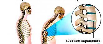
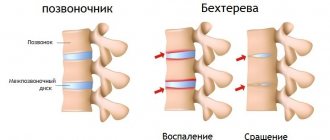
Spondylitis often progresses to ankylosis - a fixed formation consisting of bone and fibrous tissue. Ankylosis leads to complete immobility of the spine. The pathology is more common in rheumatic inflammation, including ankylosing spondylitis. With a long-term and chronic inflammatory process, a column of bone tissue is formed in the ridge, completely covering the vertebrae. In this area, inflammation goes away, because it is a protective reaction to the pathological course of the disease, but the vertebra completely loses flexibility.
Spondylitis on MRI
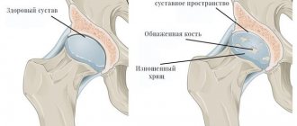
If the entire spine is covered with bone tissue, then it becomes like a bamboo stick and the patient takes a forced “proud” or “petitioner” pose (the head is always raised up or tilted down). If the bone growth is not surgically removed, the patient will not be able to assume a different body position. The second serious complication is a compression fracture. To create the outer bone frame, calcium is pulled from the spine, which leads to osteoporosis. As a result, the patient often receives compression fractures, which are dangerous to health and life.
Important! There are no conservative methods for eliminating ankylosis. It is necessary to perform a complex surgical operation to remove bone growths manually by doctors. In order to agree to perform such an operation, it is necessary to have serious indications, including a strong fusion that interferes with life.