Gonarthrosis is a pathological condition that is characterized by degenerative processes occurring in the knee joint.
Gonarthrosis of the knee joint is characterized by destruction of cartilage, as well as deformation of the joint, accompanied by disruption of its functions. Most often, this disease affects older women.
You can undergo a course of treatment for gonarthrosis at the CELT multidisciplinary clinic! Anyone can become our patient—metropolitan registration is not required.
At CELT you can get a consultation with a traumatologist-orthopedic specialist.
- Initial consultation – 3,000
- Repeated consultation – 2,000
Make an appointment
Causes
In accordance with the reasons for its appearance, it is customary to distinguish two main types of gonarthrosis:
- primary - the cause of its appearance has not yet been established; as a rule, this is bilateral gonarthrosis, which occurs in elderly patients;
- secondary - occurs against the background of an existing injury or pathology of the knee joint at any age; As a rule, this is unilateral gonarthrosis.
In more than 20% of cases, the pathology develops after intra-articular fractures of the tibia, ligament rupture or meniscal deformation. Often gonarthrosis develops 5 years after the onset of the pathology, but in rare cases it appears after 3 months.
The appearance of the disease can be triggered by high load on the joint. People over the age of 40 who regularly work out at the gym are at risk. Excessive stress when performing heavy exercises can lead to degenerative changes. Not only training, but also stress on joints due to excess weight can lead to similar consequences. Therefore, you need to follow a diet and control your body weight. Genetic predisposition often causes gonarthrosis. If a person has weakened ligaments and is susceptible to arthritis, then he is at risk and should avoid excessive stress on the joints.
Symptoms
The development of the pathology is slow and at the initial stage, people notice mild pain in the joints while walking or climbing stairs. There is tightness and stiffness in the knee area. The main symptom is sharp pain when getting out of bed or when starting to walk after a long journey. After warming up the joint, the pain syndrome disappears. The disease does not manifest itself in any way externally except for swelling, as fluid accumulates. This leads to the formation of slight swelling and change in shape.
The second stage is characterized by severe pain during moderate physical activity and when walking. In a calm state, the pain disappears, after which it reappears with loads in the anterior-internal part of the joint. With sudden movements, a crunching sound may occur. The shape of the joint changes and becomes spherical due to the accumulation of fluid.
The third stage leads to acute pain during exercise and at rest. A person not only cannot sleep due to severe pain, but also wakes up at night and cannot fall asleep.
Due to impaired mobility of the knee joint, a person cannot bend the leg normally. The size of the joint increases, the shape of the legs changes (becomes O-shaped or X-shaped). This leads to a disturbance in gait, and in the most severe cases, the need for support (the person moves with the support of a cane).
Examination by a specialist at the first stage often does not lead to the detection of gonarthrosis due to the absence of external manifestations. At the second and third stages, there is a decrease in mobility, curvature of the limbs, and joint deformation. As synovitis develops, the shape becomes rounded and smoothed, and during palpation the specialist detects a fluctuation.
Symptoms of the disease
As with most arthrosis, the insidiousness of this pathology is due to the fact that at the very beginning of their development, they do not have pronounced symptoms, as a result of which their diagnosis is difficult.
A person should pay attention to symptoms such as heaviness in the knee and aching pain while walking. Many patients report discomfort in the knee joint in the morning after waking up or after resting in a lying position. To feel relief you need to walk a little.
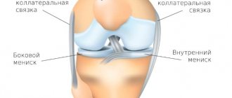
One of the signs of arthrosis is changes in the knee joint
Over time, the pain intensifies and is felt more on the inside of the knee. Often a crunching sound occurs while driving. Gradually, the process of bending or straightening the leg becomes more difficult. Progressive unilateral or bilateral gonarthrosis is manifested by lameness. At an advanced stage, a person can only move with the help of crutches. At first, the pain decreases when the person lies down, but over time it bothers him even at night.
With primary gonarthrosis, external changes in the knee joint are not observed. As the pathology develops, the deformity becomes noticeable, and a crunching sound is heard when the knee moves.
When palpating the knee, you can detect a painful area, which is usually located on the inside, but can be to the right or left of the joint.
Each degree of gonarthrosis has its own signs:
- Grade 1 gonarthrosis is characterized by: rapid fatigue of the legs and minor discomfort. Movements are accompanied by moderate crepitus, and the joint itself experiences limited mobility. There are no changes in the bone and only x-rays make it possible to identify the narrowed gap.
- Arthrosis of the knee joint 2 degrees is characterized by increased pain, especially after prolonged stress on the leg (walking, standing, etc.). More often there is a crunch and problems when moving the knee joint. Atrophy of the quadriceps femoris muscle is possible. It is difficult to fully bend or straighten the knee. As a result of radiography, the joint space is already noticeably narrowed, osteophytes are visible.
- With advanced grade 3 gonarthrosis, pain occurs even at rest. In the area of the damaged joint, there is an increase in temperature and swelling. When osteophytes break off, symptoms of blockade appear, which are characterized by acute pain. It is difficult for the patient to move, the joints are deformed. X-ray shows a significant change in the axis of the leg. The joint is practically motionless.
Stages and their clinical manifestations
It is customary to distinguish three stages of development of this disease, which are manifested by different symptoms and dysfunction of the knee joint:
Stage I gonarthrosis
This stage includes clinical manifestations characteristic of the onset of the disease.
As a rule, this is a dull pain localized inside the knee joint and occurs after a long walk. From time to time the knee joints swell, but there is no visual deformation.
Stage II gonarthrosis
This stage is characterized by an increase in existing clinical manifestations:
- the pain becomes prolonged and becomes more pronounced;
- when you move your knee, you can clearly hear a crunch in the joint;
- in the morning there is stiffness in the joints;
- there is a slight limitation in extension and flexion of the joint;
- the size of the joint increases.
Stage III gonarthrosis
At this stage, all the symptoms are fully manifested:
- pain bothers you constantly - both during exercise and at rest;
- restrictions on joint extension and flexion are clearly expressed;
- gait is disturbed;
- the knee joint is noticeably enlarged and deformed.
Gonarthrosis 3 degrees
IV stage of gonarthrosis
Grade 4 gonarthrosis is one of the stages of the disease, which is characterized by the development of degenerative processes in the knee joint.
Gonarthrosis 4 degrees
Degree of development of gonarthrosis
With grade 1 gonarthrosis, rapid fatigue of the limb and slight discomfort occurs. Moderate crepitus may be observed during movement. Sometimes there is a barely noticeable restriction of movement in the joint.
The bones of the knee at this stage do not undergo significant changes. On an x-ray you can see a slight narrowing of the joint space.
The second degree is characterized by the occurrence of pain, especially after you stand or walk for a long time. The characteristic crunch becomes more pronounced. There are problems with extension and full flexion of the limb. The so-called starting pain occurs.
Atrophy of the quadriceps femoris muscle may occur. The angle of full flexion and extension of the limb at the knee joint is significantly limited. X-rays clearly show significant narrowing of the joint space and proliferation of osteophytes. The edges of the bones are “flattened”.
At stage 3, the nature of the pain changes - it intensifies and can manifest itself at rest. Swelling and a local increase in temperature often occur in the joint area. Due to the possible presence of a “joint mouse” (fragments of broken osteophytes) in the joint, symptoms of joint blockage may occur. They manifest themselves as acute pain, accompanied by a feeling of jamming.
In this case, the patient may be completely deprived of the ability to move independently. Deformation of the knee joints occurs with a change in the axis of the limbs, which is clearly visible on x-rays. Joint instability may occur.
Anatomy and pathological changes in the joint with gonarthrosis
The knee joint consists of the surfaces of the tibia and femur. In the anterior zone there is the patella, sliding along the depression between the condyles of the femur. In this case, the fibula does not affect the functionality of the knee joint. The surfaces of the femur and tibia have a cartilaginous layer, the thickness of which is 5 mm. It absorbs shock and reduces friction during movements.
The disease begins to develop with a deterioration in blood supply in small intraosseous vessels through which blood enters the cartilage tissue. This causes the cartilage to become dry and thin, leading to a loss of shock-absorbing function. The second stage of pathology develops against the background of compensatory changes in bone tissue. The subchondral part becomes denser, and osteophytes (bone growths in the form of spines) form along the edges of the joint.
The joint capsule and synovium become dry and distorted, and the joint fluid thickens (impaired joint lubrication). Due to the lack of transport of nutrients to the cartilage tissue, destruction occurs. After the destruction of cartilage, degenerative changes accelerate, which provokes the development of the third stage of the disease. At a severe stage, indentation and change in the shape of the bones occur, which causes decreased mobility and acute pain in the knee joint.
Classification of pathology
There are many diagnostic methods, but their effectiveness can only be greatest with an integrated approach.
Examination by an orthopedist
An examination by an orthopedist is the first and very important diagnostic measure. This includes:
- Palpation of the joint;
- Linear bone measurements;
- Anglometry (determining mobility in the affected joint at different angles).
Tests for gonarthrosis include:
- Study of blood count and erythrocyte sedimentation rate (ESR);
- Determination of the level of fibrinogen, urea and other biochemical parameters of blood and urine.
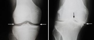
The main method for diagnosing gonarthrosis is to examine the diseased joint using X-rays. At the initial stage of development of the disease, an X-ray examination may not show anything, and if it does, it will only show minor changes. In the later stages, narrowing of the joint space, cartilage sclerosis, bone damage, and salt deposition are detected.
Ultrasound examination of a knee joint affected by arthrosis gives the best results, but it cannot completely replace radiography. Therefore, all people suffering from degenerative-dystrophic joint diseases should definitely take an x-ray.
This is the most advanced diagnostic method, which allows you to examine all parts of the joint layer by layer and determine the earliest changes in cartilage tissue. The disadvantage of the method is the fairly high cost of examining each segment. And the undoubted advantage is high accuracy: it often turns out that gonarthrosis is far from the only disease of the musculoskeletal system in a particular patient.
Diagnostics
Before starting treatment for gonarthrosis of the knee joint, CELT doctors carry out diagnostics that make it possible to identify the cause and extent of the disease, as well as determine subsequent treatment. To do this, an examination is carried out by a specialist, anamnesis is taken, an X-ray examination, computed tomography and magnetic resonance imaging are performed. An integrated approach allows you to study pathological changes in bone structures in detail, as well as determine changes in soft tissues.
Our doctors
Samilenko Igor Grigorievich
Traumatologist - orthopedist, doctor of the highest category
24 years of experience
Make an appointment
Zubikov Vladimir Sergeevich
Traumatologist-orthopedist, Doctor of Medical Sciences, doctor of the highest category, professor
44 years of experience
Make an appointment
Marina Vitaly Semenovich
Traumatologist-orthopedist, head of the minimally invasive traumatology and orthopedics service
Experience 36 years
Make an appointment
Poltavsky Dmitry Ilyich
Traumatologist-orthopedist
Experience 28 years
Make an appointment
Treatment
It is currently impossible to completely cure gonarthrosis, but the development of the disease can be significantly slowed down. This is exactly what the orthopedists at the CELT clinic focus all their efforts on. The earlier the course of treatment is started, the longer the development of pathological processes is delayed. Drug treatment consists of taking non-steroidal anti-inflammatory drugs, which can not only minimize pain, but also relieve inflammation. In addition, physiotherapeutic treatment and exercise therapy are used.
As for surgical treatment at the CELT clinic, it allows one to achieve positive results and is carried out only if there are indications. One of the low-traumatic but effective methods of surgical intervention is arthroscopy. It is an intra-articular procedure aimed at eliminating mechanical problems in affected joints in elderly patients.
Do you want to lead a normal lifestyle and not limit yourself to anything? Contact CELT - and we will make every effort to restore your health.
How to treat Hoffa's disease of the knee
Hoffa's disease or lipoarthritis is the degeneration of fatty tissue (Hoffa's fatty bodies) located around the knee joint. Its manifestation is loss of normal joint mobility, constant crunching, discomfort, swelling, pain in the knee area and lameness. According to statistics, this disease most often occurs in women over 45 years of age.
The method of treating gonarthrosis differs little from the methods of treating arthrosis of other joints.
For this purpose the following are traditionally used:
- NSAIDs are nonsteroidal anti-inflammatory drugs that are prescribed intramuscularly or intravenously. Medicines in the form of injections provide a longer-lasting and stronger pain-relieving effect. These include drugs such as diclofenac, olfen, diclac, ibuprofen, indomethacin, ketoprofen;
- NSAIDs COX-2 are the most effective and gentle compared to NSAIDs COX-1. They can be used long-term, for several months. These are meloxicam, celecoxib and nimesulide;
- Hormonal drugs. This group of medications is used for intra-articular injections in the presence of synovitis of the knee joint (inflammation of the synovial membrane). The goal of therapy is to relieve inflammation and pain as quickly as possible. The disadvantage of use is the damaging effect on cartilage tissue, a large number of contraindications and side effects. The most commonly used synthetic hormones for gonarthrosis are: hydrocortisone, Kenalog, Diprospan;
- Antienzyme drugs. They neutralize the synthesis of certain enzymes and prevent further joint degeneration. The most well-known drugs in this group are: contrical, ovomine, gordox. For gonarthrosis they are administered intra-articularly.
For this purpose, medications are used that replace the substances necessary for the synthesis of cartilage, providing a highly specific protective effect on the cartilage tissue. They are also called chondroprotectors. Such preparations contain substances that are part of the cartilage matrix. These medications are natural, well accepted by the body and actively stimulate collagen synthesis.
The drugs justifiably used for arthrosis of the knee joint include structum, DONA, alflutop, rumalon, mucosat. All of them are slow-acting medications that need to be taken over long periods. Some of them are available in the form of injection solutions. This form of application is the most effective.
To do this, you can use various kinds of gels, ointments and creams. For the most part, they are warming and anti-inflammatory. The purpose of their use is to activate local blood circulation and relieve inflammation. The most well-known drugs in this group: apizartron, finalgon, dolobene, feloran, fastum gel, nikoflex.
Vasodilators are used to reduce intravascular muscle tone. Such medications allow you to increase internal blood flow and improve the trophism of tissues located around the joint. For gonarthrosis, Cavinton, Trental and Actovegin are recommended. To strengthen the vascular walls, upsavit or ascorutin are used.
Antispasmodics such as mydocalm, sirdalud, tizalud and drotaverine (no-shpa) can remove excess muscle tension in the damaged segment. It often occurs as a compensatory reaction of the body.
The most progressive method of treating gonarthrosis in recent years has been the inclusion of hyaluronic acid-based drugs in the treatment protocol. It is a natural component of articular cartilage and synovial fluid. Therefore, its introduction into the knee joint does not cause inflammation, rejection or other negative reactions.
At the same time, the use of drugs such as otrovisk, sinocorm or hyalual can soften movements and relieve pain caused by friction of articular surfaces. For gonarthrosis, the most recommended drug in this group is fermatron.
The sequence of treatment is determined by the doctor according to current protocols. In this case, anti-inflammatory therapy, a course of chondroprotectors and physiotherapy can be prescribed simultaneously. Hyaluronic acid preparations are allowed to be injected into the joint only when the inflammation is completely relieved. Otherwise, instead of a therapeutic effect, you can, on the contrary, aggravate the course of the disease.
Orthopedics and traumatology services at CELT
The administration of CELT JSC regularly updates the price list posted on the clinic’s website. However, in order to avoid possible misunderstandings, we ask you to clarify the cost of services by phone: +7
| Service name | Price in rubles |
| Appointment with a surgical doctor (primary, for complex programs) | 3 000 |
| X-ray of bones and joints of the limbs | 2 200 |
| MSCT of the knee joint | 7 500 |
| MRI of the knee joint (1 joint) | 7 000 |
All services
Make an appointment through the application or by calling +7 +7 We work every day:
- Monday—Friday: 8.00—20.00
- Saturday: 8.00–18.00
- Sunday is a day off
The nearest metro and MCC stations to the clinic:
- Highway of Enthusiasts or Perovo
- Partisan
- Enthusiast Highway
Driving directions