MRI diagnosis of synovial folds of the knee joint
Filisteev Pavel Anatolyevich, head of the MRI department of the MED-7 diagnostic center, doctor of the X-ray diagnostics and tomography department of the Federal State Budgetary Institution "Central Clinical Hospital with Clinic" of the Administration of the President of the Russian Federation
Synovial folds of the knee joint are part of the normal anatomy of the knee joint and can sometimes cause clinical manifestations. Magnetic resonance imaging (MRI) and MR arthrography allow non-invasive assessment of the condition of synovial folds and differential diagnosis of the cause of pain in the knee joint. Synovial folds appear on MR images as linear, hypointense bands against surrounding fluid and fat. T2- and proton-weighted pulse sequences with suppression of the MR signal from adipose tissue have been recognized as optimal for visualizing synovial folds.
Synovial fold syndrome (SPS) is accompanied by pain in the knee joint and impaired function, and should be considered by clinicians and radiologists as one of the possible diagnoses. A diffusely thickened fold, usually accompanied by local synovitis and erosions of articular cartilage, is the most common manifestation of CVS. Treatment of synovial fold syndrome consists of conservative management of the patient at the first stage; if ineffective, arthroscopic resection of the pathological fold is performed.
Introduction
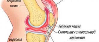
The fold of the knee joint is a duplication of the synovial membrane lining the joint cavity. The structure of synovial folds is thin, membranous, with zones of vascularization. From an embryogenetic point of view, the folds are partially vestigial synovial membranes and are found in the knee joints of healthy people. The exact function of synovial folds is unknown. A number of authors believe that synovial folds, like the eyelids, help improve the lubrication of the joint with synovial fluid.
In cases of direct trauma to the knee joint, with repeated stress or inflammation, clinical symptoms may occur due to the mechanical effect of the folds on other intra-articular structures. In particular, upon movement, the thickened and fibrotic synovial plica causes irritation of the synovial membrane at the edges of the bone condyles, which ultimately leads to reactive synovitis and thinning of the articular cartilage (synovial plica syndrome - SPL).
Changes in the mediopatellar fold occur most frequently, but an accurate assessment of the prevalence of CVS is difficult due to the lack of clear diagnostic criteria.
On MRI, intact synovial folds appear as thin, hypointense linear structures arising from the synovial lining. Their visualization is significantly improved in the presence of fluid in the joint (articular effusion, blood, or contrast agent during MR arthrography).
Main symptoms
With synovitis, most often only one joint is affected, but sometimes the disease is multiple.
The main symptoms of synovitis can be considered:
- increase in volume and smoothing of the contours of the joint;
- pain during exercise;
- pronounced swelling of soft tissues;
- difficulty moving;
- skin hyperemia;
- chills and general malaise.
If we talk about infectious synovitis, it almost always begins acutely: the patient feels severe throbbing pain, and the body temperature rises sharply. After some time, the knee joint swells, signs of general intoxication of the body appear: weakness, drowsiness, headache, nausea and even vomiting.
The non-infectious form develops quite slowly. First, the synovial membrane, which lines the joint capsule from the inside, becomes inflamed and also produces synovial fluid (synovium). The first sign is a feeling of discomfort in the area of the affected knee joint, as well as pain during exercise. Swelling gradually appears, and painful sensations increase.
If you find yourself with such symptoms, then do not self-medicate, but immediately make an appointment with a doctor.
Embryogenesis
Currently, it is widely believed that the knee joint initially consists of three compartments - medial, lateral and suprapatellar, separated by synovial septa. Incomplete resorption of these septa leads to the formation of well-developed synovial folds occupying the space between the distal parts of the epiphysis of the femur and the proximal parts of the tibia. Intrauterine movement in the fetal knee joint can simultaneously contribute to the resorption of folds and the formation of a joint cavity. This theory explains the presence of suprapatellar and infrapatellar folds, but is unfounded for the lateral and medial folds, since the patellofemoral joint is not divided into cavities in the plane of these folds at any stage of embryogenesis (that is, in the frontal one). Embryological work by S. Ogata and HK Uhthoff showed that the mediopatellar fold is not a remnant of the synovial septum. It is probably of mesenchymal origin and is associated with intrauterine lateralization of the patella, as evidenced by the predominance of mesenchyme in the mediopatellar zone.
There are no precise data in the literature on the prevalence of these persistent embryological structures in the population. In an anatomical study of the knee joints, T. Jouanin et al. in 11% of cases three main folds were present, in 10% no folds were identified. The suprapatellar and infrapatellar folds are the most common but are usually clinically intact. The lateral fold of the knee joint is extremely rare. Due to its clinical manifestations, the mediopatellar fold is the most studied to date.
Possibilities of MRI in the diagnosis of synovial folds
On MR images, synovial folds appear as hypointense cords with peripheral fluid and fat. The most informative pulse sequences in the diagnosis of synovial folds due to the high signal from the fluid are recognized as gradient T2-weighted pulse sequences, as well as T2- or proton-weighted sequences using fat suppression. MR arthrography is applicable in case of insufficient amount of intra-articular fluid and the presence of pronounced clinical symptoms of the manifesting fold. The contrast agent provides high intertissue contrast of the articular surfaces, stretches the joint capsule, which together helps to visualize all folds of the knee joint, verify or exclude their pathological changes.
The size and morphological restructuring of synovial folds are not a reliable sign of their clinical significance. In the presence of persistent clinical symptoms and the absence of other possible causes of a similar symptom complex (for example, damage to the medial meniscus or internal collateral ligament, patellofemoral arthrosis, arthrosis of the medial parts of the joint), characteristic signs of pathological fold syndrome are local synovitis, erosion of the articular cartilage of the patella and the patellar surface of the femur bones.
Possible side effects
This innovative invention, despite its minimally invasive nature and high degree of safety, is not without side effects. Analogue synovial fluid prostheses, often prescribed for arthrosis of the knee joint, rarely cause negative reactions. But they cannot be called completely harmless, if only because the main category of products sold by pharmaceutical companies has not been subjected to clinical trials.
Based on the minimal experimental clinical database and personal experience of patients who encountered problems after intra-articular use of hyaluronic acid, conclusions were drawn about the possible likelihood of developing the following consequences:
- feeling of heat, itching at the injection site;
- burning and irritation of the skin;
- nettle fever;
- swelling and redness of soft supra-articular tissues;
- painful signs in nearby muscles;
- local infectious and inflammatory reaction;
- numbness, tingling, feeling of “pins and needles” in the limbs;
- anaphylactic shock.
As we see, with all their positive qualities, innovative drugs produced as “synovial fluid” can also harm health. Efficiency is also not 100% guaranteed. Do not forget that each organism has its own individual characteristics, so there is no absolute guarantee of a successful outcome. In addition, it is unlikely that such therapeutic measures will produce results in pathofunctional changes in articular elements at terminal stages. Hence the conclusion: far from cheap treatment may well be ineffective.
Suprapatellar fold
In the literature, there are several synonyms for the suprapatellar fold: suprapatellar septum, superior fold, patellar fold.
The suprapatellar fold is localized between the suprapatellar bursa and the cavity of the knee joint, has an obliquely descending course, starting from the synovium on the anterior surface of the femoral metaphysis and ending in the posterior part of the quadriceps tendon in the area of attachment to the patella. Until the end of the 4th month of intrauterine development of the fetus, the suprapatellar fold completely separates the cavity of the knee joint from the suprapatellar bursa, and by the end of the 5th month its shape does not differ from that of an adult.
It is believed that the key factor ensuring the degree of fold reduction is mechanical. Depending on the range of motion in the knee joint, either a small perforation in the fold or its partial or complete absence may form. T. Zidorn classified the degree of “development”, or more precisely, reduction, of the suprapatellar fold into 4 groups depending on the morphological features:
MR tomogram of the knee joint in T2 V.I., sagittal plane
- type I (septum completum) - closed suprapatellar bursa - the suprapatellar fold completely separates the patellar recess from the cavity of the knee joint;
- type II (septum perforatum) - the suprapatellar fold has one or more holes connecting the suprapatellar zone with the joint cavity;
- type III (septum residuale) – a residual connective tissue cord is visualized, often on the inside;
- type IV (septum extinctum) – characterized by complete involution of the suprapatellar fold.
In type II folds, the zones of its perforation, which provide circulation of articular fluid to the area of the inversion, are called the hilum.
The suprapatellar fold persists very often - up to 89% of cases according to autopsy data. On MRI of the knee, the fold is best identified in the sagittal plane as a linear hypointense band posterior and superior to the patella.
During arthroscopy, a completely unreduced suprapatellar fold can be suspected only by a decrease in the volume of the suprapatellar recess. Sometimes the diagnosis is made by chance during joint puncture, when the injection needle ends up in the suprapatellar bursa and not in the joint cavity.
Liquid implants: price and analogues
Before we list the most commonly used HA agents intended for intra-articular administration, we draw attention to an important point: all therapeutic manipulations must be carried out by a highly competent doctor in the rheumatology department, in compliance with all aseptic standards. In addition, in technical terms, this procedure is completely different from intramuscular injections, so only a professional medical professional can ensure correct, non-traumatic and safe insertion of the needle into the joint capsule. The session is carried out under ultrasound control.
Synvisc's analogues
| Product name | 1 dose of HA / mg | Production |
| Adant | 25 | Japan |
| Fermatron | 20 | Great Britain |
| Gialgan Fidia | 20 | India |
| Sinokorm | 20 | Austria |
| Go-on | 25 | Ireland |
| Ostenil | 20 | Germany |
| Giruan Plus | 10 | Korea |
| Synvisc (3 syringes) | 16 | USA |
| Hyalurome CS | 60 | Romania |
| Visco Plus | 20 | Sweden |
| RusVisk | 16 | Russia |
| Giastat | 20 | Russia |
Infrapatellar fold
The infrapatellar fold, or ligamentum mucosum, is the most common fold of the knee joint. Its formation occurs from 8 to 12 weeks of gestation and depends on the degree of involution of the primitive embryonic membrane separating the medial and lateral sections of the joint. The shape of the fold formed the basis for its arthroscopic classification by S. Kim and W. Choe:
- 0—no fold;
- 1 - complete persistence of the fold, the fold is separated from the anterior cruciate ligament;
- 2 - complete persistence of the fold, the fold is separated from the anterior cruciate ligament and has several bundles;
- 3 - vertical fold - complete persistence of the fold, which is associated with the anterior cruciate ligament and divides the joint cavity into medial and lateral sections;
- 4 - fenestrated vertical fold - a vertical fold with a hole/defect.
The infrapatellar fold begins in the anterior parts of the intercondylar recess of the femur, expands forward and downward, and attaches to the lower pole of the patella. The thickness of the fold varies widely from submillimeter to the size of the anterior cruciate ligament.
The infrapatellar fold is easily identified on sagittal MR images as a linear, hypointense structure running through the Hoffa fat pad in a plane parallel to the anterior cruciate ligament. In some cases, especially in patients with complete rupture of the anterior cruciate ligament, a well-developed infrapatellar fold can simulate its preserved bundles. It is also necessary to carry out a differential diagnosis of the infrapatellar fold with local nodular synovitis, postoperative changes and a loose body in the area of the Hoffa fat pad. The awareness of radiologists about the existence of this anatomical structure allows in most cases to make the correct diagnosis.
Orthopedics and traumatology services at CELT
The administration of CELT JSC regularly updates the price list posted on the clinic’s website. However, in order to avoid possible misunderstandings, we ask you to clarify the cost of services by phone: +7
| Service name | Price in rubles |
| Appointment with a surgical doctor (primary, for complex programs) | 3 000 |
| MRI of the elbow joint (1 joint) | 7 000 |
| Ultrasound of soft tissues, lymph nodes (one anatomical zone) | 2 300 |
All services
Make an appointment through the application or by calling +7 +7 We work every day:
- Monday—Friday: 8.00—20.00
- Saturday: 8.00–18.00
- Sunday is a day off
The nearest metro and MCC stations to the clinic:
- Highway of Enthusiasts or Perovo
- Partisan
- Enthusiast Highway
Driving directions
Mediopatellar fold
The mediopatellar fold is also called the medial fold, synovial patellar fold, pterygoid fold, patellar meniscus, and synovial protuberance. The mediopatellar fold begins in the area of the medial wall of the knee joint, goes obliquely downwards and is woven into the synovium around the Hoffa fat pad. It can be connected to the suprapatellar fold, but more often it has a separate attachment. If overdeveloped, the mediopatellar fold may extend to the medial surface of the femoral trochlea and the medial facet of the patella.
MR tomograms, axial plane, proton-weighted mode with signal suppression from adipose tissue
J. Sakakibara, based on the shape and size, identified 4 types of mediopatellar folds:
- type A – the fold is represented by a linear rope-like protrusion of the synovial wall;
- type B – the fold is represented by an elongated linear cord with uneven contours, but does not reach the medial condyle of the femur;
- type C – the fold is elongated, thickened, with uneven fringed contours, extending to the area of the medial condyle of the femur.
- type D – the fold extends to the area of the medial condyle of the femur, is thickened, uneven, and has a central defect (fenestrated fold).
This classification has received universal recognition and approval, as it is easy to use and clinically relevant. Types A and B of the mediopatellar fold are asymptomatic. Types C and D may impinge between the medial femoral condyle and the patella with subsequent thickening and induration, damaging the cartilage at the patellofemoral joint. The main mechanism of action of the fold is compression of the articular cartilage of the medial facet of the patella during flexion and the patellar surface of the medial femoral condyle during extension. A number of authors are of the opinion that the fenestrated mediopatellar fold (type D) more often causes damage (impingement) of the articular cartilage of the medial parts of the patellofemoral joint.
For visualization of the mediopatellar fold using MRI, T2- or proton-weighted images in the sagittal and axial planes with or without fat suppression are considered the most informative. The mediopatellar fold has a low MR signal and is easily recognized in a typical location against the background of a small amount of intra-articular fluid. On the other hand, one should always pay attention not only to the size of the fold and its location relative to the medial condyle, but also to the condition of the articular cartilage in the medial facet of the patella, femoral condyle, and the patient's complaints. With massive intra-articular effusion, lateral displacement of the fold may occur, complicating the differential diagnosis of types B and C. The large size of the mediopatellar fold may make it difficult to examine the medial parts of the joint during arthroscopy.
Anatomy
The synovium is located around joints and ligaments, providing them with protection. This occurs due to the release of a small amount of fluid (lubricating secretion), which reduces friction and provides shock absorption.
The exact cause of hip synovitis is unknown, but some theories include a history of trauma to the femur or a recent viral illness such as an upper respiratory tract infection, bronchitis, or otitis media.
One of the reasons for the development of inflammation of the hip joint is infection in the synovial fluid. This occurs against the background of any other infectious disease. In this case, fever, a significant increase in body temperature and general poor health are added to the classic symptoms. In this case, it is necessary to first deal with the source of infection.
Main reasons:
- sports injury;
- stretching;
- blood diseases;
- metabolic problems;
- endocrine pathologies;
- dystrophic joint pathology;
- overweight;
- allergy;
- bruises.
Among the infectious pathogens that most often cause synovitis are pneumococci, streptococci, staphylococci, Koch's bacillus (tuberculosis) and STIs (sexually transmitted infections).
Lateral patellar fold
Schematic representation of the anatomical structures of the lateral recess of the knee joint (normal)
The lateral patellar fold is the most rarely encountered fold of the knee joint. Most often, it has a longitudinal shape, is very thin and is located 1-2 cm lateral to the patella. The lateral patellar fold originates on the lateral wall and is attached to the infrapatellar fat body. S. Ogata and HK Uhthoff hypothesized that rare persistence of the lateral patellar fold is associated with lateral displacement of the patella, leaving no room for it in the lateral recess. The lateral fold may make it difficult to perform knee arthroscopy through the anterolateral approach.
The lateral patellar fold should not be confused with the more commonly encountered pterygoid, superolateral, and transverse arcuate folds.
The arrows indicate: in Figure A - lateral patellar fold (white thin arrow), lateral pterygoid fold (black thin arrow), transverse arcuate fold (black thick arrow); in Figure B - superolateral fold (white thick arrow).
The lateral pterygoid fold is located close to the patella along the outer wall, the superolateral fold is a type of suprapatellar fold dislocated outward. A transverse arcuate synovial fold is often found at the junction of the vertical and anteroposterior portions of the lateral recess.
Pathogenesis and clinical manifestations
Synovial fold syndrome (shelf-, plica-syndrome or their most common variety - mediopatellar fold syndrome) describes a complex of painful dysfunctions of the knee joint, in which the only finding is a thickened and fibrous-changed fold.
MRI scan of the knee joint
Patient A., 18 years old. MR imaging of the knee, proton-weighted fat suppression (axial plane) and arthroscopic slides. Complaints of pain in the internal parts of the knee joint. There is elongation, thickening, and fibrillation of the medial patellar fold, which reaches the medial femoral condyle (type C according to Sakakibara). The arrow indicates the area of “cracking” and chondromalacia of the articular cartilage of the medial facet of the patella.
The onset of pathology is usually an injury to the knee joint (direct blow, repeated load, twisting, leading to stretching of the fold), less often inflammatory and degenerative changes. Post-traumatic, inflammatory swelling of the fold leads to loss of elasticity, thickening and clinical manifestations if it reaches the patellofemoral joint during movements. The result of fibrosis and hypertrophy is reactive mechanical synovitis, as well as erosion of the articular cartilage of the femur and patella. In the early stages of the disease, the cause of pain is often the fold itself, while in a later period, chondromalacia of the articular cartilage and synovitis come to the fore. It should be remembered that the size, shape, and rigidity of the folds of the knee joint directly depend on the hereditary and constitutional characteristics of a particular person, however, most experts agree that a thick fold manifests itself clinically much more often than a thin one.
Mediopatellar fold syndrome is more often diagnosed in adolescents than in adults due to the rarer occurrence of concomitant lesions of the menisci and ligaments. The most typical occurrence of symptoms is after blunt trauma to the knee. The main complaint of patients is pain in the medial parts of the joint, which can occur both with load and at rest, and intensifies with repeated flexion/extension. The pain is localized medially from the patella, above the line of the joint space of the knee joint. Nonspecific signs include crepitus, popping, clicking, pseudojamming, and effusion. It is most often necessary to differentiate CVS from a medial meniscus tear and patellar instability. If a dense, painful cord is palpated medial to the patella, the diagnosis of mediopatellar fold syndrome becomes more likely.
There are two main mechanisms for the pathological effects of the suprapatellar fold. The first is compression of the knee joint cavity by the suprapatellar bursa with impingement of the articular cartilage of the superomedial parts of the femur during flexion. The second is inflammation of the suprapatellar bursa.
MRI scan of the knee joint (sagittal plane)
MR tomogram of the knee joint (sagittal plane) in T2-weighted mode with fat suppression before contrast (A) and T1-weighted after intravenous administration of a contrast agent (B). A completely unreduced suprapatellar fold is identified with the formation of a closed suprapatellar bursa. There is thickening of the synovial membrane of the bursa (signs of bursitis), prolapse of the lower pole of the bursa into the upper parts of the patellofemoral joint with compression phenomena.
As a rule, both pathological conditions occur with a completely unreduced fold (type I) with the formation of a closed suprapatellar (subquadricipital) cavity.
The lateral patellar fold is extremely rare and is usually asymptomatic. Clinical and MRI signs of lateral CVS are identical to those of mediopatellar fold syndrome. Manifestations of lateral CV joint may include pain in the lateral area of the joint and clicking. A palpable tourniquet-like painful cord of appropriate localization is a pathognomonic symptom.
Morphology and metabolism of synovial joints
Synovial joints are a complex multicomponent organ-specific system, including both elements of the internal environment (articular cartilage, synovial fluid, synovial membrane) and the subchondral bone adjacent to the articular cartilage, through which the energetic, plastic and mechanical functions of the articular cartilage are carried out to a certain extent (Pavlova V N., 1989). All of these components are closely functionally related and, depending on the nature of the interaction and the characteristics of their organization, provide locomotor function.
Articular cartilage is devoid of vessels and nerves; therefore, the nature of the supply of substances and the performance of their functions affects the morphological organization. Thus, this structure is characterized phenotypically, and according to some researchers, genotypically, by a heterogeneous population of chondrocytes that have a vertical position-specific arrangement throughout the thickness of the articular cartilage and specific metabolic characteristics. Chondrocytes occupy 5% of the volume of cartilage and provide the biosynthesis of proteins and carbohydrates necessary for the formation of a complete matrix that can withstand mechanical loads. In the superficial zones of the contacting surfaces of the joints, chondrocytes are located in 1-2 layers, have oval nuclei, surrounded by cytoplasm, elongated in the marginal sections. The very location of the chondrocytes corresponds to the direction of the collagen fibers that form the cell-free part of the superficial zone (Lamina splendens), that is, parallel to the surface. Often these cells do not form capsules, but according to electron microscopic characteristics they resemble fibroblasts: they have a nucleus with a compact chromatin arrangement, and their cytoplasm contains a developed endoplasmic reticulum (Fig. 1-1.). In these cells, there are practically no accumulations of glycogen, which is one of the markers of cytodifferentiation of chondrocytes. The most extensive area is in the middle zone, in which chondrocytes are located in capsules individually or in the form of isogenic groups consisting mainly of two cells. According to electron microscopic characteristics, they differ in the organization of the cytoplasm: chondrocytes with a developed endoplasmic reticulum are found, which indicates a high level of protein biosynthesis in them, mainly collagen. Some cells in the cytoplasm have a complex organization of the Golgi complex and contain a large number of secretory granules, which indicates an intensification of the synthesis of carbohydrate-containing compounds.
Glycogen is found in almost all cells in the form of small accumulations diffusely located throughout the cytoplasm. The cells of the deep zone of articular cartilage are distinguished by a columnar organization, including two or more chondrocytes. For the most part, they have an organized cytoplasm with a developed endoplasmic reticulum, Golgi complex, a large number of mitochondria and lysosomes, and extensive accumulations of glycogen. The presence of the latter in these cells is interpreted as a signal for ossification. If we consider the cell population of articular cartilage as a whole, it should be noted that the complication of the ultrastructural organization of cells proceeds from the superficial zone to the deep zone (Brighton S. T., Kitajimana T., Hunt R. M., 1984). In the zone of calcified cartilage, the cells are located in expanded capsules at a considerable distance from each other, have dense nuclei, poorly organized cytoplasm with a large number of inclusions in the form of glycogen, lipids and dense osmiophilic particles. Chondrocytes adjacent to the ossification zone have pyknotic nuclei, often surrounded only by fragments of cytoplasm. The cells are located in capsules, the sizes of which are larger than those of chondrocyte capsules in other parts of the cartilage.
Rice. 1.1. Options for the structural organization of chondrocytes:
- chondrocyte of the superficial zone with developed membrane organelles of the cytoplasm, uv. 12000;
- fragment of a middle zone chondrocyte with developed smooth endoplasmic reticulum and accumulations of glycogen in the cytoplasm, uv. 16000;
- chondrocyte of the middle zone with a large number of mitochondria, uv. 12000;
- chondrocyte of the middle zone with developed granular endoplasmic reticulum, uv. 12000;
- chondrocyte of the deep zone, vacuolization of the cytoplasm, uv. 12000;
- deep zone chondrocyte, single tubules of the endoplasmic reticulum, microfilaments and microtubules of the cytoskeleton, uv. 12000; contrasted according to Reynolds, EMV-100BR.
Currently, the idea of chondrocytes as metabolically inert cells has been proven untenable; Data have been obtained indicating that this type of cell is characterized by a high intensity of metabolic processes (Pavlova V.N. et al., 1988).
Chondrocytes of all zones of articular cartilage are characterized by certain similarities and differences in their metabolic profile. Thus, the enzymes of the Krebs cycle, hexomonophosphate shunt, and cytochrome oxidase progressively increase in the cytoplasm of chondrocytes located in the direction from the superficial to the deep zones of cartilage. Glycolytic enzymes are present in chondrocytes of all zones of articular cartilage, the same situation is typical for lipids (Sampson N. W., Cannon M. S., 1986). These data confirm the previously expressed point of view about the presence of two populations of chondrocytes, in this case differentiated by enzymes - anaerobes and aerobes - oriented in the thickness of the articular cartilage in accordance with the specifics of the supply of nutrients to it.
With age, articular cartilage undergoes certain changes. The density of chondrocytes per unit area decreases in them. This leads to the fact that single chondrocytes cannot metabolically provide large areas of the matrix and catabolic processes begin to prevail over anabolic ones. The emerging situation can be considered as a “weak link” in the articular cartilage system. Due to the reduction of membrane organelles in cells and the proliferation of cytoskeletal elements in them, the ultrastructural organization of chondrocytes is simplified. However, even in the articular cartilage of people aged 60-80 years, chondrocytes were identified, based on the ultrastructural organization of which it can be concluded that the processes of biosynthesis of fibrillar bits and proteoglycans are activated in them. But the bulk of chondrocytes have signs of involutive atrophy (Vinogradova E.V., Mikhailov I.N., 1983).
Cartilage cells are the subject of close attention of researchers, since ultimately the biochemical reliability of the matrix depends on their reactivity and plastic function.
It has been established that cell differentiation and age-related changes are accompanied by characteristic transformations in the macromolecular organization of the matrix. In general, in cartilage tissue, as well as in other types of connective tissue, there is a clear relationship between cellular and noncellular components. That is, the cell reproduces matrix macromolecules: collagen, glycoproteins, proteoglycans and non-collagen proteins, and macromolecules, forming the microenvironment of the cell, influence its cytodifferentiation.
The named components are in the matrix in a clear relationship with each other, and in ontogenesis, a transformation of their structural and spatial order occurs. Let's look at these processes in more detail.
It is known that at the stages of chondrogenesis, the biosynthesis of collagen, one of the main components of the matrix, is heterogeneous. Using biochemical and immunological research methods, it was shown that precartilaginous cells synthesize type I collagen (Modis L. et al., 1986). The specific function of chondrocytes is the biosynthesis of type II collagen. However, even when studying mature articular cartilage, it was revealed that chondrocytes of the superficial zone can synthesize type I collagen, and in the cellular territorial matrix of chondrocytes of other zones, pericellular collagens (type Y collagen) are determined, forming the support of cells like an exocytoskeleton. In addition, a large number of minor collagen components were identified in cartilage tissue. Research in recent years has shown that the normal process of collagen metabolism in cartilage tissue is long, with the half-life of macromolecules ranging from 50 to 300 days. According to theoretical calculations made on the basis of isotope studies, it has been established that its complete exchange in the cartilage matrix in humans can take place within 120-360 years (Maroudas A., 1980), that is, the matrix system has a large margin of safety. In general, in the cartilage matrix, collagen makes up 50-70%, while most of it is contained in collagen fibers, and its strength and functional properties are associated with their orientation and ordered arrangement in the thickness of articular cartilage. In the matrix of human articular cartilage, the anisotropy of collagen in the superficial and intermediate zones is almost at the same level, and in the deep zone it is slightly higher. With aging, under conditions of a pathological process and physiological changes, the synthesis and typical composition of collagen may change. It has been established that the birefringence of collagen in all zones of articular cartilage remains at the same level until a certain age. So, in the surface zone - up to 70 years, in the middle zone - up to 60 years, in the deep zone - up to 50 years, and then decreases by 27, 18 and 24%, corresponding to the above zones and ages (Dedukh N.V., 1988).
With aging, the length of collagen fibrils and their intertwining increases, which makes a certain contribution to the theory of “structure fatigue” and contributes to disruption of the organization of the matrix: unmasking of collagen fibers, their destruction and, accordingly, disruption of the relationship with proteoglycans (Hough AJ, Weber RJ, 1986).
Due to the fact that the process of fibrillogenesis is based on the participation of proteoglycans and glycoproteins in it, fundamental research is devoted to assessing the role of these macromolecules (Serov V.V., Shekhter A.B., 1981, Pavlova V.N. et al., 1988) . Thus, it has been shown that proteoglycans can be present in the matrix either in the form of complexes or minor components. An important structural unit of proteoglycan complexes are glycosaminoglycans (GAGs). To date, 8 types of GAGs have been identified, 6 of which are sulfated forms. In general, GAGs present in articular cartilage can be grouped into three functional groups: 1) hyaluronates, 2) chondroitin and chondroitin sulfates, 3) keratan sulfates. It has been established that precartilaginous cells are characterized by a high level of synthesis of hyaluronic acid, which plays a certain role in cell proliferation and migration. Cell differentiation is associated with a disruption in the tissue ratio: hyaluronic acid/hyaluronidase. As cells differentiate, the synthesis of hyaluronic acid decreases, and the content of hyaluronidase and chondroitin sulfates increases. Chondroitin sulfates are distributed fairly evenly throughout the thickness of the articular cartilage, while the content of keratan sulfates increases towards the deep zone (Williams I. M. et al., 1988). If we compare the data on the content of GAGs and collagen in cartilage, the following picture emerges: the deepest layers of articular cartilage contain 2 times more chondroitin sulfates and 6 times more keratan sulfates compared to collagen. With an increase in sulfated forms of GAG, reflecting a higher level of matrix organization, an increase in collagen biosynthesis and its orientational ordering is also recorded (Foldes I. et al., 1980). Based on the dynamics of identification and data on the organization of these macromolecules in the matrix, researchers have postulated the point of view that sulfated forms of GAGs are a matrix for orientationally ordered collagen (Serov V.V., Shekhter A.B., 1981, Modis L. et al. , 1986). In recent years, methods for visualizing proteoglycans using electron microscopy have been developed (Fig. 1.2.).
Rice. 1.2. Proteoglycans in the matrix of articular cartilage (contrasted with Alcian blue 8GS.EMB-100BP):
- granules in cellular and intercellular matrix, uv. 16000;
- variants of proteoglycan granules, uv. 44000;
- proteoglycans and collagen fibers, uv. 45000.
It is known that the proteoglycan complex includes 5% chondroitin sulfates, 2-7% keratan sulfates and 7-12% non-collagenous proteins (Hadley S., Phelps S., 1982; Hardingham T., 1981). However, at the stages of ontogenesis, the ratio between these macromolecules does not remain constant (Gongadze N. R., 1986; Kosyagin D. V., 1983). Thus, if chondroitin sulfates predominate in the articular cartilage of children, then keratan sulfates predominate in adults (Elliott RI, Gardner DR, 1979; Triphaus G., Schmidt A., Buddecke E., 1980). In addition, the ratio of chondroitin-4-sulfates to chondroitin-6-sulfates changes with age, due to a sharp decrease in the first component and a slight increase in the second (TGreilind N., 1976). A change in the ratio and density of proteoglycan fractions with age has been established (Roughley R. I., 1987). In addition, they disrupt the connection between protein and hyaluronic acid (Mitrovic DR, 1987), as well as the protein/hyaluronic acid ratio due to an increase in the content of the first component and a decrease in the second (Kosyagin D.V., 1983). Degradation of proteoglycans in articular cartilage occurs throughout human life, but increases with age. This is manifested by increased fragmentation of proteoglycan complexes due to their constituent protein chains, hyaluronic acid and glycoproteins, which can accumulate in free form in tissues (Roughley R. I., 1987). However, proteoglycans remain capable of aggregation with hyaluronic acid, practically without changing their shape. Other changes in proteoglycans are associated with an increase in histidine alanine sites and a decrease in the molecular weight of proteoglycans (Hough AJ, Weber RJ, 1986).
Using polarization microscopy methods, it was proven that in the superficial zone of articular cartilage, the GAG refraction remained at the same level for up to 50 years, and then progressively decreased and reached 34% of the initial values. In the age period from 41 to 50 years in the intermediate zone, a decrease in GAG refraction was recorded by 37%, and in the group 71 years and older - by 50%. In the deep zone, the peak of GAG birefringence was detected in the age group of 31-40 years, a further decrease was observed in all other groups, reaching 40% by the age of 71 years and older (Dedukh N.V. et al., 1988). With age, the degree of refraction of the GAG in the territorial matrix decreased, but always remained higher than in the interterritorial matrix (Fig. 1.3.). Differential analysis of GAG refraction showed that aging is accompanied by a decrease in the anisotropy of chondroitin sulfates and an increase in the refraction of keratan sulfates in all zones of articular cartilage, reaching 91% of the total composition in the deep zone of the age group 71 years and older. An increase in the proportion of keratan sulfates in articular cartilage from 60% and above was accompanied by pronounced impairments in the degree of collagen refraction, which manifested itself primarily in the deep zone. It should be noted that qualitative changes in collagen fibrils with age were identified precisely in this zone using electron microscopy methods (Kopyeva T.N. et al., 1983). The above-described changes in the structural organization of the cartilage matrix lead to a violation of its elastic qualities.
Rice. 1.3. Glycosaminoglycans in the matrix of articular cartilage:
- pronounced anisotropy, uniform distribution in the matrix, uv. 80;
- violation of the organization. Local anisotropy around cells, uv. 90. Polarized light. "Polmy-A".
Another important component of the matrix that takes part in its organization are glycoprotein macromolecules - the object of close study in connection with the identification of their role in fibrillogenesis, tissue calcification and some other functions (Shubich M. G., Mogilnaya G, M, 1979; Shubich M G., 1980; Serov V.V., Shekhter A, B, 1981; Hughes R., 1985; Anderson J., 1876). We consider it necessary to dwell on certain aspects of their participation in chondrogenesis. Summarizing the available literature data on the formation of the matrix, Shubich M. G. (1980) notes that at the stages of cell convergence and aggregation, the properties of the cytolemma change due to changes in the mobility of their constituent glycoproteins, which in turn is a stimulus for the release of hyaluronidase , which breaks down hyaluronic acid on the outer surface of the membranes. At the same time, the level of cAMP in the cell increases and, as a result of all transformations, enzymes for the synthesis of proteoglycans and collagen are activated. Another important function of glycoproteins is their participation in the aggregation of proteoglycans to ensure their interaction with hyaluronic acid (Fig. 1.4).
In addition, it is believed that structural glycoproteins, like proteoglycans, can act as a “matrix” for collagen deposition and play an important role in the organization of proteoglycans and collagen fibers (Robert R., Robert B, 1984; Modis L. 1974). In general, it has been shown that glycoproteins not only regulate the growth and orientation of collagen fibers, but also contribute to their stabilization (Serov V.V., Shekhter A.B. 1981). With age and under pathological conditions, the content of glycoproteins decreases, which leads to destabilization of collagen fibers (Anderson C, H, Salter S. 1971). In humans, an increase in the birefringence of glycoproteins was noted in the age group of 41-50 years, and then a decrease was observed, which was most clearly expressed in the intermediate and deep zones of articular cartilage. It is in this age group that the processes of unmasking (fibrillation) of collagen fibers begin to actively manifest.
Rice. I. 4. Scheme of the macromolecular organization of the matrix of articular cartilage.
It has been established that cartilage tissue contains a special type of glycoproteins - chondronectin (Kevitt A. T. et al, 1980). It is assumed that it is an intermediary for the connection of chondrocytes predominantly with type II collagen.
A component of proteoglycans and glycoproteins are non-collagenous proteins. In proteoglycan complexes they form the axial structure (protein - core) (Romhanyi G. et al., 1975; Shipp D., Bowness J., 1975). In addition, they form the structure of glycoproteins that ensure the interaction of proteoglycans with hyaluronic acid (link) (T. E. Hagdinghman, 1977, 1981, 1982).
Some of the non-collagen proteins in the matrix are in a free state. The composition and structure of non-collagen proteins changes with age and under pathological conditions (Szirmai JA et al., 1967; Heinegard D., Oldberd A., 1989).
The attenuation of biosynthetic processes is reflected in the relationship between collagen and GAGs, which contributes to a decrease in reparative potency and leads to the appearance of local destructive changes in the matrix, in particular in the collagen framework.
Due to the fact that the trophism of articular cartilage is ensured by the diffusion-deformation mechanism, there is a pronounced dependence of articular cartilage on the composition and intensity of synovial fluid exchange. It has been proven that the area of diffusion of synovial fluid into articular cartilage is from 3 to 5 mm, but the value of this indicator is affected by its viscosity. Moreover, the higher the viscosity, the greater the lubricity of the synovial fluid, but the less fluid penetrates into the articular cartilage and vice versa. Synovial fluid has certain similarities with blood plasma, but differs in the presence and content of some protein components, in particular the ratio of albumins to globulins. (3:1). The content of a-globulin in synovial fluid is almost three times less than in plasma. Synovial fluid contains transferrin, haptoglobulin, but lacks fibrinogen, as well as proteins with a molecular weight of more than 160,000. It contains GAGs, including hyaluronic acid, which acts as the main component that binds water. In addition, synovial fluid contains up to 30 enzymes. Its functions are varied. In addition to nourishing articular cartilage and regulating metabolic processes in it, it provides lubrication of articular surfaces, which facilitates their friction and sliding.
The joint capsule has two layers - synovial and fibrous. Synoviocytes, the cells lining the synovial layer of the capsule, take part in the formation of synovial fluid. By organization, synoviocytes are a specific cellular component of the integumentary layer. These are large cells containing large oval nuclei surrounded by fine cytoplasm. Synoviocytes belong to polar cells - the nucleus and organelles in them are located along the length of the axis and perpendicular to the lumen. The apical end of synoviocytes often has numerous filopodia and vacuoles (Pavlova V.N., 1980).
According to their ultrastructural features, three types of cells can be distinguished among synoviocytes, conventionally designated as A, B and C. Cells A are macrophage-like synoviocytes, contain a large number of lysosomes, they are capable of active pino- and phagocytosis. These cells are the most numerous, have a developed Golgi complex, vacuoles, mitochondria and secretory material identified as hyaluronic acid. B cells are synoviocytes of the fibroblastic type. They produce protein and polysaccharide substances. This is a small population of cells with a developed endoplasmic reticulum, predominantly of the smooth type. C cells combine the features of A and B synoviocytes.
The remaining cellular elements of the integumentary layer - fibroblasts, mast cells, lymphocytes and macrophages, plasma cells - have a typical structure characteristic of connective tissue cells. The intercellular substance of the synovial layer is rich in loosely packed reticulin fibers, aggregates of proteoglycans and glycoproteins. Thanks to this organization of the synovial membrane, it performs the functions of transporting substances “joint cavity - blood - joint cavity”.
The fibrous layer of the capsule is represented by collagen and elastic fibers, which provide processes of stretching and compression when moving the articular ends.
The articular capsule has a complex vascular bed, in which capillaries approach directly the synoviocytes of the integumentary layer, as well as a nervous regulation system consisting of numerous efferent endings of various structures (lamellar bodies, bush-like free endings), acting as proprioceptors, pain sensitivity receptors, chemoreceptors and baroreceptors . Effector pathways are represented by sympathetic soft fibers, which end in nerve endings on vascular smooth muscle cells and regulate their permeability. A more detailed analysis of the organization of the synovial membrane and synovial fluid can be found in the monograph by V. N. Pavlova (1980).
Age-related changes in the capsule are not fundamentally different in nature from those occurring in other tissues of the joint and consist of a decrease in cellularity, the presence of involutive (atrophic) changes in cells, and an increase in the volume occupied by collagen fibers in the intercellular substance. With increasing age, the polymorphism of collagen fibers in the capsule increases and the content of elastic fibers decreases.
BIBLIOGRAPHY
- Vinogradova E. V., Mikhailov I. N. Electron-histochemical characteristics of proteoglycans of human hyaline and elastic cartilage // Structure, function and development of cartilage in normal and pathological conditions: Proc. report 3 symposium - M., 1983. - P. 20.
- Gongadze L. R. Mucopolysaccharides of aging human articular cartilage // Archives of Anatomy. - 1974. - T. 67, no. 7. - pp. 99-103.
- Gongadze L. R. Basophilic line of articular cartilage in normal conditions and in some pathological conditions // Archives of Anatomy. - 1986. - T. 79, no. 1. pp. 52-57.
- Grandfather N.V. Morphological aspects of the effects of hormones on articular cartilage in ontogenesis: Abstract of thesis. dis. Doctor of Biology Sci. - M., 1988. - 32 p.
- Dedukh N.V., Kulish N.I., Cherevatova S.Kh. et al. Functional state of some endocrine glands in patients with coxarthrosis // Age-related, adaptive and pathological processes in the musculoskeletal system: Proc. report 7th school in muscle biology. - X., 1988. - P. 191-192.
- Kopyeva T.I., Muldilrov P.Ya., Belskal O.B., Pastel V.B. Structure of articular cartilage in the elderly // Archives of Anatomy. - 1983. - T. 85, No. 10.-S. 60-67.
- Koslgin D.V. The ratio of protein and carbohydrate components in monomers and aggregates of proteoglycans of human articular cartilage // Structure, function and development of cartilage in normal and pathological conditions: Proc. report 3 symposium - M., 1983. - P. 26.
- Pavlova V. N. Synovial environment of joints. - M.: Medicine, 1980. P. 296.
- Pavlova V. N., Kopieva T. N., Slutsky L. I., Pavlov G. G. Cartilage. - M.: Medicine, 1988. - 320 p.
- Pavlova V. N. Components of the internal environment of joints and their functional interaction // Advances in modern times, biology. - 1989. - T. 107. Issue. 2. - pp. 238-242.
- Serov V. 3., Shekhter A. B. Connective tissue. Functional morphology and general pathology. - M.: Medicine, 1981. - 312 p.
- Shubich M. G., Mogilnaya G. M. Glycoproteins and proteoglycans: principles of their histochemical analysis // Archives of Anatomy. - 1979. - T. 71, No. 8. - P. 92-95.
- Shubich M. G. The mechanism of cartilage formation in embryogenesis // Nature. - 1980. - No. Z.-S. 26.
- Hughes R. Glycoproteins. - M.: Mir, 1985. - 140 p.
- Anderson I. Glycopriteins of the connective tissue matrix //Intern. Rex Connect Tissue Res. - 1976. - Vol. 7. - P. 252-322.
- Anderson S. H., Salter S. IV. The fine structure of bovine nasal cartlage //J. Cell. Biol. - 1971. - N. 49. - P.650-663.
- Brighton S. T. The zonal analysis of cytoplasmic components of artcular cartilage //Artritis Rheum. - 1984. - Vol. 37. - P. 1290-1299.
- Greiling H. Biochemische Untersuchungen zur medicamentosen ther; pie der Arthrosen. Meniscuslasionen und posttraumatische Arthrose //Unfa heilung. - 1976. - Bd 128. - S. 87-97.
- Elliott R., Gardner DR Changes with agein the glycosaminoglycar of human articular cartilage //Ann. Rheum. Dis. - 1979. - Vol. 38. - 371-377.
- Foldes L., Modis L., Antalety J., Adam J. Ultrastructure of extracellularity; matrix of embryonic chick limb bud and cartilage //Acta Biol. Acad. Sci. Hung. 1980. - Vol. 31. N3.-P.81-95.
- Hadley C., Phelps C. The in vitro biosynthesis of chondroitinsulfa neonatal rat epiphyseal // Biochem. - 1972. - Vol. 126. - P. 427-432.
- Hardingham TE Cartilage proteoglycans: structure, interactions ar organization //Med. Sci. Upsala. - 1977. - N 82. - P. 67-68.
- Hardingham TE The role of link-proteins in the structure of cartilage proteoglycan aggregates //Arthritis Rheum. - 1981. - Vol. 711, N 1. - P. 28-29.
- Hardingham TEProteoglycans: their structure, interaction and molecular organization in cartilage //The Sixteenth Colworth Medal Lecture. - London, 1982. - Vol. 67. - P. 489-497.
- Hevitt AT Identification of adhesive factor for chondrocytes // Prc Nat. Acad. Ski. USA. - 1980. - Vol. 77. - P. 355-388.
- Hough AJ, Weber RJ Aging Phenomena and Osteoarthritis: cause coincidence? //Annals of clinical and laboratory science. - 1986. - Vol. 16. N. 6.-P.502-510.
- Maroudas A. Metabolism of cartilaginous tissues: a quantitative a (roach // Stud. Joint Disease. - Cambridge, 1980. - P. 59-86.
- Mitrovic DR, Aprile F., Front Ph. Age-related changes in the structure of bovine articular cartilage proteoglicans //Joint destruction: 16th Symposii of the European Society of Osteoartrology. - 1987. - P. 6-17.
- Modis L. Topo-optical investigations of mucopolysaccharides (ai glycosaminoglycans) //Handbuch Histochemie. Polysaccharide. Stuttgr 1984. - Vol. 11. Part 4. - P. 164.
- Modis L„ Adany R., Szekanecz Z., Raystijk G., Nagy I. Differentiation of the extracellular matrix in cartilage: 21st Symposium of the Polish Histochecal fnd Cytochemical Society. - Poland: Krakov, 1986. - P. 46.
- Robert S., Robert B. Structural glycoproteins of connective tissue, their role in morphogenetics and immunopathology // Connective Tissue, Biochemistry and Pathophysiology. - Berlin, 1974. - P. 240-256.
- Roughley PI Structural changes in the proteoglycans of human articular cartilage during aging //J. of Rheumatology. - 1987. - Vol. 14. - P. 14-15.
- Romhanyi G., Deak G„ Fisher G. Aldehyd-bisulfite-toluidine blue (ABT) staining as a topo-optical reaction for demonstrating the linear order of vicinal OH-groups in biological structures //Histochemie. - 1975. - Vol. 43. - P. 333.
- Salter RB, Gross A., Hamilton-Hall I. Hydrocortisone artropathy: An experimental investigation //Can. Med. Assoc. J. - 1967. - Vol. 97. - P. 374.
- Sampson HW, Cannon MS Zonal analysis of metabolic profiles of articular - epiphyseal cartilage chondrocytes: a histochemical study / Histochemical J. - 1986. - Vol. 18. - P. 233-238.
- Shipp D., Boweness I. Insoluble noncollagenous cartilage glycoproteins with aggregation subunits // Biochem. Biophys. Acta. - 1975. - Vol. 279. - P. 282-394.
- Szirmai JA, Tyssonak £., Gardell S. Microchemical analysis of glycosaminoglycans, collagen, total protein and water in histological layers of nasal septum cartilage // Biochem. Biophys. Acta. - 1967. - Vol. 136, N 5. - P.331 - 340.
- Triphaus G., Schmidt A., Buddecke E. Age-related changes in the incorporation of S sulfate into two proteoglycan populations from human cartilage //Hoppe-Seylers Z. Physiol. Chem. - 1980. - Vol. 361, N 12. -P. 1773 - 1779.
- Williams l. M., Kafz R., Dchilds M. et. al. Keratan sulfate content in the superficial and deep layers of osteophytic and nonfibrillated human articular cartilage in osteoarthritis //Calcif. Tissue Int. - 1988. - Vol. 42. - P. 163-166.
Treatment
The first stage of treatment for CVS usually involves conservative techniques such as restriction of physical activity, massage, physical therapy, stretching of the hamstring muscles, and strengthening of the quadriceps femoris muscle. Drug therapy consists of cutaneous, oral or intra-articular administration of non-steroidal anti-inflammatory drugs and corticosteroids.
If conservative therapy is ineffective, arthroscopic resection of the synovial fold is performed. Radical surgery involves complete removal of the fold from the base. With a non-radical operation, for example, when dividing a fold with scissors, its independent repair may occur with the recurrence of symptoms.
An interesting study by DS Tearse et al., who, based on data from 3000 arthroscopy of the knee joint, found lateral folds in 21 patients. Of these, 14 were thickened, fibrous and correlated with the severity of complaints. In 13 patients, resection of the fold was accompanied by complete recovery.