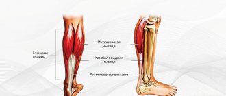
Joint examination
When examining joints, patients are often surprised by the choice of method that their doctor recommends. This happens because the joint is a complex multicomponent structure, and it is even more difficult to “look inside” here than in diseases of many other organs.
What, what are our joints made of?
In the formation of a “typical” joint - wherever it is located, in the head or in the leg - formations of different composition and functions are involved. Firstly, these are at least two (sometimes more) bones. Their contacting (articular) surfaces are covered with cartilage and together are surrounded by a kind of “coupling” - an articular capsule. Inside it, the bones are washed with joint fluid to reduce friction.
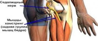
Secondly, these are ligaments - elastic “fixators” on different sides of the joint, giving it elasticity, flexibility and preventing it from falling apart. A joint also includes the muscles that move it and the tendons that attach these muscles to different parts of the joint. (Anatomically, this assignment is not entirely accurate, but for examination it makes sense, since tendon injuries often lead to pain in the joint.)
Joints, ligaments, tendons
Joint diseases are one of the most frequently diagnosed diseases today. Statistics say that about 30% of the world population
suffer from failures and dysfunctions of the joints.
Roughly speaking, every third person
suffers from the above ailments.
Until recently, people of the older generation were at risk, but now young people are increasingly seeking medical help. This is related to modern life
. After all, a person rarely experiences uniform loads, but spends the whole day without moving or works at full capacity 24 hours a day. If we talk about sports activities, they should be done in a moderate mode, without injuring or overloading the joints of the body. If you are overweight, heavy loads are strictly contraindicated, because they can significantly overload already stressed joints.
Why do such diseases appear?
- Injuries
that are accompanied by dislocations, fractures, bruises, ligament damage or frequent minor injuries. Recent injuries occur in a number of professions, from miner to professional athlete. - Accompanying illnesses
. They are metabolic disorders, endocrine or autoimmune disorders. Sometimes joint problems are associated with impaired blood flow in the extremities. - Genetics
. Scientists have proven that some joint diseases can be inherited from relatives. - Physical aging
. Over time, the cartilage tissue in the joint loses its ability to resist stress, which leads to the development of various problems with it. However, advanced age is not a direct cause; it only increases the risk of developing certain forms of illness. - Hypothermia
. For example, wearing clothes or shoes out of season, or getting wet in the rain or snow.
How do joint diseases manifest in the early stages?
A specific ailment, naturally, has its own symptoms (just like the treatment of joint diseases), but most of them are expressed by similar symptoms:
- Painful sensations in the joints
. This is the most common symptom that indicates problems. It often occurs at night or closer to dawn. In the initial stage, pain is insignificant and increases in the absence of adequate treatment. Sharp and sudden pains are faithful companions of gout and some types of arthritis. - Knee pain when going up or down flights
of stairs may indicate problems in the corresponding joints. These sensations can be aggravated by excess body weight. - Stiffness in the joints after a state of rest
. If after several hours of rest it is difficult to move, and after a short time this goes away, then this may not be a good sign. Over time, as a rule, the situation worsens. - A cold accompanied by joint pain
may indicate the presence of rheumatoid arthritis. - Hard formations or compactions on the phalanges of the fingers
indicate the first signs of osteoarthritis.
The most common types of joint diseases
Arthritis
unites a huge group of ailments. They are manifested by swelling, pain, and inflammatory reactions of the joints. Pain in the joint is usually accompanied by swelling of the entire joint area and changes its appearance. The disease can develop gradually and not quickly in chronic forms, and in acute cases - unexpectedly and suddenly.
Arthrosis
is premature wear of the cartilage tissue located inside the joint. The main reason is the aging of the smallest cells of the cartilage mass. Often the joint suffers from this disease due to injury.
Bursitis
can manifest itself with excessive loads on the joints, causing an inflammatory reaction in the bursa (periarticular bursa). Quite often, bursitis develops in athletes or in people who suffer injuries.
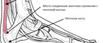
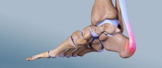
Heel spur
is a disease that is associated with the formation of a bone growth in the area of the tubercle on the heel bone. This is accompanied by an inflammatory reaction, as well as degenerative changes in nearby tissues. In advanced phases, osteophytes or salt impregnation of the damaged area with calcification appear.
Gout
acts as one of the subtypes of arthritis and is associated with metabolic disorders, namely with an excess of purines. Over time, the buildup of uric acid turns into deposits that accumulate in the joints, causing severe pain.
Rheumatoid arthritis
It is considered a systemic disease that affects connective tissue and supporting connective tissue.
The disease has not been fully studied. . Women over 45 years of age
are more susceptible to the disease . Out of 10 cases, only one is affected by a man. The pathological process affects small joints in the feet and hands. If rheumatoid arthritis is not treated promptly, the supporting connective tissue is destroyed. The patient may lose his ability to work, and even become disabled.
Goals of therapy
Treatment of rheumatoid arthritis is aimed at achieving several goals:
Reducing pain, swelling, and other clinical manifestations of pathology.
Preventing deformation and destruction of bone and cartilage tissue, preserving the functional characteristics of the joint, reducing the likelihood of disability, improving the quality of life of patients.
What is the treatment?
- Anti-inflammatory drugs without steroids;
- Glucocorticosteroids;
- Basic anti-inflammatory drugs.
- Anti-inflammatory drugs and anesthetics. Available in the form of an ointment or gel. The products are applied 1 to 2 times in 24 hours to the area affected by inflammation, rubbing in a circular motion.
- Antibiotics. The prescription of drugs depends on the sensitivity of pathogenic bacteria to the main components.
All chemical drugs have a lot of serious side effects
, from provoking stomach ulcers to cirrhosis of the liver.
What should you watch out for?
Postmenopausal osteoporosis
– osteoporosis associated with decreased production of female sex hormones.
Senile osteoporosis
– osteoporosis, associated with general aging and wear and tear of the body, a decrease in the mass and strength of the skeleton after 65 years.
Corticosteroid osteoporosis
– occurs with prolonged use of hormones (glucocorticoids).
Secondary osteoporosis
- occurs as a complication in diabetes mellitus, cancer, chronic renal failure, lung diseases, hyperthyroidism, hypothyroidism, hyperparathyroidism, calcium deficiency, rheumatoid arthritis, ankylosing spondylitis, chronic hepatitis, Crohn's disease, long-term use of aluminum preparations.
Treatment of osteoporosis
Treatment of osteoporosis is a very complex problem, which is dealt with by immunologists, rheumatologists, neurologists, and endocrinologists.
Need to achieve:
- stabilization of bone metabolism indicators,
- slow down bone loss,
- prevent fractures,
- reduce pain syndrome,
- expand physical activity.
Etiological therapy - it is necessary to treat the underlying disease that led to osteoporosis.
Pathogenetic therapy
– pharmacotherapy of osteoporosis.
Symptomatic therapy
– pain relief.
Preparations that stimulate bone formation: fluoride, calcium, strontium salts, vitamin D3, bioflavonoids.
cure
perhaps
impossible
to detect
osteoporosis completely .
You can only improve the condition of the skeletal system with drugs that affect the absorption and absorption of calcium, and with calcium preparations themselves
.
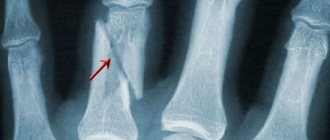
Complications of osteoporosis
Most common fractures
vertebral bodies, femoral neck, radial bones.
According to WHO, hip fractures place osteoporosis in 4th place among all causes of disability and mortality. Osteoporosis reduces
life expectancy
by 12–20%.
the risk of repeated spinal fractures
by 4 times and the risk of hip fractures by 2 times.
Prolonged bed rest contributes to the development of pneumonia, bedsores, and thromboembolism
.
Solutions
Natural products of the Coral Club
Anti-inflammatory, relieving swelling:
Ant tree bark, Coral Boswellia, Licorice, Coral Burdock Root, Papaya, Garlic, Alfalfa, Coral Magnesium.
Products that promote the regeneration and restoration of bone and cartilage tissue:
Inflacore, Flexicor, MSM, Spirulina, Daily Delicious Beauty Shake, Over 30, Calcium Magic, Bi-Luron.
Protectors, bioavailability enzymes and lubricants:
Artichoke, Lecithin, Omega, Assimilator, DigestAble.
Locally externally:
Tea tree oil, Cool relief ointment, Healing balm, Emu oil, body gel.
How they work
The bony surfaces of true joints are covered with special hyaline cartilage, and the joint itself is enclosed in a dense capsule formed by fibrous connective tissue from dense and strong fibers, ligaments and tendons of nearby muscles. This capsule is called the joint capsule. It protects the joint from various external damage (ruptures and injuries). This is the most innervated part of the joint, and therefore has great pain sensitivity. In addition to the protective function, the joint capsule is designed to ensure sufficient sliding of the articulating surfaces of the bone elements relative to each other.
The internal cavity of the joint is lined with a synovial membrane, the cells of which produce a special fluid that “lubricates” the joint and facilitates movement. In the articular cavity of the knee joint there are menisci - cartilage pads that provide it with special shock-absorbing properties.
Joints also have ligaments - strong, dense formations that, on the one hand, make the connections between bones stronger, and on the other, limit the range of motion in the joints, preventing them from becoming loose and loose.
Periarticular tissues located in the immediate environment (muscles, tendons, ligaments, blood vessels and nerves) play an equally important role. They are sensitive to any internal and external negative influences; disturbances in them immediately affect the condition of the joint.
Forewarned is forearmed
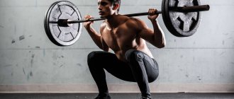
Matveev A.A.: “ They shout about the beneficial effects of sports on every corner, but as a doctor, I consider it fair game to give you counter-examples from our practice.
There are a lot of athletes among our patients. There is a common belief that playing sports is always accompanied by pain, and injuries at the initial stage usually give minimal discomfort and do not interfere with continued training. As a result, athletes dismiss the problem as a normal part of the training process .” As a result of excessive stress, our joints and their “immediate environment” suffer. There are different types of injuries. They can affect ligaments, bones, cartilage, joint capsules and nerves - individually or in combination. Soft tissues are damaged by excessive stretching, twisting, bending, or impact forces and respond with inflammation. The nerves that serve the tissues in turn signal the damage with pain—an unpleasant but absolutely necessary bodily defense mechanism that alerts us to the onset of a serious problem.
But not everyone immediately pays attention to the problem.
This applies primarily to athletes who consider slight discomfort to be a normal part of the training process.
All the moving parts of the body that are used during training are subjected to repeated stress over long periods of time and eventually fail. Grueling, exhausting training, nervous and physical stress, premature aging due to extreme loads, numerous injuries and a whole bunch of occupational diseases. All this is the price of success in sports. Over time, repeated trauma and inflammation take their toll and cause degeneration of the affected structures, leading to chronic pain and dysfunction. At this point, the tissues are no longer restored naturally. Irreversible changes occur in them, they degenerate, and the damage becomes incurable. If the joint is affected, then at the final stage, non-surgical treatment methods can no longer relieve the pain, so often the only option is joint replacement surgery. Overuse injuries include bursitis, strains, impingement (impingement syndrome), inflammation, joint hypermobility, stress fractures, and tendonitis.
Some injuries in athletes have specific names: “runner's knee”, “tennis elbow”. Cyclists face problems in the hip joint, the reason being poor circulation in the hip area. Football players and skiers are more likely than others to experience damage and rupture of knee ligaments. This list can be supplemented and supplemented...
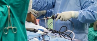
Every second case in our practice is a rupture and damage to the ligaments of the knee joint. In such cases, we perform arthroscopy to repair the knee ligaments.
Muscles and tendons. How to Improve Sports Performance and Avoid Injuries
Translator: Tatyana Arkharova
Editor: Veronica Rees
Source: Bas Van Hooren
Tendons transmit force from our muscles to our bones, and proper interaction between muscles and tendons is very important to an athlete's performance and injury prevention.
When a muscle contracts (tenses), the tendon stretches and remains that way as long as the muscle contracts. When a strong muscle pulls on a “weak” tendon, it can become very stretched (Figure 1). This, in turn, can lead to microtraumas and ruptures in the tendon fibers.
When a tendon is pulled frequently and does not have time to heal, it can eventually lead to injuries such as tendinopathy . As a muscle becomes stronger and larger, the tendon must “adjust” to prevent overexertion and associated damage. Increasing the stiffness of the tendons allows them to stretch less and serves as a protective mechanism.
Strong muscles need tough tendons.
Fig.1 Top images. Left: Imbalance between muscle and tendon. A muscle that strongly stretches a tendon. Right: Balance between muscle and tendon. Contraction of the muscle reduces tension in the tendon. Below: Microscopic images of stretched rat tendons. A - tendons without stretching with smooth, parallel collagen fibers; B - slight tendon sprain characterized by some deformation of the fibers; C - moderate stretching, expansion of the space between the fibers is observed; D - strong stretching, unevenness of fibers, increased space between fibers.
Imbalance due to training
Muscles and tendons adapt to mechanical stress and are sensitive to mechanical stress. The process by which a mechanical stimulus is converted into a biochemical response is called mechanotransduction .
, adaptation occurs . But the timing of adaptation and the mechanical stimuli that cause these adaptations may differ between muscle and tendon tissues. Recent in vivo experiments have shown that high-intensity training leads to adaptations in tendon tissue. It was also shown that a moderate duration of exercise (3 seconds + relaxation) led to better adaptation, as opposed to a shorter duration (1 second + relaxation) or a longer duration (12 seconds).
Therefore, training, especially plyometric low intensity training , can lead to an imbalance between muscles and tendons, and ultimately lead to injury.
Is there evidence of an imbalance?
In a recent cross-sectional study, Mersmann et al found that volleyball players had greater imbalances in the strength of the knee extensor and patella muscles compared to otherwise active people of the same age. The authors suggested that this imbalance may contribute to patellar ligament injury resulting from plyometric training.
A tendon that is too “weak” in relation to a stronger muscle can lead to a tendon injury, but a tendon that is too tight in relation to a weaker muscle can also lead to injury. A stiff tendon is less likely to stretch, such as the Achilles tendon during running.
Sports performance
A “weak” tendon can lead not only to injury, but also to poor athletic performance, as performance decreases due to faster contraction of muscle fibers. As a result, strength indicators are worse. A tendon that is too tight can also lead to poor performance. Therefore, finding the “golden mean” not only reduces the risk of injury, but also has a positive effect on the athlete’s performance.
What is needed for balance?
Imbalances can be avoided with regular strength training. For exercises to be effective for muscles and tendons, they must meet several criteria:
1. Mechanical load:
In vivo experiments indicate that a stretch of about 5% is optimal for training tendon stiffness. These results are consistent with other recent work in which commensurate stretch resulted in the largest increase in phosphorylation.
In both in vivo and in vitro experiments, lighter loads (with less load weight) resulted in less adaptation/phosphorylation. To obtain sufficient stress on the tendon, the muscle must contract strongly. Using a weight greater than 85-90% of the maximum voluntary contraction results in a strong muscle contraction and enough stress (~5%) on the tendon to cause adaptation.
2. Load duration:
With a short duration of loads, for example, as with plyometric training, the adaptation process in the tendon tissues is reduced. In vivo studies show that contraction durations of approximately 3 seconds with a rest period of 3 seconds result in tendon adaptation, indicating efficient mechanotransduction (the process through which forces and other mechanical signals are converted into cellular signals).
Shorter (1 second) and longer (10 second) contractions resulted in decreased phosphorylation.
3. Rest period:
Unfortunately, no in vivo studies have been conducted to determine the optimal rest period between sets. Only in vitro experiments have examined the effects on tendons of repeated training without rest and with a rest period of about 6 hours. Evidence suggests that a minimum of 6 hours of rest is required between tendon workouts.
4. Other factors:
Although the type of contraction—concentric, eccentric, or isometric—is not of primary importance when it comes to tendon adaptation, it is important to consider some of the advantages and disadvantages of different types of training.
With dynamic - concentric-eccentric - training, the tendon experiences heavy loads only for some time. Therefore, it is recommended to increase the duration of the exercise to approximately 6 seconds so that the stimulus is sufficient for effective mechanotransduction. You can also do exercises that put a lot of stress on the tendons, such as bending your knee to 60 degrees while performing a squat.
The advantage of isometric training is that the duration and intensity are easier to control compared to dynamic exercises. The exercises can also be more easily modified to avoid injury to the tendons. Isometric exercises are recommended to be performed 3 times a week with approximately 2 minutes of rest between sets (Fig. 2).
Figure 2. Tendon training
It has been suggested that low mechanical load training, such as calf raises, may lead to an imbalance between muscle and tendon strength because low mechanical load has a greater impact on the muscle than the tendon. A recent systematic review found that high-intensity strength training has potential benefits over eccentric exercise for Achilles tendinopathy, although the effect is small .
Several studies have used relatively long durations of muscle contraction to treat tendinopathy. For example, Rio and colleagues found that isometric muscle contractions reduced pain over the long term in people with patellar . However, recent studies have not found the same effect in patients with Achilles tendinopathy.
Tendinopathy can cause injury, and when the injured tendon is stressed, the healthy tendon tissue “protects” the weaker and injured tissue. Since healthy fibers are strained more, damaged ones do not receive incentives to adapt. This can be solved using the so-called “stress relaxation”. As the intact collagen fibers slowly relax, the damaged tissue becomes more “stressed” and thus adapts.
Gelatin
Collagen fibers under a microscope
Recently, it was shown that taking 15 g of gelatin in combination with ~225 mg of vitamin C one hour before exercise leads to an increase in collagen synthesis compared to placebo. This can be used for injury prevention or during rehabilitation in combination with the previously described exercises.
A recent study of 18 people found that exercise therapy for Achilles tendinopathy produced better results with 2.5 g of gelatin taken 30 minutes before exercise.
We also remind you that hydrolyzed collagen has greater bioavailability. 15 g of collagen hydrolyzate per day is equivalent to 15 g of gelatin, and even more due to better absorption.
Additionally: SMT - Scientific approach produces collagen hydrolyzate of excellent quality in different tastes. One serving contains 5 g of the necessary substance - 3 servings can replace the gelatin with vitamin C described above. You can order collagen at home using the link.